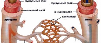
Carotid angiography is an invasive X-ray imaging procedure of the carotid arteries that is performed through a catheter inserted into a blood vessel in the arm or leg. A contrast agent is injected through the catheter, which is opaque to X-rays and therefore shows the internal lumen of the carotid arteries. This procedure is the gold standard for visualization of the carotid and cerebral vessels.
Preparing for the study
- Preparation for the study involves shaving the access site (usually the groin area).
- You should tell your doctor about any allergic reactions you have.
- If you are diabetic, tell your doctor so your medications can be adjusted on the day of the test.
- Tell your doctor if you are allergic to anything, especially iodine, shellfish, X-ray contrast, latex, or rubber products (such as rubber gloves).
- It is advisable to drink enough fluids before the examination to reduce the possible effects of contrast on the kidneys.
In what cases is cerebral angiography contraindicated?
Among the contraindications to this study:
- allergy to iodine-containing contrast agents;
- individual intolerance to the composition of the contrast drug;
- exacerbation of mental illness;
- renal failure;
- acute heart attack;
- chronic decompensated pathologies of internal organs;
- children's age (up to 2 years);
- pregnancy;
- breast-feeding;
- bleeding disorders.
Preparation for cerebral angiography of cerebral vessels
To carry out diagnostics, written consent from the patient is required. The procedure is carried out on an empty stomach after 12 hours of abstinence from food. To relieve tension and anxiety in the patient, a tranquilizer or sedative may be administered.
After research
Tell your doctor or nurses if you feel:
- Itching, dry throat
- Nausea
- Chest discomfort
- Vision problems
- Weakness in one or more limbs
At the end of the study, you will be transferred to a ward. It is necessary to remain in bed for several hours. The doctor will periodically examine the access site and assess the general condition. If there are no problems, then discharge can be made the next day.
How is cerebral angiography of cerebral vessels performed?
The procedure is carried out under the supervision of a team of doctors: a radiologist, an anesthesiologist and a cardiac resuscitator. First, the site of puncture of the vessel is prepared - treatment with an antiseptic and local anesthesia. Then the vessel is pierced with a special needle and a catheter is inserted. After this, a contrast agent is supplied into the vascular bed.
The next step is to perform a series of x-rays or CT scans. The obtained data is interpreted and deciphered by the vascular surgeon together with the radiologist.
Cerebral angiography allows you to determine the location, structure and pathological changes of cerebral vessels.
Complications are unlikely. Usually they concern the introduction of a contrast agent not into the vessel, but into the surrounding tissues. With sufficient experience of the doctor and careful implementation of the study protocol, this risk is minimized.
In the State Clinical Hospital named after. A.K. Eramishantseva widely uses this research method. You can discuss the possibility and necessity of undergoing it with the doctors of the neurosurgery department.
Possible complications of carotid angiography
Complications are rare - the risk is 0.5%. The most dangerous are complications from the brain. They can develop as a result of the effect of contrast or when pieces of plaque are torn off during catheter insertion. Complications include:
- Transient ischemic attack - short-term disturbance of cerebral circulation
- Ischemic stroke is a persistent disorder of cerebral circulation
- Bleeding from the arterial access site
- Carotid artery dissection with thrombosis
- Allergic reaction to contrast
Timely diagnosis of complications allows you to minimize their consequences.
Cerebral angiography of cerebral vessels
The procedure is invasive and involves inserting a catheter into the femoral artery, through which it is advanced to reach the carotid artery. To perform cerebral angiography, a contrast agent is injected into the vessels of the brain. After it is distributed through the arteries and veins, X-rays are taken to show the flow and flow of blood.
Depending on the scope of the study, cerebral angiography can be:
- survey (contrast is injected into the main artery responsible for the blood supply to the entire brain);
- selective (contrast affects a specific area of the brain).
What is it for?
Angiographic research is used to clarify the diagnosis and prescribe the most effective treatment. Also, such an examination is carried out in preparation for surgery, making it possible to determine the location and condition of the vessels. Angiography is combined with endovascular interventions: angioplasty and stenting. Sometimes the decision about the need to implant a stent into a vessel is made immediately during an angiographic examination - in this case, stenting is carried out simultaneously with angiography.
What allows you to diagnose SCA?
SCA allows you to diagnose various circulatory disorders and accurately determine their location.
Let's consider the main pathologies of cerebral vessels, symptoms, treatment with traditional methods, as well as effective folk remedies.
List of the most important disorders in the circulatory system:
- atherosclerosis;
- stenosis;
- arteriovenous malformation;
- aneurysm;
- carotid-cavernous anastomosis;
- dural fistula.
Atherosclerosis
What is cerebral atherosclerosis, its symptoms and methods of treatment.
The formation of fatty and cholesterol deposits on the inner surfaces of arteries, thickening of the vessel walls and narrowing of the lumen, up to complete disappearance, is called atherosclerosis.
Initial signs of the disease:
- headache;
- noise in ears;
- memory loss;
- sleep disturbance;
- psycho-emotional changes.
As the disease progresses, objective symptoms appear:
- movement coordination disorder;
- impaired skin sensitivity;
- ischemic stroke.
In the treatment of atherosclerosis, lipid-lowering drugs are used to help cleanse blood vessels of cholesterol plaques.
All these funds are divided into three main groups:
- Reducing the absorption of cholesterol in the intestines.
- Lowering cholesterol levels in the blood.
- Correcting cholesterol synthesis in the body.
A specialized diet and periodic cleansing of cerebral vessels with folk remedies play an important role in the prevention of atherosclerosis.
This is interesting! Natural honey is very effective. It strengthens blood vessels, stimulates lipid metabolism and promotes the production of high-density lipoproteins, which transport cholesterol from peripheral tissues to the liver.
Vascular stenosis
Stenosis is a narrowing of the vascular lumen. It often develops as a consequence of atherosclerosis, leading to a significant decrease in blood flow speed, restriction of blood supply to certain areas of the brain, stroke or heart attack.
Symptoms that suggest stenosis of cerebral vessels, diagnose and prescribe treatment:
- dizziness and headaches;
- paresis and paralysis of the limbs;
- motor coordination disorders;
- intermittent blindness.
Treatment involves stenting the deformed arteries . In this case, a metal wire tube is inserted into the affected vessel, inflated with a special balloon and expands the walls of the vessel.
Arteriovenous malformation
Arteriovenous malformation (AVM) is a pathological interweaving of intracranial arteries and veins. Characterized by neurological disorders and epileptic seizures.
The walls of the vessels of the malformation are abnormally thin; their overflow with blood in 50% of cases causes rupture and hemorrhage into the surrounding brain tissue.
Treatment is surgical and consists of radical removal of the AVM or complete endovascular exclusion of it from the bloodstream ; Radiosurgical treatment is also possible.
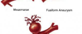
Aneurysm
An aneurysm is an enlargement of a small section of a blood vessel.
By appearance they are distinguished:
- saccular;
- lateral;
- fusiform.
Based on the size of the aneurysm, it is considered:
- small if its diameter is less than 11 mm;
- medium - up to 25 mm;
- giant - over 25 mm.
The danger of an aneurysm is that in the almost complete absence of preliminary symptoms, there is a high probability of its rupture and hemorrhage in the brain.
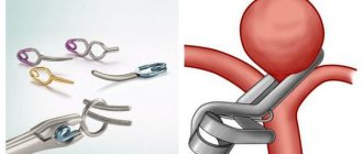
Treatment is surgical. The aneurysm is switched off from the bloodstream in one of the following ways:
- clipping - pinching the neck with a spring clip;
- occlusion - blockage of a cavity with special microspirals or a flow redirection stent .
Carotid-cavernous anastomosis
Carotid-cavernous anastomosis is the flow of arterial blood into the venous system of the cavernous sinus.
More often it has a traumatic origin - the carotid artery is damaged by sharp fragments of bone tissue during a traumatic brain injury. As a result, the venous blood of the sinus mixes with the arterial blood, the pressure in the sinus increases sharply, and venous stagnation occurs.
Treatment is endovascular occlusion of the anastomosis .
Dural fistula
A dural fistula is an abnormal connection of an artery and vein of the dura mater.
The exact etiology is unknown. The following factors are considered as reasons:
- traumatic brain injury;
- inflammation of the epidural space;
- thrombosis of the venous sinuses.
A fistula is dangerous due to vascular thrombosis, rupture and hemorrhage.
Treatment is endovascular embolization .
When is angiography performed?
Angiography is performed before surgery, as a postoperative study and as a diagnostic method for suspected vascular pathologies. If the patient comes with complaints of:
- regular loss of consciousness
- headache,
- frequent dizziness,
- hearing loss,
- numbness of the limbs,
- spontaneous hematomas, MRA cannot be avoided.
The procedure is also prescribed after:
- traumatic brain injury,
- with a sharp drop in visual acuity,
- after a stroke,
- if you suspect vascular disorders of a diabetic nature.
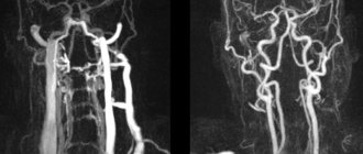
How is MR angiography done?
The procedure takes 40-45 minutes.
Scanning steps:
- The patient is placed on the tomograph conveyor, the limbs and body are fixed with special rollers. Random movements during an MRI session lead to distortion of MR angiography results. The device may produce noise when operating; use headphones for protection.
- After briefing, medical personnel are located in an adjacent room. Communication with the patient is maintained using an intercom. For emergency communication, the patient can use the panic button.
MRI of the neck with MR angiography on a closed tomograph
- For more accurate scanning, a gradient coil is placed above the area of interest, creating an alternating electromagnetic pulse. The table with the subject is moved into the tube of the apparatus, where the constant force field generator is located.
- After a series of native images, the patient is injected with a gadolinium solution. Scanning continues after filling the vascular bed with a contrast agent.
- During an MRI session, angiography of veins and arteries may require holding your breath for 10-20 seconds.
The shooting is carried out in axial, sagittal and frontal projections. Based on the results of MR angiography, a three-dimensional image of the arteriovenous network can be reconstructed. The 3D model helps to clarify the localization of the pathologically changed area. The thickness of the scanned section is from 1 mm.
MRI angiography of vessels is performed using open and closed type devices. The first ones do not have a tunnel module, the power of the tomograph is a maximum of 0.8 Tesla. Such devices are recommended for MRI in patients with claustrophobia and overweight patients.
Closed tomographs provide a magnetic field strength of 1.5 Tesla.
MRI of the neck with contrast MR angiography
High power allows you to make high-quality images of the studied area.
Benefits of MRA
Benefits of MRA
- This diagnostic procedure allows specialists to obtain a clear and detailed image of the blood vessel without the use of catheterization. This means that the risk of arterial injury and complications after the procedure is completely eliminated.
- The time required for non-contrast MRA is significantly shorter than the period for CT or radiography, since no prior patient preparation is required.
- Non-contrast MR angiography costs less than CT examination of blood vessels due to the absence of the need to purchase a contrast agent.
- The method is absolutely safe, while CT and X-ray examinations expose the patient to radiation. The procedure takes place without pain or unpleasant physical sensations, so it can be used for children and people with a low pain threshold.
Methodology for conducting SCA
SCA refers to an invasive test where a radiopaque contrast agent is injected into an artery.
General scheme of conducting SCA
- The arterial puncture site is treated with an antiseptic and numbed.
- A catheter is inserted through the puncture, which is passed into the target vessel, and a contrast agent is supplied into it.
- A series of x-rays are taken.
- The catheter is removed, the puncture site is treated with an antiseptic, and a pressure bandage is applied.
Scheme of selective cerebral angiography
Carotid artery catheterization
Direct carotid angiography involves catheterization of the carotid artery.
The patient is laid down, the head is turned in the opposite direction and thrown back. The puncture point is determined by the pulsation of the carotid artery - it is located between the upper level of the thyroid cartilage and the inner side of the sternocleidomastoid muscle.
Currently practically not used.
Catheterization of the vertebral artery
With direct vertebral angiography, catheterization of the vertebral artery can be performed in different ways.
- Direct puncture of the vertebral artery. An angiographic needle is inserted at the level of the IV–V cervical vertebrae, advanced to their transverse processes and the vertebral artery is punctured between them.
- Puncture of the subclavian artery. In the area of the clavicle, the pulsation of the subclavian artery is palpated, it is punctured with a needle, and a catheter is inserted to the site of the branch of the vertebral artery.
- Puncture of the brachial artery is performed in the axillary fossa. A catheter with a side hole and an internal plug is inserted into the lumen of the needle and advanced to the mouth of the vertebral artery. Then a catheter of smaller diameter is inserted through it on a flexible metal string.
Currently, these accesses are practically not used.
It is important! Since the lumen of the vertebral artery is smaller than that of the femoral and carotid arteries, the contrast agent must be supplied more slowly.
Femoral or radial artery catheterization
More often, selective angiography is performed using an indirect method. In this case, catheterization of the femoral or radial artery is performed. This method makes it possible to diagnose four arteries at once: two paired carotid and two paired vertebral arteries.
For this:
- Prepare the puncture site.
- The femoral artery in the medial third of the inguinal ligament or the radial artery in its distal segment is palpated.
- After inserting the main catheter, it is advanced along the vessel to the aortic arch.
Arterial catheterization technique
When performing SCA, arterial catheterization is performed using the following technique:
- An angiographic needle punctures the skin at an acute angle.
- Move the needle forward until the artery pulsates.
- They pierce the artery with a sharp push, trying not to damage its opposite wall.
- After a stream of blood appears, a conductor is inserted into the needle, secured, and the needle is removed.
- Using a guidewire, an introducer-dilator is inserted into the vein. It is necessary for atraumatic installation of the catheter and is equipped with a three-way stopcock to prevent blood loss.
- The guidewire is removed and the catheter is inserted into the introducer lumen.
On a note! Advancement of the catheter along the vascular bed is absolutely painless, since the inner walls of the vessels are devoid of pain receptors.
Use of contrast agents
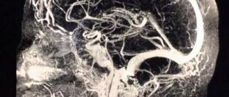
Contrasting brain vessels with 3D reconstruction
Radiocontrast agents are used in angiography to visualize blood vessels.
These are iodine-containing drugs that are used to fill the bloodstream using a catheter, thereby increasing the clarity of the X-ray image of vascular formations.
Iodine-containing preparations are ionic and non-ionic.
- Ionic ones can provoke an allergic reaction, causing an increase in the osmotic concentration of blood plasma.
- Non-ionic ones are much safer, but their cost is higher.
Table 1. Iodine-containing contrasts.
| Compound | Drug name | Active substance | Osmolarity level |
| Ionic | Vizotrust Urografin Verografin Trazograph Diatrizoate | Sodium amidotrizoate | High |
| Ionic | Hexabrix 320 Ioxaglat | Ioxagloic acid | Short |
| Nonionic | Iopamidol Yopamiro Scanlux Tomoscan | Iopamidol | Short |
| Nonionic | Yomeron | Yomeprol | Short |
| Nonionic | Omnipack Iobriz Introvise Iohexol | Iohexol | Short |
| Nonionic | Oxylan Telebrix | Yoxitalamic acid | Short |
| Nonionic | Yopromide Ultravist | Yopromide | Short |
| Nonionic | Iodixanol Visipak | Iodixanol | Short |
| Nonionic | Optiraeus | Ioversol | Short |
One of the mandatory points in preparation for angiography is an iodine sensitivity test . It consists of slowly injecting 2 ml of contrast intravenously and observing the body’s reaction for three to four hours. At the moment, with the use of modern non-ionic contrasts, this procedure is not required.
The examination is canceled if the following symptoms appear:
- skin redness;
- rash;
- itching;
- swelling;
- nausea and vomiting;
- headache;
- cough;
- suffocation.
Before introducing an iodine-containing drug into the catheter, it is heated to body temperature. The rate of administration should be commensurate with the speed of blood flow.
Possible unpleasant sensations include a metallic taste in the mouth, a rush of blood to the face, a feeling of heat throughout the body, especially in the pelvic area.
Remember! All radiopaque agents are nephrotoxic. If renal function is impaired, strict monitoring of creatinine and urea levels is necessary.
Tools needed to conduct SCA
The following set of instruments is used to perform angiography:
- puncture angiographic needle;
- standard conductor with Teflon coating;
- introducer-dilator;
- catheter, sometimes of slightly different diameters.
Angiographic installation
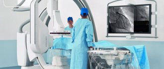
The angiography procedure is performed in the department of X-ray surgical methods of diagnosis and treatment.
The angiographic installation consists of the following complex:
- A table that transmits x-rays and moves in a horizontal plane.
- X-ray tube with electron-optical converter.
- Image recording and playback system.
- Monitor.
A series of x-rays is taken in frontal and lateral projections, at a speed of one or more images per second. Simultaneous recording allows, at the end of the study, to examine each image in detail and decipher it.
CT angiography: what is it?
Multislice computed tomography is a modern method of radiological diagnosis of the pathology of organs and their systems. During the study, several processes take place simultaneously:
- Forward movement of the table with the patient on it;
- Movement of the X-ray tube in a spiral around the subject being examined;
- Registration of an X-ray image by a sensor located on the opposite side and transferring it to a computer screen.
As a result, the doctor receives a series of layer-by-layer images of the examined area of the body, after which the information is processed by a computer and a two- or three-dimensional image is built. It often happens that after a standard study, contrast enhancement of the vessels of the pathological focus is necessary to determine the nature of its blood circulation - then CT angiography of the vessels appears in the diagnostic plan. This study is aimed at obtaining reliable data on the patency of blood vessels, the direction of their course and disturbances in blood flow.
The medical diagnostic center “Medicine of the Northern Capital” uses a modern multislice tomograph Siemens Somatom installed in 2016.
What does this diagnostic method provide?
- Time-of-flight diagnostics allows one to obtain sections of vessels perpendicular to the direction of blood flow.
- The phase contrast method makes it possible to determine the speed of blood flow.
- MPA with contrast enhancement allows you to study the condition of the vessel in more detail by filling it with a contrast agent, and obtain amplitude information about the speed of blood flow.
Deciphering images and making a diagnosis can only be carried out by a specialist who has the necessary knowledge. In the image, tissues and organs of different densities are expressed in different shades of a black and white palette. Seals will appear as white spots in the pictures, and liquids will appear as black spots. The specialist assesses the condition of the vessels and identifies the presence of pathologies, based on the location of the blood branch, the density of individual sections of the area under study, and shape.
The images allow the doctor to identify deviations from the norm such as:
- changes in the diameter of blood vessels, their narrowing as a result of developing arteriosclerosis or vascular spasm;
- deficiency of blood flow, indicating the development of intracranial hypertension;
- change in the position of blood vessels, indicating neoplasm or tissue swelling;
- changes in the structure and structure of the carotid artery;
- expansion of the vessel wall during an aneurysm.
MRA images allow not only to identify pathologies and dysfunctions, but also to determine the extent of the defective area.
How to prepare for the test
Before the study, be sure to inform your doctor about the presence of:
- allergies to strawberries, shellfish, seafood or iodine-containing substances;
- history of bleeding of unknown origin;
- an allergic reaction to a contrast agent in the past;
- pregnancy.
Before the study, it is necessary to exclude food and liquid intake for 4-8 hours. Before the examination, you need to remove all jewelry, decorations and costume jewelry to prevent their contours from interfering with the resulting image.
Indications for prescribing angiography
Despite the significant information content when examining vessels of a particular location, strict indications are needed in order to perform CT angiography. All indications for angiography can be divided into:
- General, which determine the choice of research method;
- Particular ones, characteristic for the study of a certain vascular bed, which we will discuss below.
General indications for angiography include the need to:
- determining the exact location of the pathology;
- clarification of its nature: narrowing, blockage, compression of the vessel, aneurysm;
- calculating the effective lumen of the vessel;
- selection of the best treatment in this particular case: surgical or medicinal;
- assessing the effectiveness of the blood flow bypass.
Contraindications for angiography
Contraindications to manipulation are divided into relative and absolute.
However, if manipulation is necessary for health reasons, almost any contraindication can be circumvented. Limitations to diagnosis are associated with the presence of chronic or acute pathology, which may be aggravated by the administration of a contrast agent. Before angiography, you should tell your doctors if you know you have the following diseases or conditions:
- Acute or chronic kidney pathology;
- Increased thyroid function;
- Pregnancy at any stage;
- Weight more than 150 kg;
- Compounded allergic history;
- Pathology of blood clotting;
- Neurological diseases that do not allow a person to remain motionless while an angiography procedure is performed.
Angiography of coronary vessels
This is a method for assessing blood flow in the coronary vessels, or coronary angiography. Angiography in this case allows you to assess the patency of the arteries that supply the heart. The positive aspect is the possibility of performing diagnostics without hospitalization of the patient and the low invasiveness of the procedure. Angiography of the coronary vessels is possible using computed tomographs that perform 64 slices or more. Indications for CT coronary angiography:
- IHD;
- Various types of arrhythmia;
- Preparation for AV ablation or interventional treatment for additional pathways of electrical impulses in the myocardium;
- Significant disturbance of the electrolyte composition of the blood;
- Malignant arterial hypertension;
- Suspicion of endothelial dysfunction.