Treatment under the compulsory medical insurance policy is possible!
Submit your application
Follow the news, subscribe to our social networks
Details
Aneurysm embolization is a modern method of high-tech treatment of pathological vascular protrusion. Aneurysms are sac-like or spindle-shaped dilation of a vessel with thinning of its wall. The thin vascular wall can rupture and cause massive bleeding, which without the help of surgeons would likely kill the patient. The aneurysm can be cut out, disconnected, or closed. The deliberate occlusion (closing) of the lumen of a blood vessel using a substance (embolus) specially introduced for therapeutic purposes is called embolization.
Embolization is a minimally invasive procedure that is an alternative to surgery. It is indicated in cases where open surgery is very traumatic or fraught with complications. In neurosurgery, endovascular embolization of cerebral aneurysm is increasingly replacing operations with craniotomy.
The goal of aneurysm embolization is to prevent blood flow in the aneurysm sac by filling its cavity with special coils. This should prevent rupture and bleeding. Endovascular embolization of a cerebral aneurysm does not restore areas of already damaged brain.
Materials for occlusion are spirals, special sponges or histoacrylic. To treat an aneurysm, coils or an embolic agent are usually used.
Spirals come in different configurations, lengths and diameters. Structural coils are made of platinum, which, when inserted into the vessel, folds into a three-dimensional shape to fill the aneurysm cavity.
Additional devices, such as a stent, may be needed to help hold the coils inside the aneurysm. Stent intervention involves permanently placing a stent in the vessel adjacent to the aneurysm to provide support that holds the coils inside the sac. The most complex technologies are used for embolization of cerebral aneurysms, because the risks of the disease are very high, and access to them using the open method is very difficult.
Symptoms of cerebral aneurysm
A small protrusion of blood vessels (less than 11-12 cm) has virtually no symptoms. The patient experiences discomfort only when the protrusion of the cerebral arteries has increased in size.
| Type of intervention | Price |
| Endovascular embolization of cerebral aneurysm | 450,000 - 600,000 rub. |
Medium (12-26 mm) and large (from 26 mm) aneurysms are characterized by a number of symptoms.
Among them:
- deterioration of hearing and vision,
- pupil dilation,
- partial numbness of the facial muscles.
Pronounced symptoms of brain damage appear when an aneurysm ruptures.
The patient experiences:
- nausea,
- acute pain in the temples, occipital and frontal parts of the head.
Often the patient cannot speak or swallow. When an aneurysm ruptures, the functioning of the vestibular apparatus is disrupted and vital functions of the body are lost. In addition, spasms and convulsions occur. The patient may fall into a coma. These symptoms are also associated with brain damage.
Important!
Treatment of an aneurysm through surgery should not be delayed. It should be understood that the pathology is very dangerous. The patient can die at any minute. It is extremely difficult to save him, especially if there is no way to quickly get medical help.
If you are diagnosed with a cerebral aneurysm, you should immediately contact a specialist for treatment.
Department of X-ray surgical methods of diagnosis and treatment
What are cerebral aneurysms?
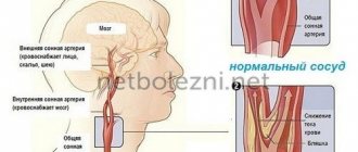
Arterial aneurysms of cerebral vessels are a protrusion of the wall of a cerebral artery in a small area.
Cerebral aneurysms occur in approximately one person in 10,000. Most often, they occur spontaneously and are not congenital. The average age of patients is 40–60 years, but aneurysms can appear at any age.
Why aneurysms are scary The most dangerous complication is aneurysm rupture . This condition is called hemorrhagic stroke , or subarachnoid hemorrhage . Before rupture, aneurysms are most often asymptomatic and are discovered during examination of the patient due to other reasons. The risk of aneurysm rupture is up to 6% per year.
An aneurysm (both before and after rupture) can put pressure on brain structures and cause double vision, facial pain, headaches, and blurred vision. In rare cases, aneurysms can lead to the development of a mini-stroke.
Blood clots sometimes form inside large aneurysms, small parts of which break off and travel through the bloodstream into small vessels of the brain, clogging them. As a result, a small area of the brain is no longer supplied with blood and an ischemic stroke develops.
How are aneurysms diagnosed? To diagnose cerebral aneurysms, computer and magnetic resonance imaging of the brain in angiographic mode are currently used. The accuracy of these methods reaches 96%. For a detailed assessment of the condition of the cerebral arteries, it is necessary to perform an angiographic study (X-ray examination of blood vessels). This method is the most informative for diagnosing an aneurysm. This procedure is carried out in clinics equipped with special X-ray equipment - an angiography unit.
Aneurysm. Results of magnetic resonance angiography
Angiographic examination is performed under local anesthesia. The surgeon performs a puncture of the femoral artery, that is, he makes a small puncture on the thigh and inserts a catheter into the vessel - a thin flexible tube that does not injure the walls of the vessel. By advancing the catheter through the vessels, the doctor inserts it into the vessels supplying blood to the brain. Then a contrast agent is injected into the vessels through a catheter, allowing the arteries and changes in them to be seen under X-rays. The doctor takes pictures of the arteries in several projections.
How are aneurysms treated Once an aneurysm is diagnosed, it is necessary to decide on treatment, since there is a risk of it rupturing. Therapeutic treatments, such as medications, cannot prevent aneurysm rupture.
Currently, there are two main methods of treating cerebral aneurysms: microsurgical clipping and endovascular (intravascular) aneurysm embolization. The goal of both interventions is to turn off the aneurysm from the bloodstream: blood stops flowing into the aneurysm itself, while the blood flow through the remaining vessels of the brain is not disturbed. Once the aneurysm is removed from the bloodstream, it becomes impossible for it to rupture.
Which method to choose is decided individually, depending on the location of the aneurysm, its shape, size, surgeon experience, availability of consumables and necessary equipment.
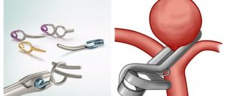
The microsurgical method for treating cerebral aneurysms involves placing a special metal clip on the neck of the aneurysm. This method is reliable for aneurysms located in areas accessible to the surgeon and aneurysms of complex anatomical shape. When localizing aneurysms in hard-to-reach places, it is preferable to choose the intravascular method.
Clipping
Intravascular embolization
Intravascular embolization of cerebral aneurysms is carried out as follows. As with diagnostic angiography, but under general anesthesia, a special catheter is inserted into the vessel carrying the aneurysm. Special microcoils are placed through a catheter into the cavity of the aneurysm. In the aneurysm cavity, the coils coil up, filling it. The number of coils depends on the size of the aneurysm. The operation is performed under X-ray guidance, and the surgeon performing it can evaluate the results from the image on the monitor.
After intravascular embolization of an aneurysm, it is necessary to monitor the degree of thrombosis. To do this, diagnostic angiography is performed after 6 months, 1 and 3 years. If the aneurysm is partially thrombosed or grows, repeated surgery may be necessary.
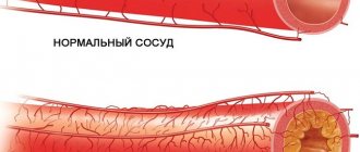
An aneurysm is a characteristic sac-like expansion of a blood vessel (an artery or, less commonly, a vein), resulting from overstretching and thinning of its walls.
Most often, atherosclerosis, arteriosclerosis (a chronic disease characterized by thickening and hardening of arterial walls), hypertension, late stages of syphilis, as well as vascular injuries, infections and congenital defects of the vascular wall lead to weakness of the vascular wall. As a rule, aneurysms do not manifest themselves in any way (asymptomatic) and are discovered accidentally during various studies for other diseases. In other cases, an aneurysm is discovered when a patient presents with symptoms resulting from compression of adjacent organs and tissues by the aneurysm.
Types of Aneurysms Aneurysms can be acquired or congenital. Acquired aneurysms develop as a result of various diseases that disrupt the structure of the vascular wall (atherosclerosis, trauma, vascular infection, etc.). Congenital aneurysms, as a rule, are already present at the time of birth of the child or develop after birth, mainly due to some defects in the structure of the vessel walls.
Based on their location, the following types of aneurysms are distinguished: Cerebral aneurysm is the most common and dangerous form of the disease, characterized by local dilation of the arteries of the brain. Cerebral aneurysms are most often congenital and appear between the ages of 16 and 60 years. According to US researchers, every 17 people are carriers of a cerebral aneurysm. An aortic aneurysm is characterized by an expansion of a certain section of the aorta 2-4 times its original size. An aortic aneurysm can affect any part of this vessel. The diagnosis of “aortic aneurysm” is made to approximately 50 thousand people a year after appropriate studies and 7% of those who die from other pathologies. Just like a brain aneurysm, an aortic aneurysm is asymptomatic in the early stages, but in the later stages, patients complain of pressing pain in the chest or abdomen (depending on the location of the aneurysm).
Development and prognosis of an aneurysm As mentioned above, the symptoms of an aneurysm are determined by its location, size, and relationship to neighboring organs and tissues. However, it often happens that for a long time the aneurysm does not manifest itself in any way and is detected only after the development of complications. Aneurysm rupture is a serious and often fatal complication. The most dangerous are the rupture of an aortic aneurysm and the rupture of an aneurysm of the cerebral arteries. Precursors of rupture of a cerebral aneurysm can be migraine-like headaches and epileptic seizures. When an aneurysm ruptures, the patient experiences a sharp, stabbing headache, which may be accompanied by nausea and vomiting (usually one-time), and the patient may often lose consciousness during an attack.
Most patients who suffer a rupture of a cerebral aneurysm die if they do not receive the necessary help. And 50% of survivors remain disabled for life. An aneurysm is often misdiagnosed as a brain tumor. In such cases, angiography of cerebral vessels is performed to differentiate the two diseases.
Treatment of an aneurysm Treatment of an aneurysm consists of preventing its rupture. If a rupture does occur and the patient remains to live, then prevention is aimed at preventing a second rupture, which, as a rule, is incompatible with life.
Traditionally, vascular aneurysms are treated surgically - clipping (application of special clips to the aneurysm immediately after its separation from the vessel). This treatment method is quite effective and is available in most specialized surgical clinics.
Another method of treating aneurysms is endovascular embolization. Modern studies have proven that endovascular aneurysm embolization is less invasive than the clipping method and allows for better results in certain groups of patients with aneurysms. The essence of the method is to insert a special catheter (a thin tube with a diameter of no more than 2 mm) into the patient’s femoral artery, which is passed to the required vessel (up to the vessels of the brain). Through a catheter, a thin spiral-shaped platinum thread is inserted into the aneurysm cavity, which fills the aneurysm cavity and blocks blood flow in it, which in turn reduces the risk of primary rupture of this aneurysm.
Risk group. Danger of aneurysm
An aneurysm is a disease of the arteries of the brain that is found in various people, regardless of age.
The risk group includes:
- smoking,
- alcohol abusers,
- patients with hypertension, hereditary vascular pathologies.
The insidiousness of an aneurysm is that in the early stages the symptoms of brain damage are almost invisible. It is not easy to notice them, especially for a constantly busy person. At the same time, a relatively small number of people undergo comprehensive examinations of the brain and blood vessels.
Many patients do not pay attention to headaches and nausea. Usually these conditions are associated with chronic fatigue and age.
Important! 25% of patients suffering from protrusion of the wall of a cerebral vessel die.
Request a call back Get a free consultation
Causes
A complete theory of the occurrence of a-me has not yet been constructed, but the factors contributing to the disease have been well studied.
The following factors are distinguished:
- congenital defects of the muscular layer of the cerebral arteries (type III collagen deficiency), in most cases occur at the junction (bifurcation) of the arteries, with strong bends - where the arteries have a complex shape. As a result, the disease is often accompanied by other pathologies: polycystic kidney disease, hypoplasia of the renal arteries, coarctation of the aorta, etc. This factor is hereditary.
- artery damage
- bacterial, mycotic, tumor embolism
- effect of radioactive radiation
- atherosclerosis, hyalinosis of the vascular wall.
The development of arterial cerebral aneurysms can be caused by hemodynamic disorders - high blood pressure, uneven blood flow (change from laminar flow to turbulent flow). This is especially clearly manifested in places where arteries are divided into smaller ones, when impaired blood flow has a constant or periodic effect on an already deformed vascular wall. Constant hemodynamic impact over time leads to thinning, aneurysm formation and rupture of the vessel wall.
Treatment of pathology
Emergency treatment for patients with a ruptured brain aneurysm includes restoring deteriorating breathing and reducing intracranial pressure.
There are 2 main treatment options.
They allow you to fasten the intracranial protrusion of cerebral vessels.
- Surgical clipping.
- Endovascular embolization.
If possible, treatment is given within the first 24 hours after bleeding to close the ruptured aneurysm and reduce the risk of recurrences that affect the blood vessels.
Let's consider the features of both methods.
After the procedure
First, the operated patient is transferred to the intensive care ward, where he spends a day under the supervision of anesthesiologists and resuscitators. There, the patient recovers from anesthesia with continuous monitoring of blood counts, diuresis, respiration, heart function and blood pressure.
The next day, the patient is sent to a regular ward, where he is given infusions to replenish blood loss and correct the electrolyte-water balance. Particular emphasis is placed on pain relief and antibacterial therapy.
The patient is prohibited from drinking for several hours after surgery. This helps avoid vomiting and nausea due to water entering the digestive tract. In general, the patient spends up to 4-7 days in the hospital. The full rehabilitation period covers up to 2-3 months.
Surgical clipping
The goal of the intervention is to apply a special clip to the neck of the aneurysm. This allows you to exclude it from the general bloodstream without blocking a normal vessel. If the aneurysm cannot be clipped, alternative techniques are used (wrapping, tripping, etc.).
Important! The operation to clip the cerebral vessels is performed with craniotomy. Microsurgical techniques are used to carry out the intervention. This allows you to free the aneurysm (its neck) from the feeding vessels. Complications after surgery arise from a previous rupture. Eliminating all its consequences is not so easy. When treating unruptured aneurysms, complications occur less frequently (in 4-10% of cases).
Request a call back Get a free consultation
Why is the procedure necessary?
This intervention helps prevent such a dangerous phenomenon as aneurysm rupture. If you ignore timely treatment, this can lead to death. According to statistics, in 40 out of 100 patients, rupture occurs during the first year of illness.
Consequently, resection saves lives and protects against various complications. In particular:
- prevents digestive tract disorders;
- relieves portal hypertension syndrome;
- prevents organic changes in the digestive system;
- does not allow intestinal ischemia to occur, etc.
Embolization
This technique has become widespread in the last 15 years. It allows you to exclude the damaged vessel from the cerebral circulatory system without opening the skull. A special catheter is inserted through one of the veins or arteries. It moves through the circulatory system until it reaches the aneurysm. After this, using special instruments, the vessel is disconnected from blood circulation.
Embolization is the least traumatic way to eliminate aneurysms. That is why it has become widespread. Efficiency and advantages of treating cerebral aneurysm using modern endovascular techniques Embolization is a method that has proven to be highly effective.
That is why it is often used to disconnect a cerebral vessel from the circulatory system when:
- Inaccessibility of the aneurysm.
- Risk of serious complications with direct intervention.
- Carrying out operations on elderly people.
- Treatment of patients in serious condition.
Intervention is also prescribed when the aneurysm cannot be clipped.
Over the past few years, a large number of interventions have been performed. Moreover, surgery on cerebral vessels was prescribed even for gigantic aneurysms. The majority of patients did not require a long stay in the intensive care unit and were discharged from the clinic within 2-3 days.
There was no damage to any major healthy cerebral vessel. Thanks to this, patients were able to lead a normal life and did not experience any restrictions.
In some patients, the aneurysm was not completely excluded from the bloodstream during one intervention. In this case, another operation was performed. Repeated intervention on the cerebral vessels did not lead to a worsening of the patient's condition.
Main advantages of the method
- Low probability of need for repeated interventions. The operation is performed a second time in approximately 1 case out of 300.
- Possibilities for intervention to stop a cerebral vessel in patients who are contraindicated for extensive surgical clipping.
- Short duration of the procedure. Usually the operation takes no more than 2-3 hours.
- Opportunities for full recovery of health. An aneurysm will not bother you.
5. Immediate improvement. After intervention on a cerebral vessel, patients recover as quickly as possible. Moreover, embolization of cerebral vessels does not prevent them from leading a normal life.