Cerebral aneurysm - symptoms and treatment
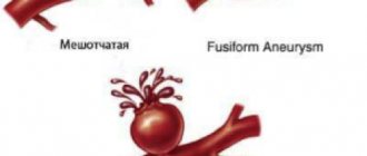
Having an idea of the protrusion of the walls of the cerebral arteries, it is logical to assume that the main principle of treating a cerebral aneurysm is its exclusion from the general blood flow. It sounds quite simple: close the lumen of the aneurysm, thus eliminating the threat of rupture. But in reality everything is much more complicated.
The vessels of the brain are located deep in the skull, dividing into branches, penetrating the brain and enveloping its surface. In combination with the high functional significance of the cerebral arteries, this factor significantly complicates and sometimes makes it impossible to completely shut off the aneurysm, especially in complex forms of the aneurysmal sac.
There are two fundamentally different methods of surgical treatment of patients with cerebral artery aneurysms: open or direct (i.e., through craniotomy) and endovascular (from inside the artery under X-ray control). Both options have their advantages and disadvantages.
In the case of open surgery, the first stage is to dissect the soft tissues of the cranial vault and perform trepanation (opening the cranial cavity). In patients in the acute, acute and subacute periods of SAH, the size of the trepanation window, as a rule, reaches large sizes. In patients with “silent” and “cold” aneurysms, when more than three weeks have passed since the rupture, it is permissible to use low-traumatic keyhole approaches (literally “keyhole”) with a trepanation size of up to 2.5-3.0 cm [11] .
Penetrating into the cranial cavity, the neurosurgeon, using an operating microscope and micro-instruments, opens the membranes of the brain, empties the subarachnoid cisterns (areas of expansion of the subarachnoid space in the area of divergence of the arachnoid and pia mater), washing out the cerebrospinal fluid along with blood clots. As a result, the severity and prevalence of vasospasm decreases.
Subsequently, the CSF flow paths are cleared, which reduces the risk of developing hydrocephalus. Then begins a delicate dissection (exposure) of the cerebral arteries and a step-by-step approach to the aneurysm along the artery on which it is located. In the case of a saccular configuration of the aneurysm, its neck (i.e., the very base) is highlighted. The final stage of the operation is the application of a vascular clip, which compresses the lumen of the aneurysm and stops the blood flow in it. Vascular clips are made from a medical grade titanium alloy and are clamps similar to small clothespins.
A description of the open method of surgical treatment is given in general terms. In practice, each operation on such patients is unique in its own way and requires the surgeon to use a large number of skills and techniques. The advantages of open surgery are visual control and the ability, in most cases, to completely block the aneurysm without leaving cervical areas (zones of growth of a new aneurysm). Also, during the intervention, bloody cerebrospinal fluid is removed, and it is possible to remove intracerebral hematomas if they are present. All this makes the postoperative period easier. The disadvantages of open surgery are traumaticity and the risk of inflammatory complications [2][4][5][10][13].
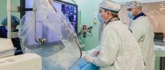
With the endovascular method, the femoral artery is punctured (pierced), an introducer (port) is installed into it, through which conductors are inserted to further advance them exactly to the location of the aneurysm. At all stages of such an operation, an X-ray contrast agent is supplied to the artery, due to which the position of the conductors and the anatomy of the arteries are monitored on the screen of an angiograph (special X-ray machine). Having reached the aneurysm, the x-ray angiosurgeon inserts platinum microspirals into the lumen of the aneurysmal sac, which, twisting, form a ball and tightly fill the aneurysm. Vascular stents fixed on balloons are also used in such operations. The stent is fixed inside the vessel and “switches off” the aneurysm from the bloodstream, taking on the blood pressure.
The advantages of this type of surgical treatment:
- low invasiveness (no need to perform traumatic craniotomy);
- the ability to reach an aneurysm of almost any location.
Disadvantages: high cost of consumables (coils, stents, etc.), lower percentage of radical exclusion of the aneurysm compared to the open method, inaccessibility of this type of operation (carried out mainly in large neurosurgical clinics at the federal level) [3][4][5 ][13].
Medicines
There is no drug treatment for cerebral aneurysm. An aneurysm is, in most cases, a sac-like protrusion of the vessel wall. It is not possible to eliminate this wall defect with any of the drugs. Another thing is to take measures to reduce the risk of aneurysm rupture (provide adequate treatment for hypertension, eliminate bad habits, be observed by specialists, conduct follow-up examinations, etc.).
Principles of first aid for a ruptured aneurysm
If a person experiences symptoms of subarachnoid hemorrhage (which were described earlier), you should immediately call an emergency medical team and briefly describe the main symptoms to the operator. While waiting for the ambulance to arrive, the patient must be laid on a flat surface in a position with the upper part of the body elevated 30 degrees (so that the head is above the level of the heart) - to improve venous outflow from the brain. You should also ensure maximum oxygen flow (open your airways, unfasten your clothes). Apply cold to the area of projection of the carotid arteries (the anterolateral surface of the neck) (any cooled objects at hand, including from the refrigerator) to ensure reflex vasoconstriction and accelerate blood clotting processes. If the patient is unconscious and vomiting occurs, turn him on his side and prevent vomit from entering the respiratory tract.
Is it possible to completely cure a cerebral aneurysm?
Modern methods of surgical treatment of cerebral aneurysms (both endovascular and direct surgical operations) in the overwhelming majority of cases make it possible to completely exclude the aneurysm from the bloodstream.
Rehabilitation after surgery
Rehabilitation of patients who have undergone surgery to disconnect a cerebral aneurysm is carried out to varying degrees and depends on whether the patient was operated on in the acute period of hemorrhage or outside the rupture, whether there were complications during the operation or in the early surgical period.
Depending on the severity of neurological disorders (speech disorders, motor disorders, etc.), classes are prescribed with a speech therapist, exercise therapy instructor, massage therapist, etc. Simultaneously with these activities, medications from the group of nootropics are usually prescribed, vascular-metabolic complex in order to ensure maximum improvement and restoration of lost brain functions.
Can a cerebral aneurysm develop again?
Unfortunately, even after a successful operation to exclude a cerebral aneurysm from the bloodstream, there is a risk of a new aneurysm developing in the same place. This is due to the fact that the wall of the vessel on which the aneurysm formed is sometimes changed not only in the area of the protrusion (aneurysm) itself, but has a defective structure in the sections adjacent to the aneurysm. The degree to which the aneurysm is turned off during surgery is also important, since preservation of even a small fragment of the aneurysm neck can subsequently lead to the formation of a protrusion in this obviously weak spot of the vessel.
Are folk remedies used?
Folk remedies for the treatment of cerebral aneurysm are ineffective. Lack of timely adequate treatment for a ruptured aneurysm can lead to the death of the patient.
Causes
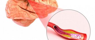
The main link in the pathogenesis of the disease is the thinning of the layers of the vascular wall. The wall cannot resist and adapt to blood pressure and begins to stretch. The stretching accelerates due to the filling of the cavity with blood, and a vortex blood flow is formed.
The reasons that trigger the process can be various diseases:
- inflammatory diseases of the membranes of the brain, meningitis lead to the destruction of the layers of the artery wall, it becomes less plastic;
- damage to the skull and brain;
- the presence of a focus of infection, which may not be located in the brain, for example, syphilis, infective endocarditis (the pathogen enters the skull with the general bloodstream);
- high blood pressure leads to thinning of arterial vessels;
- diseases with impaired immune response - systemic lupus erythematosus;
- dyslipidemia, which leads to the formation of atherosclerotic plaques in the body;
- neoplasms that can grow into the head arteries, but may not be located near the brain and damage the vascular wall with their decay products.
About the prognosis of the disease
If it is not possible to perform the operation, the prognosis will definitely be unfavorable. Although there is data about patients who lived long and prosperous lives with an aneurysm and died from other diseases. Single congenital aneurysms may disappear on their own over time, however, the risk of re-formation remains high.
The most favorable prognosis can be considered in the presence of a single formation, small in size, and also when an aneurysm is detected in a young patient. The prognosis is worsened by the presence of concomitant diseases and the presence of congenital connective tissue pathology. The overall postoperative mortality rate is 10-12%.
Diagnosis of aneurysm
In our department of vascular surgery, experienced phlebologists perform high-quality diagnostics of all forms of aneurysms using modern instrumental techniques:
· ventriculography and angiography are invasive research methods that involve the introduction of contrast into the blood vessels in the affected area. Such studies can detect vascular aneurysms, their dissection, signs of rupture or thrombosis;
· ECHO of the heart, ultrasound of the arteries – used to diagnose aneurysms located outside the brain;
· Ultrasound examination of blood vessels, Dopplerography;
· CT with contrast - allows you to identify signs of aneurysm dissection.
Signs of bleeding or arterial thrombosis can also be detected by laboratory blood tests.
Preoperative preparation
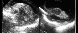
cerebral aneurysm in the picture
During planned aneurysm clippings, specialists have time to thoroughly examine the patient and prepare him for the intervention. As conservative therapy, antihypertensive drugs are prescribed, drugs that normalize heart rhythm in case of arrhythmias, and correction of the lipid spectrum is carried out in the presence of abnormalities.
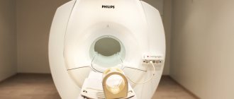
Before planning the operation, the patient undergoes all kinds of examinations, including blood tests, urine tests, coagulogram, cardiogram, etc., as with other surgical interventions. To localize and clarify the nature of the vascular formation, CT, MRI with contrast, angiography, and ultrasound with Doppler are performed.
In the case of ruptured aneurysms, the patient is admitted to a hospital with a clinic for acute subarachnoid or intracerebral hemorrhage and is sent to the neurosurgical department; there is virtually no time for examinations, so one has to limit oneself to the minimum that allows one to determine the location of the malformation.
Both trepanation and endovasal surgery require general anesthesia, although in the latter case local anesthesia may be used. Before the operation, the patient talks with the surgeon and anesthesiologist (except in cases of coma and acute bleeding), does not eat for the next 8 hours before the operation, and tries to get enough sleep. The hair at the trepanation site is shaved.
Rehabilitation of patients
If the patient suffered a rupture of an aneurysm and survived, or when he underwent surgery to remove it, he needs to undergo a rehabilitation course.
It includes three areas:
- Treatment by position using special splints. This rehabilitation method is necessary for paralyzed patients. It is carried out in the early stages.
- Massage performed by rehabilitation specialists.
- Heat treatment. In this case, applications with clay and ozokerite are used.
It is possible to supplement the rehabilitation course with physiotherapeutic procedures, which are selected individually and largely depend on the patient’s condition.
Postoperative period
After surgery, the patient may experience consequences from removal of a cerebral aneurysm:
- noise in ears;
- loss of vision or loss of visual fields;
- incoordination;
- dizziness and frequent headaches;
- anxiety, malaise, increased irritability;
- fast fatiguability.
The most common problem in the postoperative period is thrombus formation. After applying a clip or suture to a vessel, atherosclerotic plaques may settle, which will close the lumen of the vessel and lead to cerebral ischemia.
Also, in the early period after surgery, the sutures may come apart, which threatens hemorrhage into the medulla or subarachnoid space.
Symptoms
At the initial stages, the pathology may not manifest itself in any way. We are talking about the so-called asymptomatic aneurysm. As it increases, a characteristic neurological picture appears:
- headache;
- loss of sensation in the lower extremities;
- violation of fine motor skills, diction;
- numbness of the face, weakening of the facial muscles, loss of control over facial expressions.
Impaired cerebral circulation changes character and behavior. The person becomes restless and emotionally unstable.
When an aneurysm ruptures, a severe headache occurs, which is accompanied by nausea and vomiting, and pallor. The patient experiences convulsions and develops photophobia. Due to impaired cerebral circulation, a person loses orientation and falls into an unconscious state. If there are signs of aneurysm rupture, you should urgently call an ambulance and take the victim to the neurological department.
