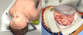
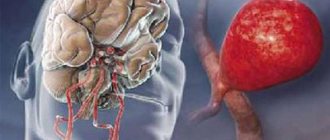
Plastic surgery of cranial bone defects is a surgical intervention used to correct and eliminate congenital and acquired anomalies of the skull. Such operations not only eliminate cosmetic defects, but also prevent the development of severe brain disorders. Their implementation is justified in the first months of a child’s life or in adulthood, if indicated. Depending on the situation, plastic surgery may involve excision or fragmentation of the cranial bones, displacement of individual blocks to ensure the correct shape of the skull and the position of the bony structures of the facial skeleton.
You can undergo diagnostics and plastic surgery of skull bone defects in Moscow at the neurosurgery department of CELT. Our multidisciplinary clinic has been operating in the capital's paid medical services market for more than two decades and offers treatment in accordance with international standards. By contacting us, you can be sure that you will be in the hands of leading domestic specialists who will help solve your problem.
Main symptoms and indications for surgery
Seeking professional help is important for any type of defect.
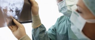
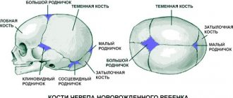
They are classified by size (small - up to 10 sq. cm, medium - up to 30 sq. cm, large - up to 60 sq. cm, extensive - more than 60 sq. cm) and by localization (occipital, parietal, frontal, temporal, combined ). In the Department of Neurosurgery of the City Clinical Hospital named after. Eramishantsev (Moscow) cranioplasty is prescribed only after a comprehensive diagnosis using CT, MRI, radiography, and determining the nature of the problem. The main symptoms indicating the possible need for surgical intervention include:
- headaches and local pain in the area of the defect, which arise as a result of changes in weather and even air temperature;
- protrusion of the contents of the skull during physical exertion, sneezing, coughing or sudden movements of the head;
- memory and sleep disturbances, emotional agitation, difficulty performing intellectual tasks and concentrating;
- disorders of the cardiovascular system and problems with blood vessels of the neuro-endocrine system.
Experts include the elimination of cosmetic defects and sealing of the cranial cavity as indications for cranioplasty. In the second case, at least two goals are pursued: to normalize cerebral hemodynamics and liquorodynamics, to protect the brain from the effects of external factors.
Plate after craniotomy
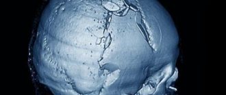
On November 20, 2021, as a result of an accident (through no fault of my own), I received a “TBI.” Depressed comminuted fracture of the vault and base of the skull. IF, SCHYA on the left. Moderate brain contusion. SAC. Contusion of the soft tissues of the head. 11/21/16: resection craniotomy, removal of fragments of a depressed fracture, acute epidural hematoma, focus of a crushed brain contusion. Syndrome of aphasic disorders, right-sided pyramidal insufficiency with severe impairment of communication function.” After which he underwent rehabilitation there in KGB No. 3.
After discharge from the hospital, he underwent outpatient treatment at the local clinic (City Clinical Hospital No. 6) and was observed by a neurologist.
In February 2021, MSEC assigned disability group 3 (working). Due to the injuries I received and my state of health, I was unable to resume my previous job after rehabilitation.
In July 2021, an operation was performed to install a plate (elimination of a defect in the cranial vault).
Constant visits to doctors and various examinations also continued.
In February 2021, MSEC was re-appointed, as a result of which the 3rd (working) disability group was removed, and Kuzmichev I.A. recognized as fully operational, i.e. disability has not been established.
In October 2021, the plate was removed due to the occurrence of a “fistula”.
In March 2021, MSEC assigned disability group 3 (working).
On October 23, 2021, a repeat operation was performed to install a plate (elimination of a defect in the cranial vault)
Constant visits to doctors and various examinations also continue.
The psychosis has not decreased; I see a psychotherapist on an ongoing basis. In connection with all of the above, it is not possible to get any job, especially in your specialty.
Visually: the facial nerve is disturbed (internal discomfort on the left side of the face, copious discharge of mucous fluid from the nose, especially noticed during emotional changes, dryness of the left eye)
impairment of cognitive functions (deterioration of memory processes and concentration);
partial memory loss
congestion and tinnitus
deterioration of the respiratory system (congestion in the nasal vessels, the bronchi do not cough up mucus, a feeling of stagnation of mucus accumulated both in the chest and in the back symmetrically from the spine)
development of constant chronic headaches accompanied by congestion of the same mucous fluid flowing from the nose
frequent heart pain turning into numbness on the left side of the body (left hand up to 3 fingers “from the middle to the little finger” and the vascular vein of the left leg)
uncomfortable pain under the cheekbone when opening the jaw, spreading to the temporal part with a feeling of curvature of the left lower jaw, when opening the left eye closes
Please tell me what legal norms or law can preserve the disability group when undergoing the next re-examination at MSEC?
Features of the operation
Surgical intervention should not be started when inflammatory processes are detected in the cranial cavity, in the meninges or brain. Contraindications also include noticeable mental abnormalities of the patient, the presence of a piece of bone tissue or a foreign object in the cranial cavity, and bulging of the brain in the problem area. If visual examination and diagnosis do not reveal any of the listed points, surgery is prescribed.
Skull plastic surgery after trephination and other similar manipulations are called cranioplasty. It is aimed at restoring the integrity of the skull after decompression and stabilization operations, gunshot wounds, falls from a height, and mechanical damage. The choice of intervention technology depends on the timing of the plastic surgery, and it can be primary (1-2 days after injury), primary delayed (up to 2 weeks), early (up to 8 weeks), late (more than 2 months).
There are several rules that must be followed during any type of cranioplasty surgery:
- maximum possible preservation of bone tissue elements;
- use of separated fragments as an autograft;
- strict correspondence between the plate size and the size of the closure area;
- absence of sharp and too thin parts of the skull.
Elimination of a skull defect is a rather complex and responsible operation, the first step of which will be excision of the meningeal scar. All manipulations are performed as carefully as possible to avoid trauma to the brain tissue. For good adhesion of the skull bones and the implant, the edges of the entire problem area are exposed, and the material is necessarily installed “flush” with the skull bones. To prevent such a negative phenomenon as graft displacement, reliable fixation is carried out. Conventional soft tissue suturing is not enough.
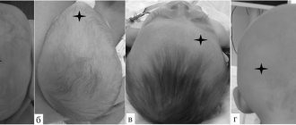
A flat back of the head, sloping forehead, and flattening of the skull can be the cause of dissatisfaction with one’s own appearance. Can plastic surgery change and improve what is given by nature? The answer is obvious: having a beautiful head shape has now become possible thanks to aesthetic cranioplasty. The cherished dream of many people was solved with the help of three-dimensional technologies used in Dr. Guryanov’s VIP-Studio 3D surgery.
Now, thanks to precise three-dimensional modeling and minimally invasive plastic surgery technologies, it has become possible to correct the shape of the skull, changing the shape of the forehead or back of the head, and get rid of asymmetries.
Aesthetic cranioplasty allows you to:
- change the shape of the forehead, back of the head
- eliminate flattening on the surface of the skull
- eliminate moderate asymmetry of the skull in different areas
- correct deficiencies after reconstructive cranioplasty
Restorative (reconstructive) cranioplasty is designed to help people who have suffered a skull injury. The main function of such an intervention is to save the patient’s life. However, after reconstruction, a person may experience undesirable changes in his appearance in the area of the operation. Aesthetic cranioplasty can solve these problems: it allows you to eliminate irregularities, achieve symmetry in the shape of the skull and restore lost details of the bone structure.
Dissatisfaction with one's own appearance can cause numerous complexes and a decrease in the quality of life. Women especially suffer from this, for whom aesthetic cranioplasty can be a real salvation.
It often happens that attempts to disguise a sloping forehead or an ugly back of the head with a hairstyle simply do not work. For example, in men who suffer from lack of hair on their heads. This is where aesthetic cranioplasty comes to the rescue. The tiny incision through which the flexible silicone implant is installed is invisible even on a hairless head.
Significant changes in the shape of the skull are now possible without major osteoplastic surgery with minimal incisions and minimal rehabilitation after surgery by installing a custom-made silicone implant that will take into account all your wishes regarding the shape of the head.
During a consultation with Dr. Guryanov, together with the artist, a three-dimensional computer modeling of the desired shape of the forehead or back of the head is performed, then, based on the data obtained and a computer tomogram, a virtual three-dimensional model of the implant is calculated. The custom silicone implant is manufactured using high-precision 3D printing technology.
The operation lasts an hour to forty minutes. Aesthetic cranioplasty can be performed on all patients over 18 years of age. The maximum rehabilitation period is one and a half weeks.
An individual implant is made of medical grade silicone for long-term implantation. This silicone is non-toxic, completely safe and FDA approved. An individual implant is installed on the bone through a small incision in the scalp and straightened. VIP-Studio guarantees the safety of the implant after installation for 300 years!
Implants manufactured in VIP-Studio have a unique micro- and macro-texture developed by our specialists. The texture promotes integration of the implant with surrounding tissues and prevents excessive growth of the capsule around the implant. When installing the implant subperiosteally (on the bone), there is no disruption of the nutrition of the hair roots and scalp, since the vessels responsible for the blood supply are located in the soft tissue mass and cannot be damaged during the formation of the implant bed.
Aesthetic cranioplasty is not the only method for correcting the shape of the head. Our VIP STUDIO takes a comprehensive approach to achieving the patient’s desired appearance:
- pronounced brow ridges can be eliminated using
, - Recession of the temples and atrophy of adipose tissue in the forehead area can be compensated by introducing one’s own fat into the necessary areas (lipofilling).
Types of implants for plastic surgery
If you believe historical data, a description of materials for plastic surgery of a trepanation defect first appeared in the medical literature back in 1565. At that time, the choice of specialists settled on gold plates. Modern doctors have a significantly increased choice of materials. Main types of implants:
- Autografts. This involves the use of patient tissue, which is taken during the initial operation. It is possible to produce material of the required size from the bone of the cranial vault or rib. If the problem area is small, preliminary crushing of bone fragments is performed to achieve the desired size.
- Allografts. In this case, the doctor uses materials that have been canned, frozen, or decalcified. Materials refer to bones and dura mater taken from corpses and carefully processed.
- Xenografts. This type of transplant refers to materials taken from animals. This group also includes inorganic materials, implants made on the basis of hydroxyapatite (a mineral from the apatite group, an analogue of chlorapatite and fluorapatite).
When choosing, it is important to consider the following requirements: biocompatibility, plasticity, resistance to mechanical stress, no risk of infectious complications, compatibility with modern neuroimaging methods.
Today, such a service as skull plastic surgery with a titanium plate is quite popular. It is possible to manufacture a graft from titanium alloy, stainless steel, chromium- and cobalt-based alloy. But practice shows that the most suitable option is the patient’s own bone tissue. Therefore, the task of reconstructive neurosurgery specialists is to preserve skull fragments as much as possible during the primary operation.
Depending on the situation in the neurosurgery department of the City Clinical Hospital named after. Eramishantsev's methods are used: the uatoplasty method using a pedicle graft, free autoplasty, bone-saving method, adhesive osteosynthesis, grinding of fragments with subsequent placement on the dura mater.
Methodology for plastic surgery of skull bone defects
Plastic surgery is performed under general anesthesia. The technique for carrying it out is selected based on the type of defect.
| Type of plastic surgery | What is? |
| Linear craniotomy | Plastic surgery involves partial removal of bone structures along the ossified cranial suture. During the process, the neurosurgeon cuts the tissue parallel to it, spreads the edges of the wound and removes part of the bone in the form of a strip 20 mm wide. In order to eliminate the risk of damage to the sagittal sinus, the bone in its area is bitten off. A material is applied to the edges of the bones to prevent their regeneration and fusion to prevent relapse. The final stage is suturing the soft tissues. |
| Circular craniotomy | This plastic method involves cutting the scalp tissue in a circle in the frontal and occipital areas along the border of the hairline. In areas above the temporal arteries, dissection is not performed. Removal of a bone strip 10 to 15 mm wide also occurs in a circle. The edges of the bone are processed to prevent fusion, and then sutured through the wound. |
| Bilateral flap craniotomy | This method of plastic surgery involves creating an arcuate incision in the soft tissue along the border of the scalp along the midline of the head. The resulting skin flap is turned away, after which holes are made in the temporal, parietal, frontal and occipital bones, which are connected by biting off a bone strip 15-20 mm wide. Thus, the neurosurgeon forms a bone flap, which is divided into two parts. After two to three weeks, the same plastic surgery is performed on the other side of the skull. |
| Cranioplasty | If there are defects in the bones of the arch, the use of an implant is required, which can be made from the patient’s own bone tissue or from metal, methyl methacrylate. The manufacturing itself is carried out in advance or during the plastic surgery. Its installation is carried out in such a way as to guarantee maximum tight fit to the edges of the hole and its sealing. |
You can eliminate complications and achieve the best results from plastic surgery by contacting CELT. We employ leading domestic neurosurgeons. You can make an appointment with them online through our website or by contacting our operators: +7 (495) 788 33 88.
Make an appointment through the application or by calling +7 +7 We work every day:
- Monday—Friday: 8.00—20.00
- Saturday: 8.00–18.00
- Sunday is a day off
The nearest metro and MCC stations to the clinic:
- Highway of Enthusiasts or Perovo
- Partisan
- Enthusiast Highway
Driving directions
Possible complications after surgery
If the patient’s own biomaterial is used during the operation, then the risk of any exacerbations will be minimal. The recovery period in this case will last less than with the installation of alternative grafts. Possible consequences of cranioplasty after trepanation:
- bleeding (reducing blood loss is achieved through infiltration of the skin);
- development of purulent-inflammatory processes (most often due to rejection of an artificial implant);
- infection of the problem area (the main reason is insufficient treatment of the operated area with disinfectants).
The capabilities of modern neurosurgery, a wide variety of surgical methods and materials make it possible to eliminate defects in the skull of any shape, size and level of complexity without consequences.
Are you interested in the price of skull surgery? Call our clinic and make an appointment with a neurosurgeon. Contact phone number. All further actions can be learned only during a face-to-face consultation.