The sutures of the skull in newborn children are non-ossified parts of the skeleton, which consist of remnants of connective and membranous tissue. The fontanelles are characterized by increased mobility, as well as elasticity, which manifests itself under the influence of compression pressure.
The soft sutures of the cranium contribute to the safe deformation of the head bones during the physiological growth of the brain, as well as during the passage of the child through the mother’s birth canal. The presence of fontanelles in newborn children indicates the full development of the skull.
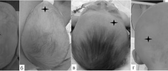
The fontanelles of a newborn baby and their location
Skull sutures in a newborn child are residual signs of preservation of the membranous skeleton, which makes it possible to compress the bones of the baby’s head without the risk of damaging brain tissue, as well as the walls of blood vessels. The table below shows the types of fontanelles, as well as their location in the overall structure of the skull.
| Type of fontanel | Location of skull sutures in newborns |
| Rear | The posterior fontanel consists of connective tissue, which is located at the junction of the parietal and occipital bones. These skull sutures heal completely within 1-2 months. after childbirth, subject to adequate nutrition and physical development of the child. In cases of birth trauma or congenital genetic abnormalities, closure of the posterior fontanel may occur over a longer period of time. After complete fusion of this suture of the skull, solid bone with a sufficient level of density and mineralization is formed at the location of the connective tissue. |
| Front | The anterior fontanel is the largest and is located directly in the area of the junction of the parietal and frontal bones. The fusion of this seam occurs by the age of 2 years of active growth of the child. In children diagnosed with cleidocranial dysostosis, closure of the anterior fontanel may occur in a later period of their physical development, or this area of the infant’s skull remains soft all the time, which is considered a severe pathology of physical development. |
| Wedge-shaped | The sphenoid fontanel is a lateral suture of the skull, which is located between the frontal, sphenoid, temporal and parietal bones. This section of the skull is shifted closer to the front of the head. |
| Mastoid | The mastoid fontanel is located slightly posteriorly at the junction of the occipital, parietal, and temporal bones. This segment of the skull helps alleviate static pressure on the back of the skull. |
Sutures of the skull in a newborn
Sutures of the skull in a newborn child are located symmetrically in relation to each other. Each of the above fontanelles has its own timing of fusion with the transformation of connective tissue into full-fledged bones of the cranial vault.
Normal size of the fontanel
The normal size of the fontanel has its own range, which varies and can be individual for each newborn. However, there are generally accepted dimensions that you can rely on.
Dimensions of a large fontanel:
| Interval | Size |
| 0 -1 month | 25 - 28 mm |
| 1 - 2 month | 25 - 28 mm |
| 2 - 3 month | 23 - 24 mm |
| 3 - 4 month | 20 - 21 mm |
| 4 - 5 month | 18 - 20 mm |
| 5 - 6 month | 18 - 20 mm |
| 6 - 7 month | 16 - 17 mm |
| 7 - 8 month | 16 - 17 mm |
| 8 - 9 month | 14 - 15 mm |
| 9 - 10 month | 12 - 14 mm |
| 10 - 11 month | 9 - 12 mm |
| 11 - 12 month | 5 - 8 mm |
Dimensions of a small fontanel
| Interval | Size |
| 1 - 3 month | 5-7 mm |
The mastoid and sphenoid fontanelles close almost immediately after birth, and in some cases, even during intrauterine development.
Functions of the soft crown
At different stages of a newborn’s life, the fontanelles of his skull perform the following physiological functions:
- soften the static pressure on the skull, which is formed when the child passes through the mother’s birth canal;
- provide elasticity to the tissues of the skull for the rapid development of all parts of the brain;
- minimize the risk of birth trauma, damage to the blood vessels of the head and the walls of the birth canal.
The main purpose of the soft crown is the safe birth of a child while maintaining the full functions of the central nervous system. At the same time, the newborn’s brain develops several times faster than the formation of bone tissue. In this regard, a sufficient amount of free space must be maintained in the cavity of the baby’s cranium at all times.
What function does the fontanelle perform?
- The fontanelles act as shock absorbers, which are located between the bones of the newborn’s skull and protect the brain from possible injuries.
- The presence of fontanelles contributes to the normal and unhindered passage of the fetal head through the birth canal. The fact is that the fontanelles allow the baby’s head to “fold” and “shrink”, this helps the child to be born without injuries and concussions.
- A large fontanel serves as an indicator of the presence of pathologies. If it is swollen or, conversely, sunken, one may suspect pathology or a developmental disorder.
- The baby’s brain actively grows throughout the first year of life, and the fontanelles help the bones of the skull cope with the load.
- The fontanelle helps to “cool” the brain when the baby’s temperature rises.
- In the first year of life, a child actively gets acquainted with the world; falls and blows are not uncommon. The fontanelle suppresses the force of shaking.
- The fontanel helps assess the physiological development of the child.
Features of the structure of the skull of newborns
The sutures of the skull in a newborn go through certain stages of their formation. At each stage of the fontanelle’s existence, qualitative changes occur in the structure of their tissues.
Cartilaginous stage
The cartilaginous stage of fontanel formation is characterized by the gradual replacement of membranous areas of the skull with denser connective tissue in the form of cartilage. These areas of the skull are still distinguished by a rather soft structure, but at the same time their elasticity is no longer so pronounced.
Depending on the type of fontanel, the transition to the cartilaginous stage of the formation of the sutures of the skull can begin at 2 or 6 months. child development. Fontanas with this structure require careful care, and the newborn must be ensured complete safety while minimizing the risk of damage to the still soft bones of the skull.
The cartilaginous stage of development of the fontanelles ensures the implementation of the following processes, covering all structural elements of the cranium:
- the skull is still quite elastic, but its seams are more protected from the negative effects of environmental factors;
- the edges of the bones of the skull become overgrown with denser connective tissue;
- Preparations are being made for the transition of the process of skull formation to a new stage with the future closure of its vaults.
Going through the cartilaginous stage of cranial development is a prerequisite for the full formation of the skeleton of a newborn child. At this stage of growth, the baby should be provided with high-quality nutrition with mother's milk or formulas containing a high percentage of calcium, phosphorus, and proteins.
Bone displacement
The function of bone displacement is a physiological feature of soft fontanels, the presence of which ensures the mobility of individual parts of the skull under the influence of static load. As the skull develops and the newborn child grows, its skeleton is formed.
The fontanelles gradually become overgrown with cartilaginous tissue, which becomes less and less elastic. At this stage, a methodical displacement of the skull bones occurs, transforming its anatomically correct shape. This process continues throughout the entire period of physical development of the infant and covers all parts of the skull.
Postnatal period
The postnatal stage of fontanelle closure continues throughout infancy and early childhood. During this period of time, the sutures of the skull go through all stages of their development from the moment when they consisted exclusively of membranous tissue to their transformation into the state of cartilage with further displacement and the formation of full-fledged bone joints.
The postnatal stage covers the entire period of extrauterine physical development of the newborn. At this stage of the child’s formation, it is necessary to periodically conduct preventive examinations with a pediatrician in order to promptly detect signs of pathological abnormalities in the process of closing the soft fontanelles.
Final stages
The sutures of the skull in a newborn go through all stages of their development, and then close with the bone tissue of the skull. This is the final stage of the formation of the skull in children, upon completion of which the child retains dense sutures of several types.
Gender characteristics
The sutures of the skull in a newborn male child are characterized by faster fusion with the transition to the stage of formation of cartilaginous tissue, as well as further closure with the main part of the skull. In girls, these processes proceed much more slowly.
This gender-specific feature of slower development of skull bones in children of different genders is explained by differences in the hormonal background of their body.
There is less estrogen in the blood of boys, which slows down the process of ossification of the sutures of the skull. In girls, the period of complete closure of the fontanelles slows down on average by 1-2 months, but there are exceptions when the closure of the sutures occurs as quickly as in male infants.
Closing dates and what they depend on
The table below shows the average timing of fontanelle closure in newborns at different stages of their physical development.
| Name of the fontanel | Time frame for complete fusion of cranial sutures from the moment of birth of the baby |
| Rear | Over a period of 1 to 2 months. |
| Wedge-shaped | Complete closure of the sutures of this part of the skull is observed after 6 months. after the birth of the child. |
| Front | The anterior fontanelle heals after 9-18 months. depending on the characteristics of the individual development of the newborn. |
| Mastoid | The cranial suture of this type heals no earlier than after 6-18 months. from the birth of the baby. |
The timing of complete closure of the soft cranial sutures depends on the following factors, which have a direct impact on the process of formation of the bones of the newborn’s skull, as well as other parts of his skeleton:
- quality of nutrition (in children who consume exclusively mother's breast milk, overgrowth of fontanelles occurs 2 times faster than in infants fed with artificial formula);
- hereditary predisposition to faster or slower closure of the sutures of the skull;
- genetic anomalies of intrauterine and postnatal development, when the full formation of the skull is impossible due to poor heredity;
- the birth of a child ahead of schedule (in this case, the time of overgrowth of the fontanel is shifted due to the early onset of extrauterine development of the baby);
- disruption of metabolic processes in the body, in which there is no complete absorption and distribution of nutrients throughout the skeletal tissues;
- chronic vitamin D deficiency;
- concomitant diseases of the digestive system, which are characterized by incomplete absorption of mother’s breast milk;
- too intense growth of the newborn child (the rapid physical development of the baby at the postnatal stage can lead to early closure of the fontanel, which is also not always a deviation from the norm).
In order to exclude the factor of abnormal development of the cranium in newborn children, it is necessary to regularly visit a pediatrician who examines the condition of the fontanelles of the cranium. In this case, the factor of the presence of individual characteristics of each child’s body is taken into account.
What should a newborn's fontanel look like?
A small dimple on the crown of the child - the fontanel - performs an important task during the birth of the baby. And even after birth, she is assigned a serious role, and along with this, special attention from mothers and doctors.
Fontanas are areas at the junctions of the cranial bones, covered instead of bone tissue with soft elastic membranes. Thanks to them, the baby's head is plastic and during childbirth can adapt to the curves of the mother's pelvis. The volume and size of the baby’s head decreases at the time of birth, which helps protect both the baby’s brain and the mother’s organs from damage.
There are six fontanelles in total, but in full-term babies at the time of birth, as a rule, only one remains open, in the area of the crown - the so-called large fontanel. Normally, its size ranges from 0.5 to 3 cm, and its shape resembles a diamond. After birth, it helps the baby adapt to a changing external environment: maintain body temperature, regulate fluctuations in intracranial pressure.
We have been involuntarily trying to get around this large fontanel all year, when we stroke the child’s head, take off his cap, and comb it. Just under the skin, thin and shiny, there is a strong but elastic membrane, which will later be replaced by bone, and beneath it a fairly large vein pulsates. It is she who swells, transmitting vibrations of the arteries and heart when the baby cries, screams or takes a deep breath.
The large fontanel overgrows gradually and finally closes between 6 and 18 months. When exactly this happens depends primarily on the characteristics of the baby’s body. Although too slow or, conversely, rapid overgrowth of the fontanel can be a sign of illness, not by itself, but together with other symptoms. So, most often the “dent” heals too slowly due to rickets. It also happens that the fontanel disappears already in the first six months of the baby’s life - the reason for this is a violation of the metabolism of calcium and phosphorus in the body.
The “hollow” does not require special care. You can touch the fontanel with your hand or with a comb - although, of course, you shouldn’t put too much pressure on it, as well as on any other part of the child’s body.
By the appearance of the fontanel, you can assess the condition of the baby. Normally, it should neither swell nor sink; touching the fontanel with your fingers, you can easily feel the pulsation.
You should consult a doctor if the fontanel becomes hard to the touch, no pulsation can be felt inside it, it swells or sinks, and the baby is worried or, conversely, looks lethargic (normally, the fontanel can swell when the baby cries, but then quickly returns to its original form). When the fontanel is pulled inward, this may indicate severe dehydration of the child: he should be seen by a doctor immediately.
Should I worry if the fontanel does not heal on time or is too large?
Cranial sutures that remain soft for a long period of time, despite all the physiological periods of their closure, do not fuse, require a diagnostic examination.
In this case, the presence of an anomaly in the extrauterine development of the child’s skeleton cannot be ruled out. Newborn children with similar symptoms should be examined by a pediatrician who monitors the growth of their body.
Long-term non-overgrowth of the fontanel may be an individual characteristic of the child’s body, or it may indicate the presence of the following diseases:
- disruption of the physiological processes of ossification of the sutures of the skull;
- congenital dropsy of brain tissue;
- increased intracranial pressure, the presence of which prevents the anatomically correct formation of the skull bones.
Too large sizes of the fontanelles, or the preservation of a soft crown for a long period of time is typical for children who were born prematurely, and also have genetic disorders of physical development.
Early closure of the fontanelle or its size is smaller than normal
Early closure of the fontanel in a newborn child is not a sign of pathology. There is a misconception that due to the rapid fusion of cranial sutures, the development of brain tissue will be disrupted.
Even after the fontanel is closed, the soft parts of the skull still contain a sufficient amount of cartilage tissue in their structure, which ensures a high degree of elasticity of the frontal, occipital, parietal and temporal bones. In this case, there is no threat to the physical and mental development of the newborn child.
According to medical statistics in the field of pediatrics, in 1% of newborns, complete fusion of the skull sutures occurs at the age of 3 months. This indicates that the baby's body has sufficient amounts of calcium, phosphorus, vitamin D and other nutrients necessary for rapid and active growth.
A sharp decrease in the size of the fontanelles with the appearance of accompanying symptoms in the form of strabismus, convulsions, lack of response to external stimuli, loss of consciousness, indicates a progressive pathology. In this case, the child should be examined by a specialist.
What does it mean when a newborn's fontanelle pulsates?
Increased pulsation of epithelial tissues located directly in the area where the fontanelle is located indicates that large vessels supplying blood supply to the brain pass near this part of the head. Such symptoms often frighten young parents who perceive an overly pronounced pulse as a sign of pathology.
Increased pulsation of the great vessels is possible in the following cases:
- the room where the child spends most of his time is too hot and stuffy;
- the baby showed increased activity, moved a lot, which led to more frequent contractions of the heart muscle, and hence an increase in blood pressure;
- the child is dressed too warmly;
- the newborn is in a state of increased psycho-emotional arousal due to the fact that he is teething, he wants to sleep, eat, or simply shows his dissatisfaction with the events happening around him.
It is recommended to observe the activity of the blood vessels of the fontanel at a time when the child is in a state of complete physical calm. If at the moment there are no signs of an overly active pulse, then there is no serious cause for concern.
When does the fontanel heal?
There is no clear answer to this question. The thing is that this process is purely individual; each baby has his own time frame for overgrowing of the fontanelles.
As mentioned above, paired sphenoid and mastoid fontanelles close up soon after birth, and some babies are born with already ossified fontanelles, both options are the norm.
The posterior or small fontanel may heal soon after birth or by three months.
When does a large fontanelle heal in a baby? This question can be answered in more detail. The anterior fontanel is always closely monitored, since its abnormal overgrowth may indicate pathology. But here, too, there are no clear time boundaries, as a rule, the time for a large fontanel to heal is between 10 and 14 months, but sometimes it can ossify by 3 months, or remain open, for example, at 18 months, but the fontanel closes until 2 years.
The specialist evaluates the condition of the fontanel, the physical development and health of the baby as a whole, so it is wrong to think about pathologies based only on the size of the fontanel.
There is an unspoken rule that in children who are breastfed, the fontanelles heal somewhat faster than in babies who are bottle-fed. However, this hypothesis does not have precise confirmation and is based on general statistics.
Protruding fontanel
A swollen and overly enlarged fontanel, which rises above the general contours of the skull, is the main sign of high intracranial pressure or an acute inflammatory process.
This is always a symptom of a pathological condition of the tissues located inside the baby’s skull. The table below lists the main diseases of newborn children in which swelling of the fontanel is observed.
| Name of the disease | Characteristics of the pathological process |
| Encephalitis | Acute inflammation of the brain caused by bacterial, viral microorganisms or toxic substances. |
| Meningitis | An inflammatory process affecting the lining of the brain with a risk of damage to the cerebrospinal fluid. |
| Hydrocephalus | Excessive accumulation of fluid in the cavity of the ventricles of the brain. |
| Down syndrome | A severe genetic disease characterized by abnormalities in the mental and physical development of a child. |
| Rickets | Disruption of the mineralization process of bone tissue, which ultimately leads to various types of deformations of the skull, spine, and limbs. |
| Clavico-cranial dysostosis | A genetic pathology that is characterized by the complete absence of the collarbones, as well as improper formation of the skull bones. |
| Achondroplasia | The disease, which is expressed in the form of metabolic disorders, causes dwarfism and underdevelopment of the bone tissue of the skull. |
A child with signs of a convex and voluminous fontanel should be immediately shown to a pediatrician. Otherwise, severe complications may develop, as well as death.
Pathologies of the fontanel
- If the large fontanel closes too early, this may indicate an increase in phosphorus-calcium metabolism.
- Early ossification of the fontanel can affect brain development, interfering with the normal process of brain development.
- Closing too early may indicate that the skull is too small.
- In case of late closure, calcium deficiency may be suspected, which is associated with insufficient intake of vitamin D. In the future, such children may develop rickets.
- If the fontanel closes early, and later episodes of increased intracranial pressure occur, the sutures may diverge.
- If the fontanel has dimensions that significantly exceed the norm, oxygen starvation may be suspected during pregnancy or after birth injuries.
- The following causes of too large fontanelle: metabolic disorders or thyroid function, infectious diseases.
- If the fontanel has a sunken appearance, we can talk about dehydration (the cause of which may be diarrhea or repeated vomiting).
- A bulging fontanelle indicates increased intracranial pressure.
- Deviations in the healing of fontanelles occur in premature babies.
- Constant increased pulsation of the large fontanel is a sign of neurological disorders.
- An enlarged fontanel combined with an excessive increase in the volume of the head indicates hydrocephalus (fluid accumulation).
How should you care for your fontanel?
The surface of the epithelial tissues of the head in the area where the fontanelle is located should be washed daily with warm water and cosmetics for the care of sensitive baby skin. When bathing the baby, care must be taken to avoid injury to the soft crown of the head.
The bathroom in which the child takes water procedures should be made of hypoallergenic plastic, and objects with sharp edges should not be placed in close proximity to the baby.
When combing a baby's hair, you should not put pressure on the still unformed bones of the skull. When a child sleeps, he must be periodically turned from side to side to prevent unilateral deformation of the soft crown.
Cranial sutures in newborns are the junctions of the main bones of the head, which ensure the safety and protection of brain tissue from the negative effects of environmental factors. Within 9-18 months. The fontanelles of the skull are soft areas of epithelial tissue that are easily injured as a result of even a minor blow or strong compression.
As the baby grows, the soft segments of the skull close with gradual ossification of the elastic cartilage. The main function of the fontanelles is to ensure a less traumatic birth, as well as accelerated development of the brain.