In this article you can find out what the head areas are, how this part of the body is structured and why did it appear during evolution in the first place? The article begins with the simplest thing - basic information about the organization.
What is meant by the skeleton of the head or, more simply, the skull? This is a collection of many bones, paired or not, spongy or mixed. The skull contains only two large sections:
- cerebral (the cavity in which the brain is located);
- facial (this is where some systems, such as the respiratory or digestive, originate; in addition, more sensory organs can be found here).
As for the brain region, it is worth mentioning that this area is divided into two:
- cranial vault;
- its foundation.
Evolution
It is important to know that vertebrates did not always have such a large head. Let's dive a little into the past. This part of the body appeared in ancient vertebrates during the fusion of the first three segments of the spine. Before this phenomenon, the same segmentation was observed. Each vertebra had its own pair of nerves. The nerves of the first vertebra were responsible for smell, the second for vision, and the third for hearing. Over time, the load on these nerves increased, it was necessary to process more and more information, which led to the thickening of these segments responsible for these sense organs. So they merged into the brain, and the union of the vertebrae formed the brain capsule (like a skull). Note that even the modern human head is still divided into the segments from which it was formed.
What is the average size of an adult's head? Length - 17-22 cm, width - 14-16 cm, height - 12-16 cm, circumference - 54-60 cm. The length of the head is usually greater than the width, so it is not round, but elliptical. It is also very interesting that the numbers (length, width and height) are not constant, they either increase or decrease. And all this depends on the location of the person.
When does the fontanel heal?
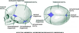
How long does it take for the fontanelle to close? The longest period is for a large fontanel: in different children it takes from 6 months to two years to close it. After childbirth, not only the configuration of the child’s head changes, but also the shape of the fontanelle, and even its size. With the gradual rounding of the head, the size of the crown will also decrease.
The small fontanel may either already be closed at the birth of the child, or it may heal within a month or two. Paired fontanelles in full-term babies rarely pulsate, and if they remain, then only the lateral ones, which close quite quickly, within a couple of weeks after birth.
Sometimes parents worry: why is it that in one child the fontanel resembles a small dot to the touch and closes soon after birth, while in others it can be felt even at the age of two? The timing of fontanelle closure can be influenced by several factors.
- Genetic features. The time for overgrowth of the fontanel and its size directly depends on the hereditary predisposition. Therefore, if you want to know how quickly your baby’s fontanel will heal, ask your parents (and your spouse’s parents) how quickly this process took place in their children.
- Child's due date. Premature babies may be slightly physically behind their full-term peers: don’t worry, by the age of two you will have forgotten about this difference. In the meantime, the fontanel in a child born prematurely will take a little longer to heal.
- The level of vitamin D and calcium in the baby's body. Long overgrowth of the fontanel may be a consequence of calcium deficiency, but an excess of this element causes the cavity to overgrow too quickly.
- Taking certain medications during pregnancy. If a pregnant woman takes calcium and calcium-containing products during pregnancy, this may also affect the timing of fontanelle healing.
Brain
Before moving on to studying the areas of the head, it is worth saying that the head is considered the most important part of the body for a reason. After all, this is where they are located:
- brain;
- organs of vision;
- hearing organs;
- olfactory organs;
- taste organs;
- nasopharynx;
- language;
- chewing apparatus.
Now we will learn a little more about the brain. What is it and how does it work? This organ is formed from nerve fibers. Neurons (these are brain cells) are able to control the functioning of the entire human body by generating an electrical impulse. In total, twelve pairs of nerves can be observed that control the functioning of organs. Signals sent by the brain reach their destination through the spinal cord.
The brain is kept in fluid all the time, which prevents it from contacting the skull when the head moves. In general, our brain has pretty good protection:
- hard connective tissue;
- soft connective tissue;
- choroid;
- cerebrospinal fluid
The liquid in which our brain “floats” is called cerebrospinal fluid. The pressure of this fluid on the organ is considered to be intracranial pressure.
It is also important that the work of the brain and organs located on the head requires large energy costs. For this reason, we can observe intense blood circulation in this area. This:
- Nutrition: carotid and vertebral arteries.
- Outflow: internal and external jugular veins.
So, at rest, the head consumes about fifteen percent of the body’s total blood volume.
If the fontanelle changes
But obvious changes in the “behavior” of the fontanel are very important, since they may indicate a problem.
A protruding fontanel in a newborn may be a sign of serious problems - intracranial hemorrhage, encephalitis or meningitis. Each of these diseases is characterized by an increase in intracranial pressure - this is what causes the fontanelle to bulge. However, do not panic ahead of time and do not give your child a serious diagnosis on your own, since bulging fontanel is not the only sign of brain pathologies. In any case, contact a specialist.
What should be a warning sign, besides a bulging fontanelle? Difficult-to-control high fever, lethargy, or, on the contrary, a loud cry, convulsions, fainting, injuries and falls from a height, the sudden appearance of strabismus or other symptoms associated with the visual apparatus. Any of these signs, accompanied by swelling of the fontanelle, should be a reason to urgently call an ambulance.
A retracted fontanelle in a newborn may be a symptom of dehydration in the baby. Loss of fluid and its serious shortage can be felt with severe vomiting, diarrhea, and very high fever. An inverted fontanelle is often accompanied by dry skin, cracks may form on the lips, and the child becomes very lethargic. With such symptoms, it is necessary to urgently call an ambulance to replenish the lack of fluid in the child’s body using medical methods. Before the ambulance arrives, try to feed your baby boiled water from a spoon.
The conditions described above are serious and critical, and a change in the condition of the fontanelle is not their only symptom. Therefore, if you suspect something is wrong, do not rush to panic and entrust the diagnosis of the fontanel in a newborn to a specialist: an experienced doctor will do this in just a few seconds. And your main task is to remain calm and monitor the development, general condition and mood of the child. If you notice changes that concern you, inform your pediatrician about them in a timely manner.
Skull and muscles
The skeleton of the head (skull) has an equally complex structure. Its main function is to protect the brain from mechanical damage and other external influences.
The entire human skull is formed by 23 bones. They are all motionless except for one - the lower jaw. As mentioned earlier, two departments can be distinguished here:
- cerebral;
- facial.
Bones related to the facial section (there are 15 in total) can be:
- paired - upper jaw, palatine bone, lacrimal, inferior nasal concha;
- unpaired - lower jaw, vomer, hyoid.
Paired bones of the medulla:
- parietal;
- temporal
Unpaired:
- occipital;
- frontal;
- wedge-shaped;
- lattice.
The entire brain section consists of a total of eight bones.
The cervical region, to which the skull is attached, allows the head to move. Movement is provided by the muscles of the neck. But on the head itself there are also muscle fibers that are responsible for facial expressions, one exception is the masticatory muscles, which are considered the strongest in this area.
Head areas
The entire head is conventionally divided into 13 regions. There they also distinguish between paired and unpaired. And so, six of them are classified as unpaired regions.
- The frontal area of the head (attention is focused on it in the next section of the article).
- Parietal (detailed information will be presented to your attention later).
- Occipital (discussed in more detail in a separate section of the article).
- Nasal, which completely matches the contour of our nose.
- Oral, also corresponds to the contour of the mouth.
- The chin, which is separated from the mouth by the geniolabial groove.
Now we move on to listing the seven paired areas. These include:
- The buccal region is separated from the nose and mouth by the nasolabial groove.
- Parotid-masticatory (contours of the parotid gland and muscles responsible for the chewing reflex).
- The temporal region of the head (the contours of the scales of the temporal bone, located below the parietal region).
- Orbital (outline of the eye sockets).
- Infraorbital (below the eye sockets).
- Zygomatic (cheekbone contour).
- Mastoid (this bone can be found behind the auricle, which, as it were, covers it).
Frontal region
Now we move on to a detailed examination of the frontal region of the head. The boundaries of the anterior section are the nasofrontal suture, the supraorbital edges, the posterior section is the parietal region, the sides are the temporal region. This section even covers the scalp.
As for the blood supply, it is carried out through the following arteries:
- supratrochlear;
- supraorbital.
They arise from the ophthalmic artery, which is a branch of the carotid artery. A well-developed venous network is observed in this area. All vessels of this network form the following veins:
- supratrochlear;
- supraorbital.
The latter, in turn, partially flow into the angular and then into the facial veins. And the other part goes into the eye.
Now briefly about the innervation in the frontal region. These nerves are branches of the ophthalmic nerve and have names:
- supratrochlear;
- supraorbital.
As you might guess, they pass together with the vessels of the same name. Motor nerves are branches of the facial nerve called temporal.
Parietal region
This area is limited by the contours of the bones of the crown. You can imagine it if you draw projection lines:
- in front - coronal suture;
- posterior - lambdoid suture;
- sides - temporal lines.
Blood supply is facilitated by arterial vessels, which are branches of the parietal branches of the temporal artery. The outflow is the parietal branch of the temporal vein.
Innervation:
- in front - the terminal branches of the supraorbital and frontal nerves;
- sides - auriculo-vesical nerve;
- posterior - occipital nerve.
Occipital region
The occipital region of the head is located below the parietal region, and is limited to the posterior region of the neck. So, the boundaries:
- top and sides - labdoid suture;
- bottom - the line between the tops of the mastoid processes.
Arteries contribute to blood supply:
- occipital;
- posterior ear.
The outflow is the occipital vein, and then the vertebral vein.
Innervation is carried out by the following types of nerves:
- suboccipital (motor);
- greater occipital (sensitive);
- lesser occipital (sensitive).
Nervous system
The article briefly talks about the nervous system of some areas of the human head. From the table you will find out more detailed information. In total, the head contains 12 pairs of nerves, which are responsible for sensations, the secretion of tears and saliva, the innervation of the muscles of the head, and so on.
| Nerve | Brief Explanation |
| Olfactory | Affects the nasal mucosa. |
| Visual | It is represented by a million (approximately) tiny nerve fibers, which are the axons of the neurons of the retina. |
| Oculomotor | Acts as muscles that move the eyeball. |
| Block | Dealt with irritation of the oblique muscle of the eye. |
| Trigeminal | This is the most important nerve located on our head. It innervates:
|
| Abductor | Innervation of the rectus muscle of the eye. |
| Facial | Innervation:
|
| vestibulocochlear | It is a conductor between the receptors of the inner ear and the brain. |
| Glossopharyngeal | Innervates:
|
| Wandering | It has the most extensive area of innervation. Innervates:
|
| Additional | Motor innervation of the pharynx, larynx, sternocleidomastoid and trapezius muscles. |
| Sublingual | Thanks to the presence of this nerve, we can move our tongue. |
Energy centers in the human body. Slavic tradition
Today the eastern system of knowledge about chakras and energy centers is very well known, but there is also a Slavic version of the structure of the human energy body with Springs of Power. Source - Esoterics. Living Knowledge
The concept of Springs of Power should not be confused with Indian ideas about chakras (chakras). At a superficial glance, there are many similarities between them, but in reality there are still more differences, both in terms of their meaning and in the methods of cleaning and exciting them. Although the foundations of Zhiva knowledge and the Hindu Vedas with their yogic practices come from the same northern roots, the goals and objectives of these systems are still more than different. Perhaps these differences were the reason for the separation of the paths of our peoples in the distant past.
Let's look at the location of the Springs of Power in the human body and their anatomical location. As you can see, diagram 1 shows the main 17 springs.
Rice. 1. Diagram of the location of the Springs of Power in the human body: 1 - Vyshen; 2 - Roof; 3 - Brow; 4 - Crown; 5 - Yavlo; 6 - Throat; 7 - Ladina; 8 - Fret; 9 - Lada; 10 - Yarlo; 11 - Lel; 12 - Lelya; 13 - Belly; 14 - Source (Vein); 15 - Womb; 16 - Zarod (16A -
Khvoy, 16B - Khorla); 17 - Viy.
Springs of Power (from bottom to top):
Viy - the point is located outside the anatomical body, located below the fetus, at the level of the middle of the thighs. Through it, communication is made with the world of Navi, the souls of ancestors. It is not recommended to work with it independently, without the guidance of an experienced magician.
The germ is anatomically compatible in men with the testicles and the nervous system of the penis (Hvoy), in women - with the ovaries and the nervous system of the vagina and clitoris (Khorla).
The womb is anatomically compatible with the prostate gland in men, and with the uterus in women. It is considered the center of physical forces. The Womb spring point is located exactly on the central axis of the body - the Thread of Life, which can be determined by mentally stretching the thread through the point in the perineum, located between the genitals and the anus (identical to the Guai-Yin point - the Gate of Life and Death in eastern acupuncture, also known as Gui- Yin, Hui-Yin), and a place on the head called the Fontana. Of course, this refers to the position of the body in correct posture, and not the crumpled position inherent in most contemporaries. So, the Womb is located in the middle of the body, both in the frontal and sagittal planes (i.e., when viewed directly and from the side). To find its projection on the body, divide the distance from the navel to the fetus of a man by half, for a woman - into 3 parts, and, retreating 2/3 from the fetus and 1/3 from the navel, we will find what we are looking for.
Source - anatomically compatible with the coccyx and coccygeal tail of the nerve endings.
The abdomen is anatomically compatible with the pancreas. The spring point of the Belly is also located on the Thread of Life; its projection on the body can be found by dividing the distance between the navel and the place where the ribs meet in half.
Lelya - anatomically compatible with the left adrenal gland.
Lel - anatomically compatible with the right adrenal gland.
Yarlo is considered the center of the soul. Anatomically located on the Thread of Life, in the center of the diaphragm, separating the pleural cavity and pericardium from the abdominal cavity. Physiologically, it is a muscular roll, penetrated by nerve endings, compressing the esophagus and arteries passing through it. We will find the projection on the body at a point if we draw a straight line between the nipples and divide it in the middle. In light of scientific evidence that this nerve node has an important influence on regulating the release of various hormones in the body, which, in turn, change our mood (in general, biochemical processes - all our emotions), then perhaps Yarlo is really center of the soul?
Lada - anatomically this is our physical heart, surrounded by connective tissue - the pericardium.
Lad - anatomically located next to Lad, but on the right side, and it can be called the lymphatic heart. True, sometimes there is a slightly different arrangement of organs and, accordingly, springs - the physical heart is not on the left, but on the right - in this case, the lymphatic heart moves to the other side.
Ladina - anatomically represents the thymus gland, thymus.
The throat is anatomically composed of the thyroid and parathyroid glands. Also located on Thread of Life.
Yavlo is anatomically compatible with the uvula and tonsils, and is also important in understanding Volkhov anatomy and physiology. Located on the Thread of Life.
The brow is a spring located on the Thread of Life, but although its projection represents the so-called “third eye” point, it has nothing to do with it or the Ajna chakra. Anatomically, the Chela point is combined with the pituitary gland, as well as the thalamus and hypothalamus. This is a very important aspect for further studies.
Crown - this spring is precisely compatible with the pineal gland, like the Indian Ajna Chakra. Look for the crown and its projection in the place where the newborn has a posterior fontanelle, which with age, just like the anterior fontanel and others, is overgrown with tendon tissue.
The roof is a spring, anatomically combined with the center of the tendon helmet on the head, which contains the closed anterior fontanel in a newborn.
Vyshen - anatomically this fontanel is not located on the body, but is located slightly above the body, approximately in the palm of the hand, and through this fontanel a person’s connection with his Kin is always maintained. Moreover, regardless of whether you know your pedigree or not, whether you deny or recognize family ties, he always acts, being responsible for our safety and security.
Rating: 3.97 (Views: 7393)
Read the section Rodnovers, Slavs on the esoteric portal naturalworld.guru.
naturalworld.guru
Circulatory system
When studying the anatomy of the head, one cannot ignore such a complex but very important topic as the circulatory system. It is she who provides blood circulation to the head, thanks to which a person can live (eat, breathe, drink, communicate, and so on).
The functioning of our head, or rather the brain, requires a lot of energy, which requires a constant flow of blood. It has already been said that even at rest, our brain consumes fifteen percent of the total blood volume and twenty-five percent of the oxygen that we receive when breathing.
Which arteries supply food to our brain? Mainly:
- vertebrates;
- sleepy.
Its outflow from the bones of the skull, muscles, brain, and so on should also occur. This occurs due to the presence of veins:
- internal jugular;
- external jugular.
What do you need to know about the crown?
From the first days of a child's birth, young parents are concerned about issues related to the crown of the newborn. Namely, whether it is not too big, not too small, when and how quickly it should close, why it is needed and what functions it performs. Despite all this, there are still a number of myths that do not allow young parents to calm down.
First, they help the baby to be born. When a baby is ready to be born, it must pass through the mother's birth canal. If the birth takes place without complications, the baby moves through the birth canal head first.
Sutures and fontanels give the bones of the skull flexibility and plasticity, the bones are able to fit into each other. As a result of such changes, the newborn's head can change shape, which is very important when passing through the birth canal.
The head decreases in size and this allows the baby to be born more easily. Thanks to the fontanelles, the bones of the skull remain mobile, and at the time of birth, the baby’s head can move along the birth canal, changing its volume, as doctors say, “configuring.” E
This makes the process easier for both the child and the mother in labor. Therefore, immediately after birth, the baby’s head has a somewhat unusual shape - elongated, egg-shaped. As a rule, within a short period of time after birth, the baby’s head takes on a normal shape.
After the baby is born, the fontanel allows the light baby head to grow vigorously.
The fontanel also serves as a natural shock absorber. It softens the force of impact in case of injury to the baby's head.
In infants, unlike adults, the thermoregulation system still does not work well.
Some of its functions are taken over by the fontanel. If the baby’s body temperature rises, then natural cooling of the brain occurs through the thin membrane of the fontanel.
The fontanel is the “viewing window” of the brain. Through it - thanks to a special study, a neurosonogram (ultrasound of the brain) - it is possible to qualitatively and reliably examine the child’s brain.
Where is the crown located?
The crown is the remnants of membranous tissue at the fusion sites of the skull bones. The front one is located slightly in front of the crown, and the back one is in the center of the back of the head. The crowns are separated from the brain by three membranes.
The small spaces between them are filled with cerebrospinal fluid. But basically, when they talk about the crown, they mean the front. Thanks to the presence of crowns, the baby's head changes its shape and passes through the birth canal more easily.
When does the crown become overgrown?
The process of overgrowth is individual for each baby and it needs to be controlled in boys; for example, the large crown closes a little faster than in girls. The sizes also differ from 0.6 to 3.6 cm.
Normal sizes of fontanelles
Conventionally, the entire period of the first year of a child’s life can be divided into 5 periods:
- 1st period - neonatal period (from the first day to the 1st month),
- 2nd period - from 1 month. up to 3 months,
- 3rd period - from 3 months. Up to 6 months,
- 4th period - from 6 to 9 months.
- Period 5 - from 9 to 12 months.
Video: Myths about the fontanel - Doctor Komarovsky
stobolezni.ru
Arteries
As already mentioned, the vertebral and carotid arteries, which are presented in pairs, supply food to the human head. The carotid artery is the basis of this process. It is divided into 2 branches:
- external (enriches the outer part of the head);
- internal (passes into the cranial cavity itself and branches, providing blood flow to the eyes and other parts of the brain).
Blood flow to the muscles is carried out by the external and internal carotid arteries. About 30% of the brain's nutrition is provided by the vertebral arteries. Basilar provides work:
- cranial nerves;
- inner ear;
- medulla oblongata;
- cervical spinal cord;
- cerebellum.
The blood supply to the brain varies depending on a person's condition. Mental or psychophysiological overload increases this indicator by 50%.
When should pathology be suspected?
There are a number of diseases in which the fontanel in a newborn closes too late. But, in addition to this symptom, there are certainly others.
With rickets, the slow healing of the cavity is accompanied by a noticeable lag in the child's physical development: his cardiovascular and musculoskeletal systems are poorly developed, and his immunity suffers. This condition is often caused by premature birth, after which the premature baby is not given prophylactic doses of vitamin D.
Hypothyroidism is a congenital pathology when the thyroid gland is unable to perform its functions. Late closure of the fontanel is accompanied by drowsiness and lethargy in the baby, developmental abnormalities, and chronic constipation.
With achondroplasia (disproportionate dwarfism), slow closure of the fontanel is accompanied by serious pathologies in the formation of bone tissue.
With Down syndrome (chromosomal pathology), the fontanelle also closes late and deviations in the mental and physical development of the child are observed.
Vienna
When considering the anatomy of the human head, it is difficult to ignore a very important topic - the venous structure of this part of the body. Let's start with what venous sinuses are. These are large veins that collect blood from the following parts:
- skull bones;
- head muscles;
- meninges;
- brain;
- eyeballs;
- inner ear.
You can also find another name for them, namely, venous collectors, which are located between the sheets of the lining of the brain. Leaving the skull, they pass into the jugular vein, which runs next to the carotid artery. You can also distinguish the external jugular vein, which is slightly smaller and located in the subcutaneous tissue. This is where blood collects from:
- eye;
- nose;
- mouth;
- chin
Generally speaking, everything listed above is called superficial formations of the head and face.
Parameters by which the condition of the fontanel is assessed during examination by a doctor
- are the child’s fontanelles open or closed, does this correspond to the baby’s age;
- how many fontanelles were there at the time of birth and their number at the moment;
- how the fontanelles have changed, how quickly they are shrinking, whether the shape of the fontanelles has changed;
- What do the edges of the fontanelle feel like? Normally, the edges should be elastic, and softening is a sign of a lack of calcium and vitamin D;
- How does the fontanel relate to the surrounding tissues? A sluggish, sunken or tense, bulging spring is always a sign of pathology.
Muscles
To put it very briefly, all the muscles of our head can be divided into several groups:
- chewable;
- facial expressions;
- cranial vault;
- sense organs;
- upper digestive system.
You can guess the functions performed by their names. For example, chewing ones make the process of chewing food possible, but facial ones are responsible for human facial expressions, and so on.
It is very important to know that absolutely all muscles, regardless of their main purpose, are involved in speech.


