The disease dropsy of the brain or hydrocephalus is a violation of the normal drainage of fluid in the brain, the accumulation of fluid in the ventricular system of the brain. The system consists of the lateral ventricles (I and II), connecting to the slit ventricle (III), which through the Sylvian aqueduct (interventricular canal) connects to the fourth ventricle of the brain. The fluid is produced in the choroid plexuses, then enters sequentially into the ventricles of the brain, and from the last fourth ventricle into the space between the dura mater and the brain, the subarachnoid space of the spinal cord and brain. This is where cerebrospinal fluid (a colorless liquid) is absorbed into the bloodstream. Impaired drainage of fluid and accumulation of fluid in the ventricles of the brain lead to their enlargement, compression of the tissues of the head, and the development of hydrocephalus. The pathology is characterized by a violation of the structure of soft tissues, pressure on the bones of the skull, deformation of the skull, and impaired brain function. Dropsy of the brain can affect not only children, but also adults. There is a congenital and acquired form of the disease. Hydrocephalus can be primary (the underlying disease), secondary (as a consequence of various diseases), closed (occlusive) or open (communicating).
Description of the disease
Today, almost every fifth newborn child can be diagnosed with increased intracranial pressure. Although in most situations it does not have any tragic consequences. But it is still worth checking the head for the presence of excess fluid in the head of a newborn. And if the diagnosis is confirmed, then you should definitely think about taking all the necessary measures for treatment.
Hydrocephalus of the newborn (or dropsy in other words) is the name of a complication due to which in the area of the brain in newborns there is an accumulation of cerebrospinal fluid, otherwise called cerebrospinal fluid. There are several variations of the disease, however, in children under two years of age, all its symptoms are very similar to each other.
The term "hydrocephalus" is derived from two Greek words that mean "water" and "head". In other words, this disease consists of excess fluid (water) in the head. This is where the second name of the pathology comes from, which sounds like dropsy of the brain. True, strictly speaking, this name is not entirely correct. The fact is that in the presence of hydrocephalus in the head of newborns, there is an excess not of water at all, but of cerebrospinal fluid, that is, cerebrospinal fluid. Liquor is a liquid that is vital for the functioning of nerve tissues. It can be found in the spinal cord. We will consider the fluid levels in the head of a baby below.
In addition, it is also present in the brain. In it, a substance such as cerebrospinal fluid is concentrated in four ventricles, which are located in the center of the skull. The upper two are located in both hemispheres, and the lower ones are located along the central cerebral axis. The ventricles usually communicate with each other through a system of pipes called the cerebral aqueduct. In addition, cerebrospinal fluid can enter the subarachnoid space, which separates the meninges with special cisterns located at the base of the skull.
Hydrocele in adults: symptoms
Symptoms of hydrocephalus depend on the level of fluid pressure, the period of formation of the disease and the mechanism of development. Severe morning headache, nausea and vomiting, pressure on the eyeballs from the inside, a feeling of discomfort in the eyes indicate the development of acute or subacute occlusive hydrocephalus.
When cerebrospinal fluid accumulates, the condition worsens - the patient becomes drowsy, visual acuity decreases, and intracranial pressure increases. Dislocation syndrome may develop, which is characterized as a life-threatening condition. It manifests itself as paresis of upward gaze, suppression of reflexes occurs, strabismus develops, and coma may occur. Compression of brain tissue leads to the appearance of certain symptoms: the voice changes, the patient cannot swallow, the functioning of the cardiovascular and respiratory systems is disrupted - the patient dies.
The chronic form of edema of the brain is characterized by gradual development, sleep disturbance, memory impairment, lethargy, asthenia, gait disturbance, increased muscle tone, and frequent urge to urinate. Progression of the disease can lead to cognitive impairment and dementia.
Types of disease
There are only three main forms of this pathology, in which fluid is observed in the head of a newborn:
- open hydrocephalus;
- closed, or occlusal form;
- hypersecretory form of pathology.
The closed type of the disease occurs in cases where there is a physical obstacle that interferes with the outflow of cerebrospinal fluid from the cranial container intended for it into the systemic bloodstream. This type is mainly caused by cysts along with tumors or hemorrhages.
The open type of the disease is usually observed when the mechanism of absorption of cerebrospinal fluid into the systemic circulation is disrupted. With this variant of the development of pathology, the cause of the disease is often previous infections. For example, meningitis or the presence of blood in the subarachnoid space.
Hypersecretory hydrocephalus is a relatively rare type of the disease in question and is observed in approximately five percent of cases. It usually occurs as a result of excessive production of cerebrospinal fluid. A similar situation can happen, for example, due to pathology of the choroid plexus.
Types and reasons
Dropsy of the brain can be either a congenital pathology or an acquired one.
In the first case, the development of the disease is influenced by unfavorable intrauterine factors: an acute infectious disease in the mother during pregnancy (most often this affects the child), developmental defects that arise due to genetic “errors.”
Acquired hydrocephalus most often affects children under one year of age who were born much earlier than expected, as well as babies who received a brain injury during childbirth.
The cause of the pathology can also be a traumatic brain injury or a previous infectious disease or brain tumor. The most dangerous combination of risk factors is if, for example, a premature baby develops meningitis, encephalitis or meningoencephalitis. The disease can also develop after surgical procedures.
Dropsy is divided into several types, depending on where exactly the cerebral fluid accumulates:
- external;
- internal;
- mixed (combined).
With external dropsy, the accumulation of cerebrospinal fluid is concentrated only under the membranes of the brain, it does not affect the deep areas. This condition usually occurs in newborns and children who have suffered birth trauma.
Internal hydrocephalus is a situation in which cerebral substance accumulates in the brain ventricles, through which it cannot flow normally. Such a lesion can be a congenital pathology, as well as acquired - in toddlers older than one year.
According to the assessment of true obstacles that interfere with the full circulation of fluid, dropsy is divided into:
- open (communicating);
- closed (occlusive).
In the reported form of the disease, there are no objective obstacles, the ventricles are sufficiently dilated, and there are no mechanical barriers to the flow of cerebrospinal fluid. Occlusive hydrocephalus occurs as a result of improper development of the cerebrospinal fluid ducts themselves, pathologies in the structure of the ventricles, tubules, tumors in this system, neoplasms, and adhesions. This form of the disease is almost never external; it is characterized by the accumulation of fluid inside the brain.
According to the time of development of pathology, three types of hydrocephalus are distinguished:
- spicy;
- subacute;
- chronic.
Acute develops rapidly, the pressure inside the skull increases literally in 2-3 days. Subacute pathology can develop up to six months, gradually, almost imperceptibly for parents. Its consequences can be more devastating. With chronic dropsy, the cerebrospinal fluid accumulates very slowly, for more than six months, which at first does not affect the baby’s well-being in any way, because the pressure also increases at a very slow pace. And only then, when it reaches a critical level, the diagnosis becomes obvious.
The child's body has very high compensatory abilities. If something is wrong somewhere, the body tries in every possible way to compensate for it using other resources. Therefore, it happens that when a diagnosis of “dropsy of the brain” is established, the child does not show any deterioration in well-being or change in behavior. In this case they talk about compensated hydrocephalus.
If all the body's forces are not enough to compensate, the child's well-being worsens, there are pronounced disturbances in his development, then they speak of decompensated dropsy.
A minor compensated disruption in the circulation of cerebrospinal fluid sometimes does not even require serious medical support, which cannot be said about decompensated disorders.
According to the degree of damage, doctors also divide the disease into stages. There are two of them:
- moderate;
- expressed.
According to the dynamics of manifestations, hydrocephalus can be:
- progressive (with noticeable deterioration of the condition);
- stable (when new symptoms do not appear, but there is no improvement);
- regressing (with a gradual decrease in symptoms).
Functions of fluid in the head
The volume of cerebrospinal fluid is, in fact, relatively small. Normally, in newborns it is, as a rule, 50 milliliters, and in adult patients - from 120 to 150 ml.
The functions of the fluid in the head of a newborn are very diverse:
- protection of nervous tissue from external mechanical influence;
- removal of harmful substances from the brain and delivery of nutrients to it;
- maintaining stable intracranial pressure values.
The fluid in the baby's head, like blood, can circulate inside the cranial cavity. Against this background, its composition is constantly updated. In adult patients, on average, this can happen three times a day, and in infants much more often - up to eight times a day. Every minute, adults produce 0.35 milliliters of cerebrospinal fluid, and approximately 500 milliliters per day. CSF pressure in adults can vary quite widely, namely from seventy to one hundred and eighty millimeters of mercury.
CSF is mainly formed in the ventricles of the brain. Two thirds of this fluid can be generated by their choroid plexus, and the rest - with the help of membrane elements and meninges. In special veins that are located inside the skull, in its occipital parietal part, namely within the venous sinuses, it is absorbed.
Consequently, if for some reason the circulation processes of the cerebrospinal fluid are disrupted, and it is formed in greater quantities than necessary, or is simply not absorbed quickly enough, then an excess of this fluid is observed in the newborn in the cranial cavity. It is this syndrome in children that is called hydrocephalus.
Excess cerebrospinal fluid can manifest itself differently in children and adults. For example, adults have hard skull bones, so excess fluid usually leads to increased intracranial pressure. The situation is completely different for young children under the age of three. They have rather soft skull bones, and therefore hydrocephalus very often manifests itself in the form of an abnormal expansion of the head circumference.
Conservative treatment
Conservative therapy is used in the following cases:
- the disease was detected at an early stage;
- hydrocephalus of newborns and older children is in acute form after an inflammatory process in the brain;
- There are errors regarding the mental and neuralgic state caused by the activity of bacteria.
The recovery rate with drug treatment is no more than 50%. There remains a high probability of psychomotor and mental retardation. For a more successful result, several methods are used aimed at non-surgical reduction of intracranial pressure, drainage of excess cerebrospinal fluid and minimization of complications. Auxiliary methods include physical education, massage, physiotherapy.
| Procedure name | Target |
| Taking diuretics | Reduced rate of cerebrospinal fluid production |
| Antibiotic therapy | Destroying harmful bacteria in excess liquid |
| Taking blood vessel stabilizers | Regulating blood flow in brain tissue |
| Taking vitamin complexes | Regulation of cellular metabolism |
| Taking absorbable medications | Removing stagnant fluid from the body |
| Physiotherapy | Stabilization of the course of the disease |
| Physiotherapy | Facilitating the removal of excess cerebrospinal fluid from the brain |
| Taking glucocorticoids | Promoting healthy brain function |
| Massage | Toning the blood vessels of the head |
Causes of fluid in the head of a baby
Dropsy in newborns can develop from simple prematurity. And in addition, from the presence or previous infectious diseases. For example, factors such as smoking, drinking alcohol and other bad habits of the mother, not just during pregnancy, but also in everyday life, can contribute to the development of this pathology in the newborn.
During the first few years of life, any head injury is very dangerous, as it can lead to an increase in the production of cerebrospinal fluid. A tumor that occurs in the brain can significantly interfere with the healthy flow of fluid in the head of a newborn baby. Which, in turn, will create excess pressure.
How does this pathology manifest clinically?
The fluid in the head of a newborn must circulate in a correct and normal manner, and if this is disrupted, this will certainly lead to hydrocephalus. The most important symptom is a change in the shape of the head over a fairly quick period of time. In this regard, it will be necessary to visit a pediatrician every month, who must measure the head, checking the condition with the normal fluid levels in the baby’s head.
In addition, the fontanel in a newborn baby is characterized by an increased size, since the sutures of the skull are not yet fully formed. Over time, the symptoms may become more pronounced: a network of veins will appear on the face, and the shape of the forehead, in turn, will become more disproportionate. Cramps may occur from time to time. Newborns with hydrocephalus tend to be lethargic and cry frequently.
Such children are noticeably lagging behind in development, their psychomotor skills are impaired. This usually manifests itself in the fact that the child’s head is very poorly supported. In addition, such children begin to crawl, walk and sit up late. In addition, newborns with this disease often spit up and even vomit. Among other things, they experience constant drowsiness. All such symptoms may indicate that the baby has increased intracranial pressure.
However, you should not rush to make a diagnosis based on individual similar characteristic signs, since usually only the appearance of the entire complex of the listed symptoms can indicate the presence of hydrocephalus. Only the attending doctor, as part of the examination of the child, will be able to make the correct diagnosis and prescribe the required treatment for the accumulation of fluid in the head of the newborn.
How does hydrocephalus manifest?
Depending on the form of development of the pathology and the age of the child, the external signs of the disease change.
For children under two years of age, in which in 95% of cases hydrocephalus is a congenital pathology, the following symptoms are characteristic:
- Severe course of the disease. A rapidly deteriorating condition caused by damage to brain structures.
- The main symptom is a rapid increase in head volume. Growth of 1.5 cm or more every month is typical, for at least three months in a row. Starting from the ninth month of life, growth drops to 8-9 mm.
- A child is born with a head whose girth is larger than the girth of the chest. If by six months the ratio does not change, and the head is still larger than the chest, there is reason to suspect hydrocephalus.
- The veins on the occipital, temporal and frontal parts of the head are clearly visible.
- At three months the child does not yet hold his head up and begins to sit up late, crawl, and walk.
- The fontanel on the top of the head is convex.
- The accumulation of cerebrospinal fluid primarily occurs in the frontal lobes of the brain. Head enlargement starts from the forehead.
- While fixating the baby's gaze, the baby's pupil twitches chaotically.
- The scalp becomes thin and has a painful shine.
- Overhang of the brow ridges over the facial bone - the eyes appear to be very deep-set.
A number of less characteristic signs can confuse the initial diagnosis of hydrocephalus in children and complicate treatment. For example, tearfulness, poor appetite, slow weight gain, frequent regurgitation, drowsiness, legs constantly bent at the knees, head tilting - all these symptoms are characteristic of hydrocephalus. But at the same time, they can be observed in dozens of other childhood diagnoses.
In the acute form of dropsy, when the disease progresses rapidly, other signs can be observed:
- convulsions;
- long cry on one note;
- loss of acquired physical skills (sitting, following other people's movements, vocal functions);
- vomit.
For children aged two years and older, other manifestations of hydrocephalus of the brain are typical:
- Headaches that begin in the morning (after sleep) and gradually subside in the evening. Nausea and vomiting often begin at the same time.
- Migraines that appear after stress, mental or physical work. Often accompanied by nosebleeds.
- A feeling of a rush to the head when bending over and bursting headaches.
- Pain in the fundus of the eye.
- Visual impairment: loss of sharpness, double vision.
- Urinary incontinence.
- Decreased muscle strength, accompanied by rapid fatigue.
- Cramps and fainting.
- Loss of coordination and uncontrolled movements of the limbs.
Basics of diagnosis and therapy in newborns
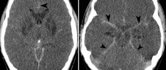
After determining the primary diagnosis, children are prescribed neurosonography along with ultrasound examination of the brain, computed tomography or magnetic resonance imaging.
If the diagnosis is confirmed, ventriculoperitoneal shunting is often performed. The essence of this operation is that the cerebrospinal fluid from the cerebral ventricles of the newborn is sucked through silicone catheters into the abdominal cavity. Less commonly, fluid may be drained into the spinal canal or right atrium.
If the operation was performed on time, then the child has every chance of further normal life, which includes attending child care and school institutions. However, it is also necessary to take into account that the size of the head after surgery most likely will not decrease, since changes in bone tissue are always irreversible.
How to identify fluid in a baby's head?
Diagnosis of the disease
There are several ways to determine the development of hydrocephalus. It is worth noting that this disease is much easier to identify in children. But in adult patients, recognition of the described disease is sometimes difficult and problematic. Previously, many adults who suffered from hydrocephalus were diagnosed with various neurological and mental disorders. However, of course, their therapy was not very effective. Only after the advent of modern diagnostic techniques the situation changed radically. And for the better.
If there is a lot of fluid in the baby's head, this is mainly detected by the pediatrician during a thorough examination of the child. Doctors can turn their attention to obvious manifestations of hydrocephalus in the form of an enlarged head, bulging fontanel, divergence of the sutures of the skull, and in addition, changes in the appearance of the skin and characteristic neurological symptoms. To facilitate the diagnostic procedure, parents are recommended to record the baby’s head circumference values at a certain time interval. If a pathology is suspected, the pediatrician can write a referral to a neurologist, pediatric surgeon or neurosurgeon.
Treatment
Recently, great progress has been made in medicine in the treatment of hydrocephalus and the presence of fluid in the head of an infant. If just a few decades ago more than half of patients with this disease died, today the mortality rate is no more than five percent.
The choice of treatment method for hydrocephalus directly depends on the etiology of the pathology, and in addition, on its form and degree of development. In some situations, etiotropic therapy is possible. However, in most cases, treatment is aimed at removing fluid from the cranial cavity. Treatment of the progressive course of hydrocephalus in children can be carried out only by surgical methods. Unfortunately, conservative therapy is ineffective.
How to remove fluid from a baby's head? The operations that are performed for closed and open forms of hydrocephalus may differ slightly. Previously, open dropsy of the brain was considered an almost incurable pathology. But in the middle of the last century, new technologies were developed that make it possible to save the majority of young patients.
To remove excess fluid from the cranial cavity, shunting is usually performed. It consists of laying a kind of pipeline through which liquor is pumped to the rest of the body cavities. Such tubes are located under the skin surface for most of their length. Typically, the fluid is drained to the peritoneum (in ninety-five percent of cases), the chest area, or the atrium. In some situations, it has to be diverted not from the brain, but from the spinal cord, from where it is sent to the abdominal cavity.
When such an operation is performed on a child, as the baby matures and grows, the catheters will require lengthening and replacement. It is worth noting that modern catheters are equipped with special valves that allow you to regulate the fluid pressure in the brain vessels. If there is no threat to life, bypass surgery is performed as planned. As a temporary measure to reduce the pressure of brain fluid, a puncture in the spinal region is used.
Closed hydrocephalus often requires rapid surgical intervention, since in this form of the disease compression of the respiratory centers can occur. Therefore, in such a situation, a temporary operation can be performed with the installation of a special container to drain the cerebrospinal fluid.
If the patient has closed hydrocephalus, all the surgeon’s efforts are aimed at eliminating obstacles that interfere with the normal circulation of cerebrospinal fluid. In some cases, such an obstacle (in the form of a vascular aneurysm, cyst, hematoma, tumor) can be eliminated. Often, an endoscopic system inserted into the ventricular cavity is used for this purpose. The surgical operation is carried out using special instruments, a laser or an electrode, which allows the functions of the brain pipelines to be restored.
However, sometimes, for example, with tumors, regardless of their benignity or malignancy, such operations are simply impossible. In this case, the surgeon lays a pipeline from the container within which the cerebrospinal fluid accumulates into an alternative container, where it becomes possible to absorb it directly into the blood.
In absolutely all cases, the main goal of the operation is to restore the balance of cerebrospinal fluid output and generation that has been disturbed for various reasons. Of course, when the disease is secondary, then the main efforts must be directed to the treatment of the underlying disease, which caused an excess amount of cerebrospinal fluid.
Treatment of hydrocephalus
The therapy is based on a set of measures aimed at activating the outflow of pathological fluid accumulation.
If therapeutic agents do not have a positive effect, the patient undergoes surgery. Endoscopic treatment demonstrates the greatest effectiveness against the background of minimal invasiveness:
- endoscopic ventriculocisternostomy of the bottom of the third ventricle,
- aqueductoplasty – arrangement of a kind of “water pipeline” through which fluid will be effectively removed from the skull.
- septostomy – removal of cysts and elimination of occlusion of the “foramen of Monroe”.
- removal of brain tumors by endoscopic method.
After such a surgical intervention, the patient very soon returns to normal life, since his brain is not subject to serious trauma. This procedure allows you to regulate the flow of fluid in the skull, however, after surgery to eliminate hydrocephalus, the patient requires constant supervision by a specialist and regular monitoring of the condition of the brain.
The prognosis for hydrocephalus depends on the cause and time of diagnosis and appropriate treatment. Children who receive treatment are able to live normal lives with few, if any, limitations. In rare cases, speech dysfunction may occur.
Complications of hydrocephalus
In the absence of treatment for the presence of fluid in the head of an infant, the disease in question can progress in most situations. This can lead to extremely negative consequences, which can also threaten the patient with death. The main complications of hydrocephalus are usually:
- the appearance of cerebral edema;
- the occurrence of epileptic seizures;
- displacement of the child's brain;
- development of coma, stroke and respiratory failure.
Not everyone knows why excess fluid in a baby’s head is dangerous. With the development of hydrocephalus in children during infancy, a slowdown and stop in the formation of new brain tissue are often observed. And this leads to a lag in the mental, mental and emotional development of the baby.
Causes of hydrocephalus
Hydrocephalus in the fetus may be associated with problems with the development of the central nervous system, or with intrauterine infection. Therefore, it is so important to undergo a comprehensive examination for various infections - herpes, cytomegalovirus, before planning a pregnancy.
Acquired hydrocephalus in most cases is a consequence or complication of diseases such as:
- Meningitis - viral or bacterial etiology.
- Meningoencephalitis is an inflammatory process that affects the membrane and medulla.
- Subarachnoid hemorrhage is a common consequence of skull trauma or occurs as a consequence of rupture of an arterial aneurysm.
- Sarcoidosis is a granulomatous lesion of the meninges.
- Intraventricular hemorrhage is a consequence of difficult and traumatic obstetric care.
Pathology prognosis
The prognosis for the development of hydrocephalus in a newborn directly depends on how quickly and, moreover, how timely the baby is diagnosed and therapy is started. Children with hydrocephalus can live a normal life, although, unfortunately, they face a number of problems associated with the maintenance of surgical shunts.
But if treatment for this disease in an infant is not started in a timely manner, its further progression threatens the baby with serious developmental delays, as well as speech impairment and irreversible changes in the brain, which will subsequently lead to disability.
Reasons for the development of the disease
They vary depending on the age of the baby.
The reasons that affect the development of the brain and nervous system of the fetus are diverse:
- chromosomal disorders (Down syndrome, triploidy, Turner syndrome and others);
- Rh conflict between mother and fetus (so-called immune dropsy develops);
- infections (tonsillitis, leptospirosis, syphilis, as well as primary maternal disease during pregnancy with herpes, toxoplasmosis, cytomegalovirus, rubella, chickenpox and measles, infection with Coxsackie viruses);
- impaired metabolism in the fetus;
- pregnancy with severe anemia, gestosis and preeclampsia, as well as against the background of uncontrolled diabetes mellitus and thyrotoxicosis;
- cardiovascular congenital diseases of the fetus and blood diseases (tachyarrhythmia, malformations of the cerebrospinal fluid tract, vascular thrombosis, leukemia, other developmental anomalies);
- during pregnancy with twins - transfusion syndrome between children (blood flow from one fetus to another);
- brain tumors;
- fetal trauma, including perinatal;
- defects of the placenta, as well as lesions of the umbilical cord;
- pulmonary, renal and intestinal diseases of the fetus;
- chondrodysplasia (impaired development of the skeletal system and cartilaginous growth zones in the child’s joints).
Malformations of the nervous system can be combined with other disorders. The most common is when the volume of brain matter is greater than the size of the posterior cranial fossa, which causes descent into the foramen magnum. Because of this, the circulation of the cerebrospinal fluid is mechanically disrupted. Stenosis, or narrowing of the Sylvian aqueduct, is common.
There may be underdevelopment or absence of openings between the ventricles and the subarachnoid space. It occurs when the IV ventricle is dilated, a cyst from the cerebrospinal fluid is formed in the posterior cranial fossa, and the cerebellar vermis cannot form normally. Dropsy of the brain interferes with the normal functioning of all systems.
In 15% of cases, the factor that influenced the development of dropsy cannot be determined. The risk of intrauterine death is quite high, so the pregnant woman is under constant medical supervision.
Hydrocephalus in a newborn is most often a consequence of an intrauterine infection, birth trauma, or intracranial hemorrhage. During childbirth, those children who were born prematurely and with low weight (up to one and a half kilograms) are more at risk. It is dangerous if the woman in labor has a narrow pelvis or the fetus experiences hypoxia or oxygen starvation during labor.
In children older than one year, the disease develops due to brain tumors, hemorrhages, inflammation of the membranes, traumatic brain injuries or genetic problems.
Advice from pediatricians
Hydrocephalus, or the accumulation of fluid in the head of a baby, is very often a congenital pathology. But is it possible to prevent such a condition long before its possible progression? In this regard, pediatricians advise, for the purpose of prevention, to examine both parents at once, and it is necessary to conduct research at the genetic level. Among other things, doctors recommend timely treatment of existing infectious pathologies and protect the mother’s body from their development throughout pregnancy.
In addition, in no case should you allow traumatic brain injury to a newborn during childbirth. It is necessary to carry out timely diagnosis and treatment of such pathological abnormalities as hydrocephalus. And it is best, according to pediatricians, for all parents to lead an exceptionally healthy lifestyle, and not just before planning a pregnancy.
The causes of fluid in the head of newborns are discussed. Thus, today every fifth newborn baby in the maternity hospital is diagnosed with increased intracranial pressure. But it is immediately necessary to reassure parents, since in ninety-nine percent of cases this diagnosis is made unreasonably and is put forward contrary to analyzes and research. But nevertheless, it is simply necessary to check suspicions of hydrocephalus. The accumulation of cerebrospinal fluid (CSF) in the brain cavity of a newborn must be treated.
The article described in detail the causes of fluid in the head of a baby. Get tested on time and stay healthy!
Useful tips
- There is no need to look for those to blame; sometimes this illness does not depend in any way on the parents and their correct or incorrect actions.
- During pregnancy, you should definitely visit an antenatal clinic. Many studies and tests that are prescribed to expectant mothers will help you learn about risk factors in advance.
- Before pregnancy, a woman should visit an infectious disease specialist at least once to find out, by donating blood, what diseases she has suffered from, and what antibodies to what dangerous infections she has in her body.
- If during pregnancy (especially in the early stages) a woman gets sick with rubella, measles or another infection, she should definitely agree to additional studies of the fetus’s condition and visit a geneticist in order to make a further (very painful) decision about bearing a child. You need to know about the risks of pathologies and treatment during pregnancy.
- If a child was born prematurely, you should not miss a single mandatory medical examination or routine visit to the doctor.
- Babies over one year old should be protected from head injuries. If you bought him a bicycle, be sure to give him a helmet. If a child is transported in a car, then it is imperative to use a car seat.
- All viral infectious diseases that infect a child cannot be treated independently - according to grandmother’s recipes, viburnum and burdock. You should definitely consult a doctor, get tested, and take medications only as prescribed by a qualified doctor.
You will learn more about this disease from the video below.