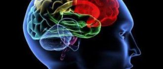
What is cerebral edema
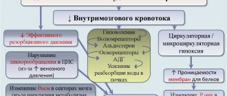
Physiologically, the maintenance of intracranial pressure is ensured by a constant volume of blood and cerebrospinal fluid. Changes in atmospheric pressure, compensation for different volumes of circulating blood are compensated by the inflow or outflow of cerebrospinal fluid. The appearance of additional formation inside the brain, hematoma, inflammatory processes, traumatic brain injury (TBI) leads to compression of cerebral structures. An increase in intracranial hypertension causes the death of parenchyma. Features of symptoms depend on the strength and location of the pathological focus.
Pathological cerebral edema leads to an increase in the size of brain tissue through excess accumulation of fluid. An increase in the amount of water is accompanied by death (if prompt decompression is not carried out in a timely manner). A gradual increase in the volume of brain tissue initially leads to neurological disorders. Psychoses and disturbances in the functioning of internal organs gradually develop.
Symptoms
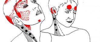
Symptoms of cerebral edema in adults are extremely variable and are caused by dysfunction of various brain structures, disorders of metabolic processes/microcirculation and a developing increase in the volume of brain tissue, especially accompanied by displacement/herniation of certain brain structures, disruption of cerebrospinal fluid dynamics/blood flow in the vessels of the brain. The localization of edema in areas of the brain also plays a significant role, which determines its effect on specific brain structures and forms, in addition to general cerebral symptoms, focal neurological symptoms depending on the affected brain structures. Clinical symptoms of cerebral edema vary significantly depending on the stage of its development, according to which the following are distinguished.
General cerebral syndrome
Clinical signs at this stage are caused by an increase in ICP (intracranial pressure), and their manifestations/severity are determined by the rate of its increase.
headache appears vomiting may occur , often without preceding nausea. The intensity of pain after vomiting usually decreases. Transient dizziness . A common symptom is congested optic discs/transient episodes of visual impairment. Changes in the cardiovascular system are noted: bradycardia , increased systolic blood pressure, decreased breathing (the so-called “Cushing triad).” Characterized by slowly increasing changes in the psyche according to the type of disinhibition: irritability, anxiety, moodiness. Objective symptoms of intracranial hypertension with a slow increase in ICP are congestion of the veins/swelling of the optic disc, radiographically - thinning of the bones of the cranial vault, osteoporosis of the sella turcica.
With a rapid increase in ICP, severe pain of a paroxysmal/paroxysmal nature appears, often bursting pain, accompanied by vomiting, which does not bring relief with the subsequent development of coma . Bradycardia , oculomotor disorders appear Against the backdrop of ICP progression, mental inhibitions are noted, which is manifested by decreased memory, severe drowsiness , non-communication of the patient, and slower speech/thinking.
Syndrome of rostrocaudal diffuse increase in neurological symptoms
Clinical signs of cerebral edema at this stage are determined by the gradual involvement of certain brain structures in the pathological process. As a rule, the pathological process first involves the cortical, later the subcortical, and ultimately the structures of the brain stem. Edema of the cerebral hemispheres is characterized by impaired consciousness and the appearance of generalized clonic seizures.
The spread of the process to the subcortical/deep structures of the brain occurs with psychomotor agitation, the development of grasping/protective reflexes, hyperkinesis , and an increase in epileptic paroxysms .
When the pathological process moves to the hypothalamic region/upper parts of the brain stem, the degree of impairment of consciousness increases sharply, manifesting as coma / stupor with initial manifestations of impaired breathing and function of the cardiovascular system. A posture of decerebrate rigidity (installation of the limbs in an extension position). The convulsions are of a stem nature ( opisthotonus / hormetonia ), mydriasis (dilated pupils) with a sluggish reaction to light is noted.
Swelling of the tegmentum of the cerebral pons causes specific breathing disorders in the form of periodic breathing, maximum bilateral miosis (constriction of the pupils), truncal gaze paresis and leads to the disappearance of the oculovestibular/oculocephalic reflexes. As the edema moves to the medulla oblongata (lower part of the brainstem), disturbances in vital functions increase, which is manifested by a slower pulse/decreased blood pressure and breathing. Neurological examination reveals areflexia of deep reflexes , diffuse muscle hypotonia , lack of pupillary response to light, immobility of the eyeballs.
Phase of dislocation of brain structures
It is based on the process of dislocation and temporo-parietal/occipital herniation of brain structures, which is manifested by characteristic focal symptoms, the main of which are brainstem symptoms ( bradycardia , decerebrate rigidity , dysphagia etc.) with damage to the oculomotor nerves ( mydriasis , ptosis , divergent strabismus ). Often there is sudden vomiting, stiffness of the neck muscles, convulsions of the extensor muscles, lack of pupillary response to light, a decrease in body temperature, a decrease in heart rate and the development of life-threatening conditions - a sharp drop in blood pressure, depression of consciousness (coma), breathing disorders (cessation).
The particular danger of displacement/herniation of supratentorial structures is determined by the high risk of developing vascular disorders and occlusion of the cerebrospinal fluid outflow pathways, which sharply intensifies the primary pathological processes, which turn from potentially reversible disorders into irreversible ones.
Classification of cerebral edema according to ICD 10
Clinical gradation of cerebral edema according to pathogenesis:
- Filtration;
- Interstitial;
- Cytotoxic;
- Vasogenic.
Depending on the location of fluid accumulation, symptoms are distinguished. Vasogenic lesions lead to disruption of the heart, vision pathology, and kidneys. Any organs that have an intensive blood supply are damaged.
The interstitial variety is characterized by compression of most structures of the central nervous system. The intervening fluid initially does not lead to tissue death, since it occupies free space. Lack of nutrients and oxygen leads to cell death. The speed of onset of the effect is determined by the degree of compression.
Classification of intracerebral edema by etiological factors:
- Postoperative;
- Traumatic;
- Malignant (tumor);
- Hypertensive;
- Ischemic;
- Inflammatory;
- Intoxicating.
Standardization of the disease according to ICD 10 allows you to correctly determine the nosological form:
- Nervous diseases – “G0-99”;
- Other nervous system disorders – “G90-99”;
- Various brain injuries – “G93”;
- Cerebral edema – “G93.6”;
- Unclassified cerebral edema – “R60”;
- Localized – “R60.0”;
- Generalized – “R60.1”;
- Unspecified - "R60.9".
Perifocal vasogenic appearance occurs due to increased permeability of the vascular wall. The penetration of blood through the wall leads to the destruction of the blood-brain barrier. The perifocal form occurs at the site of surgery, around the tumor, near the inflammatory focus.
The intoxicating cytotoxic variant develops mainly in the white matter during poisoning with carbon monoxide, cyanides, ischemic conditions, and viral infections. Viral infections provoke cytotoxic changes gradually, leading to the destruction of neurons.
The osmotic variety is formed when the osmolarity of tissues changes. Destruction of the blood-brain barrier is formed during hypoxia during childbirth, sweating through the destroyed walls of the cerebrospinal fluid. The condition is provoked by metabolic encephalopathies (for example, in diabetes mellitus).
Treatment procedure
The process is based on correct and timely diagnosis. As soon as any manifestations arise, you should immediately contact your doctor, who will carry out a careful diagnostic process and refer you for tests. Removal is prescribed only if the tumor is at the stage of its development, there is a danger of its growth and other means of influence are not effective. However, many experts say that the treatment of such lesions is based precisely on surgical intervention.
Causes of cerebral edema
If a person’s brain swells, the etiology of the disease is first established. Main causes of the disease:
- Hemorrhage inside the skull during hypertensive crisis;
- Traumatic brain injury after a fall, traffic accident, blow from above;
- Malformations of the central nervous system in newborns;
- Inflammatory diseases - toxoplasmosis, meningitis, encephalitis, purulent limited destructive cavities (abscess);
- Change in atmospheric pressure (stay above sea level over one and a half kilometers);
- Carbon monoxide poisoning;
- Birth injury;
- Ischemic or hemorrhagic stroke.
Other etiological factors are less common, but are characterized by similar pathogenetic mechanisms.
Rehabilitation course: main features
Until relatively recently, the possibilities for rehabilitation after such a condition were limited. In recent years, centers have been opened in which patients undergo recovery from TBI. This is a whole complex of events. It involves:
- neurologists who help restore central nervous system functions;
- physiotherapists who carry out procedures to restore muscle tone select treatment to eliminate headaches;
- psychologists who help the patient overcome apathy, adapt to a new stage of life and return to society. The patient’s relatives may also need the help of a psychologist;
- neurospeech therapists who restore speech function, etc.
Rehabilitation programs are developed taking into account the patient's condition. Rehabilitation is carried out in specialized centers that collaborate with highly specialized doctors. For example, occupational therapists help patients regain self-care skills, including driving a car. These doctors also conduct classes on the development of motor skills. Kinesiotherapists work to restore the functioning of the musculoskeletal system and normalize coordination of movements. The actions of all doctors are supervised by a rehabilitation specialist.
Physical therapy classes are required, including on simulators designed for this purpose. Breathing exercises play an important role, since the lungs in coma suffer as much as the brain. To restore muscle tone, swimming in the pool and various water procedures are recommended.
We carefully select a unique therapy, taking into account all the complications of the patient’s condition, and therefore show highly effective results. An individual approach to each of our patients is a distinctive feature of the Evexia center and the staff working there.
Features of cerebral edema in newborns
The structure of the brain of a newborn and infant differs from the structure of the skull of an adult. The baby has instability of anatomical structures. Mechanisms for compensating for increased intracranial pressure have not been developed; there is no redistribution of cerebrospinal fluid and blood to level out hypertension. An increase in fluid inside the skull in an infant is accompanied by acute symptoms. An increase in intracranial hypertension leads to bulging of the cartilaginous tissue of the fontanelles and soft bones.
Immediately after birth, it is necessary to exclude risk factors in children to prevent nosology. Diseases can be identified after the first signs appear:
- Muscle cramps;
- Drowsiness and inhibition of brain activity;
- Restless behavior;
- Bulging of the fontanelle when the child is resting;
- Difficulty feeding from the breast;
- Frequent gag reflex.
Early detection is necessary, since late verification of cerebral edema in a child often ends in death.
Features of intracerebral edema in alcoholism
Metabolites of ethyl alcohol lead to the destruction of the vascular wall, increasing the permeability of the wall. Alcohol abuse in adults provokes chronic vasogenic edema. The condition leads to psychosis (delirium tremens, withdrawal symptoms). The longer the abuse of ethyl alcohol, the more irreversible the changes inside the brain.
List of sources
- Pavlenko, A.Yu. Cerebral edema: conceptual approaches to diagnosis and treatment / A.Yu. Pavlenko // Emergency Medicine. - 2007. - No. 2 (9). — P. 11-15.
- Principles and methods of diagnosis and intensive therapy of cerebral edema and swelling: method. recommendations / V.I. Cherny [and others]. - Donetsk, 2003. - 49 p.
- Slynko E.I. Traumatic injuries of the spine and spinal cord / E.I. Slynko, A.N. Honda. - K.: PP Gamma-Print, 2010. - 288 p.
- Martynov V.A., Zhdanovich L.G., Karaseva E.A., Ageeva K.A., Khasanova L.A. Edema-swelling of the brain: tactics of patient management // Infectious diseases: news, opinions, training. 2018. – T. 7. – No. 1. – S. 124-13.
- Zadvornov A.A., Golomidov A.V., Grigoriev E.V. Clinical pathophysiology of cerebral edema. Part 2 // Bulletin of anesthesiology and resuscitation. – 2021. – T. 14. – No. 4. – S. 52-60.
Pathogenesis of cerebral edema
The initial stage of the pathology is accompanied by impaired microcirculation. Perifocal changes are located near the tumor, inflammatory focus, at the site of bone-destructive injuries to the skull.
A consistent increase in changes, the accumulation of copious amounts of fluid is accompanied by a disorder of vascular regulation, an increase in the size of the cerebral arteries, and subsequent intracranial hypertension.
Swelling of brain tissue, generalized swelling is characterized by circulatory and vascular edema. Multiple injuries cause massive death of neurons. The fluid squeezes out intracerebral structures, and the disposition of anatomical formations is formed. The situation increases the risk of death due to the likelihood of the brain being wedged into the foramen magnum. Death occurs due to damage to the center of thermoregulation, respiration, and cardiovascular activity.
The first signs of cerebral edema
Symptoms are determined by general and local disorders. It is impossible to determine what is primary, therefore an analysis of the initial signs of pathology is required:
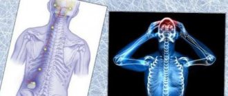
- Severe pain or increasing chronic pain in the head;
- Cloudiness of consciousness;
- Fainting;
- Meningeal syndromes are disorders of conscious activity;
- Paresis and paralysis of half the body;
- Visual pathology;
- Coordination disorders;
- Reduced blood pressure;
- Constant muscle contractions (convulsive);
- Strabismus.
Mortality due to intracerebral edema is often observed, so it is important to identify at least one sign to prescribe additional diagnostic methods - neuroimaging (MRI and SCT of the head).
Tests and diagnostics
Cerebral edema is an urgent condition that requires urgent medical care in a hospital setting (intensive care/resuscitation department), therefore, the initial diagnosis of acute brain injury should be as fast as possible and carried out during treatment. The primary guideline for diagnosing AGM is clinical symptoms, however, it must not be forgotten that at this stage they can be minimally pronounced. If OGM is suspected, a neurological/ophthalmological examination is carried out, during which pain, verbal-acoustic, behavioral reactions, ocular/pupillary reflexes, the condition of the optic nerve head, and intraocular pressure indicators are assessed. A clinical/biochemical blood test and lumbar puncture are required.
The main methods of instrumental diagnostics are computed tomography and nuclear resonance imaging, which allow one to visualize the localization/extension of the area of hyperhydration and identify signs of compression syndrome. According to indications, ultrasound of the skull in different projections, echoencephalography/electroencephalography, neuroophthalmoscopy, and cerebral angiography can be performed.
Differential diagnosis is carried out with thromboembolism of cerebral vessels , metabolic disorders , and status epilepticus .
Consequences of cerebral edema
Dangerous complications of pathology are determined by a number of pathogenetic changes:
- An increase in the amount of fluid with an increase in the degree of compression of the cerebral parenchyma, the disposition of brain tissue. Mortality occurs when available space for water storage runs out;
- Early detection, drug elimination of the pathology with subsequent resorption of free fluid is a favorable prognosis. Timely delivery of people to the intensive care unit or toxicology department allows to remove toxins and eliminate other etiological factors;
- Removal of edema after disability occurs. Meningitis, meningoencephalitis due to traumatic brain injury lead to irreversible neurological disorders.
Even after timely surgery, complete relief from the consequences of the disease cannot be guaranteed.
Neurogenic consequences of cerebral edema:
- Memory disorders;
- Strabismus divergent;
- Severe headaches;
- Motor disorders;
- Depressive state;
- Epileptic seizures;
- Pathology of speech and attention;
- Facial asymmetry.
The consequence of the condition can be constant anxiety and chronic fatigue.
Diet
Nutrition in the first 1-3 days of AGM is carried out parenterally, for which protein hydrolysates, glucose solutions, amino acids, plasma, albumin, a complex of vitamins, and special nutritional mixtures are administered. The timing of the start of enteral nutritional support through a tube is determined depending on the patient's condition, however, to restore gastrointestinal motility, it should begin as early as possible. For this purpose, special quickly digestible high-calorie nutritional mixtures are used, enriched with vitamins/microelements, a set of essential/essential amino acids in optimal dosages ( Nutrizon Energy , Nutrizon , Berlamin , Nutrizon Protein , etc.). It is recommended to use highly concentrated mixtures to reduce the volume of fluid administered, but at the same time provide the body with the necessary amount of calories. The patient can begin eating by mouth after regression of brain disorders.