Ultrasound of cerebral vessels is an absolutely painless technique that helps to identify possible pathological processes. It has been scientifically proven that ultrasonic waves do not have a negative effect on organs and tissues, therefore such manipulation can be carried out not only for diagnosis in adults, but also in children. The number of procedures and frequency of examinations are not limited - ultrasound can be performed as often as necessary. Brain examination using ultrasound is carried out both according to the doctor’s indications and at the request of the patient. Let's look at how an ultrasound is performed and how to properly prepare for it.
Medical certificate
The main components of the brain are 4 ventricles. They are represented by small cavities. They constantly produce and circulate cerebrospinal fluid. Its main purpose is to transport nutrients to cells and remove waste products. In addition, cerebrospinal fluid plays a kind of shock-absorbing role during head impacts.
Normally, each ventricle contains about 150 ml of cerebrospinal fluid. It is completely updated three times a day. An increase in its volume provokes an increase in the size of the ventricles and a change in their shape. When the lateral C-shaped curvature disappears, the cavity becomes rounded. In this case, they speak of the initial stage of lateroventriculoasymmetry of the brain.
What it is? Such a terrible diagnosis refers to a disorder characterized by an asymmetric change in the size of the ventricles. To a small extent, such changes are not considered a pathology. However, their noticeable progression can lead to severe brain disorders. Enlarged ventricles begin to put pressure on surrounding elements and structures, causing disruption of the functioning of neurons. In some cases, death of nerve cells is observed.
Reasons for the development of the anomaly
The pathology can either be congenital or be the result of other health problems. In infants, lateroventriculoasymmetry is more common than in adults. Approximately every 500th newborn is diagnosed with an increase in the size of the ventricles, and in 80% of cases it is caused by congenital abnormalities in brain development. The remaining 20% are due to birth injuries. A child may develop intrauterine anomalies due to problems with the woman’s health during pregnancy. We are talking about such diseases as toxoplasmosis, rubella and mumps.
The causes of lateroventriculoasymmetry of the brain in adults are somewhat different. An increase in cerebrospinal fluid and, as a consequence, the size of the ventricles can be provoked by:
- skull injuries;
- previous neuroinfections (for example, meningitis);
- tumors, cysts, hemangiomas;
- thrombosis of cerebral vessels.
Accurate determination of the cause of the outflow disturbance and excessive production of cerebrospinal fluid allows you to prescribe effective treatment.
Reviews
Larisa Vilkova:
After birth, the child was diagnosed with mild asymmetry. I had to do an ultrasound every month. Dilatation does not increase. The doctor said that this happens due to the position of the fetus in the stomach.
Olga Klochkova:
The labor was protracted, the waterless period lasted several hours. The result is hypoxia and the diagnosis is asymmetry of the lateral ventricles. Vikasol was prescribed, and a month later no pathology was detected by the NSG.
The ventricles of the brain are a collection of cavities of the nervous system filled with cerebrospinal fluid. The ventricles perform several functions:
In the brain are located:
- Lateral ventricles;
- Third ventricle
- Fourth ventricle.
Clinical picture
Moderate lateroventriculoasymmetry of the brain does not affect the human condition. We can talk about the development of a pathological process in the presence of pronounced changes in the lateral ventricles. In this case, the patient feels the symptoms of the primary disease, which provoked lateroventriculoasymmetry. This may involve problems with sensation, memory, coordination or thinking. Typically the patient complains of:
- pain in the head, a feeling of squeezing and bursting;
- nausea and vomiting, especially in the morning;
- dizziness;
- state of anxiety.
The patient is constantly accompanied by apathy and drowsiness. Such symptoms of lateroventriculoasymmetry of the brain indicate that the pathology is progressing. As a result of impaired outflow of cerebrospinal fluid, pressure on the brain increases, which leads to:
- oculomotor disorders;
- development of dementia;
- blurred vision;
- urinary incontinence;
- impaired motor function (gait becomes unsteady).
If treatment for the anomaly is not started in a timely manner, it can become chronic.
What it is
Ventriculoasymmetry is a pathological condition in which the lateral ventricles are asymmetrical in relation to each other. Normally, they have a symmetrical arrangement and the same volume, however, due to external or internal factors, their relative position changes.
Ventricular asymmetry is considered a pathology only if it leads to dysfunction of the nervous system, deterioration of the body’s functioning and the development of symptoms. If the violation of symmetry is minimal and does not lead to disorders, this is considered an anatomical feature of the brain.
In the medical classification there is no such nosological unit (independent disease) as ventricular asymmetry, however, their asymmetry can lead to disruption of the circulation of cerebrospinal fluid, which in turn determines the clinical picture.
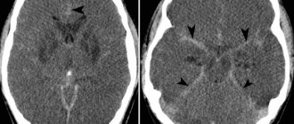
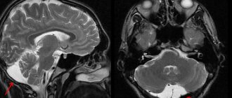
Lateroventriculoasymmetry is often latent (hidden) and does not cause symptoms. As a rule, the pathology of this system is determined by chance, by neuroimaging on computer or magnetic resonance imaging during a routine examination or after a head injury.
The change in symmetry itself is not dangerous to humans. The consequences that asymmetry can lead to are dangerous. For example, impaired liquor dynamics (fluid outflow), compression of cranial nerves, increased intracranial pressure. All consequences trigger a chain that develops its clinical picture.
Asymmetry is mainly diagnosed at an early age (up to one year) and in children.
Interhemispheric asymmetry with asymmetry of the cavities is not disturbed.
Course of the disease in children
The manifestation of the pathological process in young patients depends on age. Children are usually capricious and do not eat well. Their gaze is directed downward, but their eyes are excessively open. In children, the bones of the skull are flexible and plastic. Therefore, disorders can manifest themselves in an increase in head size. Pulsation of the fontanel and swelling of the veins are often observed.
School-age children also suffer from lateroventriculoasymmetry of the brain. Only a few know what kind of disease this is. Its development may be preceded by congenital disorders of brain formation or failures at the genetic level. At this age the disorder manifests itself:
- divergence of seams between the bones of the skull;
- slow development;
- overweight;
- sudden mood swings;
- impaired ability to move the eyeballs;
- problems with mental activity;
- decreased attachment to loved ones.
In adolescents, this disease is a consequence of cranial injuries or previous infections. It is accompanied by severe headaches, vomiting and nausea, and convulsions. Psychosis cannot be ruled out.
Severity of ventriculomegaly of the lateral ventricles
Doctors distinguish three degrees of severity of pathology:
At the first stage of development of the disease, there are no pronounced symptoms. Only a neurologist can detect it after diagnosis. Most parents notice that the child becomes excitable and irritable. Unfortunately, no other symptoms appear.
- Periodic convulsions, similar to epileptic seizures;
- Increase in head size;
- The presence of bulging veins on the forehead;
- Slow physical development;
- Mental retardation.
In addition to abnormal expansion of the ventricles of the brain, the following symptoms are present:
- Increased intracranial pressure due to the asymmetric shape of the ventricles;
- Recurrent headaches;
- Speech disorders.
Additional diseases are often added to the pathology: cerebral palsy, Down syndrome or hydrocephalus.
Diagnostic methods
Prescribing treatment and explaining what lateroventriculoasymmetry of the brain is is the task of a specialist. If symptoms of the disorder appear, you should immediately consult a therapist. After studying the clinical picture and complaints, the doctor will refer you to a neurologist. It is this specialized specialist who can confirm the preliminary diagnosis. For this purpose, he prescribes a comprehensive examination to the patient, which consists of the following activities:
- CT, MRI. These methods are considered the most informative. Using tomography, you can assess the condition of the ventricular system and determine the causes of impaired outflow of cerebrospinal fluid.
- Echoencephaloscopy.
- Electroencephalography.
- Fundus examination.
If the listed options for diagnosing lateroventriculoasymmetry of the brain give conflicting results, a lumbar puncture is prescribed. The results of cerebrospinal fluid analysis can accurately determine the causes of deformation of the lateral ventricles.
Drug therapy
Treatment of lateroventriculoasymmetry of the brain with drugs is prescribed only when the clinical picture is severe. The main direction of such therapy is the elimination of the underlying disease, which provoked a change in the size and shape of the ventricles. For this purpose, drugs from the following groups are recommended:
- Diuretics.
- Neuroprotectors.
- Vascular agents.
- Nootropic drugs.
- Sedative medications.
- Anti-inflammatory drugs.
All medications, as well as their dosage, are selected individually.
The need for surgery
When conservative therapy is ineffective, surgery is prescribed. A puncture is performed at the site of the greatest accumulation of cerebrospinal fluid. If the cause of compression of the cerebrospinal fluid outflow tract is a tumor, it must be removed.
In modern medical practice, ventricular bypass surgery has become widespread in the treatment of lateroventriculoasymmetry. During the procedure, the doctor installs a shunt, with the help of which cerebrospinal fluid begins to flow into the bladder. After the operation, the patient can return to the usual rhythm of life. Intracranial pressure gradually decreases, and other symptoms of illness disappear. Unfortunately, in half of the cases, shunting loses its effectiveness over time. The development of complications, including inflammation of soft tissues and the addition of a secondary infection, cannot be ruled out. In this case, a repeat operation with replacement of the shunt is required.
Consequences of the disease
A common question that almost all patients diagnosed with “lateroventriculoasymmetry of the brain” ask specialists: is it dangerous? It is not possible to answer this unequivocally.
If there is an unexpressed form of the disease, when changes in the lateral ventricles do not cause discomfort to the patient, specific therapy is not required. Simply observing the anomaly is enough. In case of progressive disease, treatment is necessary. It can be either medicinal or surgical. If you ignore the symptoms of the disorder and do not seek help from a doctor, the likelihood of complications increases. In addition to impaired consciousness and motor function, such a patient is at risk of coma.
Treatment
Treatment of ventricular asymmetry can be conservative or surgical. Conservative treatment is only symptomatic, since the structural disorder cannot be cured with pills. For this purpose they prescribe:
- Diuretics. Diuretics stimulate diuresis - the outflow of fluid from the body increases. This reduces intracranial pressure and suppresses symptoms.
- Neuroprotectors are agents that protect GM from metabolic disorders. Neuroprotectors also prevent metabolic disorders and irreversible changes in nerve cells.
- Vasoactive drugs are drugs that improve blood supply to the brain and restore damaged tissue.
- Nootropics are drugs that restore parameters of mental functions (speed of attention, volume of short-term memory). Nootropics act only on damaged cells and have no effect on a healthy body.
- Sedatives and anxiolytics. Used to treat agitation in children and to relieve anxiety.
If there is an objective reason, etiotropic therapy is used, aimed at eliminating the nature of the disease that led to changes in the ventricles. For example, for neuroinfections, antiviral, antibacterial and anti-inflammatory agents are prescribed. If the asymmetry is caused by impaired blood supply and acute disturbance of blood flow, drugs are prescribed that improve the rheological properties of the blood.
Surgical treatment is prescribed in the presence of tumors in the brain, complicated traumatic brain injuries, subdural hematoma (hemorrhage under the meninges that compresses the brain and displaces the ventricles).
Ventriculomegaly is a pathology that affects the ventricles of the brain. The function of these parts of the organs is to maintain intracranial pressure and ensure normal blood circulation to the human cerebral cortex.
Most often, the disease occurs in unborn babies, less often in children of preschool and school age. Ventriculomegaly in adults develops in exceptional cases, since the disease is congenital.
Let's take a closer look at what ventriculomegaly is, the causes of its occurrence and how it is treated.