Hydrocephalus is an excessive accumulation of cerebrospinal fluid in the cranial cavity, which results in an increase in the size of the subarachnoid spaces, basal cisterns, and ventricles of the brain. All conditions have been created for the treatment of patients with hydrocephalus at the Yusupov Hospital. The neurology clinic employs candidates and doctors of medical sciences, doctors of the highest category. Neurologists have the knowledge and experience to quickly diagnose the disease and provide adequate therapy.
To examine patients, doctors use the latest equipment from leading global manufacturers. When drawing up an individual treatment regimen, doctors take into account the cause, type and severity of hydrocephalus. Doctors predict the course of the disease and prevent the consequences of hydrocephalus in adults. If the therapy is ineffective, doctors collectively decide on the need to perform surgical intervention at a meeting of the expert council with the participation of professors, neurologists and neurosurgeons of the highest category.
Neurosurgeons at partner clinics use modern surgical methods to treat patients with hydrocephalus. Doctors, if indicated, perform bypass or endovascular operations. After surgical interventions, patients' symptoms of the disease decrease and their quality of life improves.
How does the disease develop and what are the consequences?
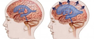
The activity of the endocrine glands, which produce cerebrospinal fluid, is disrupted. As a result, there is too much cerebrospinal fluid, and the small body is unable to ensure its normal outflow. On the left of the diagram is a normal brain, and on the right is one affected by hydrocephalus.
Hydrocephalic babies are easily recognized by their disproportionately large heads. Water cannot compress and increases the volume of the skull, and since in children under one year old rigid connections between the cranial bones have not yet formed, the increase in volume of the head occurs unhindered.
When hydrocephalus of the brain develops in an older child, excess fluid manifests itself exclusively as damage to the central nervous system.
The disease is quite dangerous: the cerebrospinal fluid does not leave the head and begins to put pressure on the nervous tissue, destroying neural connections. Without proper treatment, this usually leads to swelling of the brain and ultimately the death of the child.
Subcortical structures.
Below the cortex lies a number of important brain structures, or nuclei, which are collections of neurons. These include the thalamus, basal ganglia and hypothalamus. The thalamus is the main sensory transmitting nucleus; it receives information from the senses and, in turn, forwards it to the appropriate parts of the sensory cortex. It also contains nonspecific zones that are connected to almost the entire cortex and probably provide the processes of its activation and maintenance of wakefulness and attention. The basal ganglia are a collection of nuclei (the so-called putamen, globus pallidus and caudate nucleus) that are involved in the regulation of coordinated movements (starting and stopping them).
The hypothalamus is a small region at the base of the brain that lies beneath the thalamus. Richly supplied with blood, the hypothalamus is an important center that controls the homeostatic functions of the body. It produces substances that regulate the synthesis and release of pituitary hormones (see also pituitary gland). The hypothalamus contains many nuclei that perform specific functions, such as regulation of water metabolism, distribution of stored fat, body temperature, sexual behavior, sleep and wakefulness.
Classification of the disease
In children, there are four main types of hydrocephalus of the brain:
- External - fluid accumulates in the subarachnoid space - the cavity between the pia mater and the arachnoid mater.
- Internal - cerebrospinal fluid remains in the cerebral ventricles. The substance of the brain begins to thin out.
- Open (communicating) - all ventricular systems of the brain expand, but there are no obstacles to the movement of fluid through the cerebrospinal fluid system.
- Closed (occlusive) - disruption of the liquor flow is caused by pathologies of the ventricles: their adhesions, neoplasms.
The open form of childhood hydrocephalus can be either external or internal. But occlusal in 99% of cases is internal.
In addition, dropsy is also divided according to the form of its flow:
- Acute – a rapid increase in intracranial pressure, the baby’s condition worsens in the period from one to three days.
- Subacute - normal circulation of cerebrospinal fluid is disrupted for three to six months, after which severe brain damage occurs.
- Chronic – the development of the disease also lasts up to six months, but there is no such serious damage to the brain. Characteristic of open hydrocephalus.
Anomaly in the structure of the brain (5th ventricle)
From time to time you have to deal with an atypical structure of the brain. Such cases are of particular clinical interest.
The fifth ventricle is a slit-like hypoechoic expansion located in the anterior midline of the brain, below the corpus callosum.
This is a longitudinal medullary fissure that begins to form at 12 gestational weeks from the lamina terminalis. By 6 gestational months, this expansion begins to close, merging into one septum. From 3 months to 6 months of infancy, the septum closes completely. Extremely rarely, the cavity does not close in adulthood, forming the 5th ventricle.
Some features:
1) In childhood, expansion of more than 10 mm may indicate a disease of the cerebrospinal fluid-dynamic system;
2) The presence of the 5th ventricle on ultrasound during pregnancy indicates the normal development of the child;
3) This condition should not be confused with cystic lesions; a traumatic lesion should be excluded (can develop in boxers);
4) The cavity can be connected to the rest of the liquor system (our case) or be closed, receiving liquor from neighboring cavities through the walls (more often);
5) The prognosis is favorable, it is a feature of the structure of the brain.
MRI of the brain, 22 years old:
Do you have anything to add?!
Related articles:
Causes of pathology
As a rule, this is a congenital disease. There are also acquired forms, but they are quite rare.
The causes of cerebral hydrocephalus in children are intrauterine infections, birth injuries, genetic disorders, and central nervous system defects.
Depending on the stage at which the disease developed, its causes vary:
- Fetal hydrocephalus in the vast majority of cases develops due to defects of the central nervous system. Infectious infections are to blame for the pathology of every fifth intrauterine hydrocephalus. Only a small proportion is due to genetic disorders.
- When the disease begins to develop immediately after birth, it is most likely due to infections in the womb. Such situations account for 75%. In 15% of cases, a newly born baby begins to suffer from dropsy due to a birth injury that led to meningitis or intracranial hemorrhage. The remaining 10% are malformations of either the spinal cord or brain, as well as disturbances in the functioning of blood vessels.
- In children aged one year and older, hydrocephalus is almost equally formed as a result of one of the following reasons: hemorrhage; brain tumor (spinal or brain); consequences of TBI; consequences of brain inflammation; genetic problems; malformations of cerebral vessels.
These generalized reasons also have their own classification.
So, among the infections that cause the disease, the following are common:
- rubella;
- cytomegalovirus;
- neurosyphilis;
- herpes virus type 1 or 2;
- toxoplasmosis;
- bacteria and viruses that cause meningitis;
- parotitis.
Defects responsible for the formation of dropsy:
- Narrowing of the canals connecting the cerebral ventricles.
- Arnold-Chiari syndrome (underdevelopment of the posterior fossa of the skull, due to which the brain structures located there do not fit into it).
- Anomalies of the cerebral venous system.
- Underdevelopment of the openings through which cerebrospinal fluid drains into the subarachnoid space.
- Dandy-Walker syndrome (anomaly of the development of the cerebellar spaces and cerebellum).
Also, among oncological diseases, hydrocephalus can be caused by:
- brain cancer;
- papilloma;
- tumor of the skull bones;
- brain ventricle tumor;
- vascular plexus meningioma;
- various forms of spinal cord oncology that impair fluid circulation.
Ventricles of the human brain
The human brain is made up of an amazing number of neurons - about 25 billion of them, and this is not the limit. Neuronal cell bodies are collectively called gray matter because they have a gray tint.
The arachnoid membrane protects the cerebrospinal fluid circulating inside it. It acts as a shock absorber that will protect the organ from impact.
The brain mass of a man is higher than that of a woman. However, the opinion that a woman’s brain is inferior in development to a man’s is erroneous. The average weight of the male brain is about 1375 g, the female brain is about 1245 g, which is 2% of the weight of the entire body. By the way, brain weight and human intelligence are not interrelated. If, for example, you weigh the brain of a person suffering from hydrocephalus, it will be larger than usual. At the same time, mental abilities are significantly lower.
The brain consists of neurons - cells capable of receiving and transmitting bioelectric impulses. They are supplemented by glia, which help neurons function.
The ventricles of the brain are cavities inside the brain. It is the lateral ventricles of the brain that produce cerebrospinal fluid. If the lateral ventricles of the brain are impaired, hydrocephalus may develop.
How does the brain work?
Before moving on to considering the functions of the ventricles, let us recall the location of some parts of the brain and their significance for the body. This will make it easier to understand how this whole complex system works.
The brain is finite
It is impossible to briefly describe the structure of such a complex and important organ. The telencephalon runs from the back of the head to the forehead. It consists of large hemispheres - right and left. It has many grooves and convolutions. The structure of this organ is closely related to its development.
Conscious human activity is associated with the functioning of the cerebral cortex. Scientists distinguish three types of bark:
- Ancient.
- The old one.
- New one. The rest of the cortex, which was the last to develop during human evolution.
Hemispheres and their structure
The hemispheres are a complex system that consists of several levels. They have different parts:
- frontal;
- parietal;
- temporal;
- occipital
In addition to the lobes, there is also a cortex and subcortex. The hemispheres work together, they complement each other, performing a set of tasks. There is an interesting pattern - each part of the hemispheres is responsible for its own functions.
Bark
It is difficult to imagine that the cortex, which provides the main characteristics of consciousness and intelligence, is only 3 mm thick. This thinnest layer reliably covers both hemispheres. It is composed of the same nerve cells and their processes, which are located vertically.
The layering of the crust is horizontal. It consists of 6 layers. The cortex contains many vertical nerve bundles with long processes. There are more than 10 billion nerve cells here.
The cortex is assigned various functions that are differentiated between its different sections:
- temporal – smell, hearing;
- occipital – vision;
- parietal – taste, touch;
- frontal – complex thinking, movement, speech.
It affects the brain structure. Each of its neurons (we remind you that there are about 25 billion of them in this organ) creates about 10 thousand connections with other neurons.
In the hemispheres themselves there are basal ganglia - these are large clusters that consist of gray matter. It is the basal ganglia that transmit information. Between the cortex and the basal ganglia are the processes of neurons - the white matter.
It is the nerve fibers that form the white matter; they connect the cortex and those formations that are under it. The subcortex contains the subcortical nuclei.
The telencephalon is responsible for physiological processes in the body, as well as intelligence.
Intermediate brain
It consists of 2 parts:
- ventral (hypothalamus);
- dorsal (metathalamus, thalamus, epithalamus).
It is the thalamus that receives stimuli and sends them to the hemispheres. This is a reliable and always busy intermediary. Its second name is the visual thalamus. The thalamus ensures successful adaptation to an ever-changing environment. The limbic system reliably connects it to the cerebellum.
The hypothalamus is a subcortical center that regulates all autonomic functions. It affects through the nervous system and glands. The hypothalamus ensures the normal functioning of individual endocrine glands and participates in metabolism, which is so important for the body. The hypothalamus is responsible for the processes of sleep and wakefulness, eating and drinking.
Below it is the pituitary gland. It is the pituitary gland that provides thermoregulation, the functioning of the cardiovascular and digestive systems.
hindbrain
It consists of:
- front axle;
- the cerebellum behind it.
The bridge visually resembles a thick white cushion. It consists of a dorsal surface, which is covered by the cerebellum, and a ventral surface, the structure of which is fibrous. The bridge is located above the medulla oblongata.
Cerebellum
It is often called the second brain. This department is located behind the bridge. It covers almost the entire surface of the posterior cranial fossa.
The large hemispheres hang directly above it, separated only by a transverse gap. Below, the cerebellum is adjacent to the medulla oblongata. There are 2 hemispheres, the lower and upper surface, the worm.
The cerebellum has many slits along its entire surface, between which one can find convolutions (ridges of the medulla).
The cerebellum consists of two types of substance:
- Gray. It is located on the periphery and forms the cortex.
- White. It is located in the area under the bark.
The white matter penetrates into all convolutions, literally penetrating them. It can be easily recognized by its characteristic white stripes. In the white matter there are inclusions of gray - the nucleus. Their interweaving in cross-section visually resembles an ordinary branched tree. It is the cerebellum that is responsible for coordinating movements.
Midbrain
It is located from the anterior region of the bridge to the optic tracts and papillary bodies. There are many nuclei (tubercles of the quadrigeminal). The midbrain is responsible for the functioning of latent vision and the orienting reflex (it ensures that the body turns to where the noise is heard).
Ventricles
The ventricles of the brain are cavities connected to the subarachnoid space, as well as the spinal cord canal. If you're wondering where cerebrospinal fluid is produced and stored, it happens in the ventricles. Inside they are covered with ependyma.
Ependyma is a membrane that lines the surface of the ventricles from the inside. It can also be found inside the spinal canal and all cavities of the central nervous system.
Types of ventricles
Ventricles are divided into the following types:
- Lateral. Inside these large cavities there is cerebrospinal fluid. The lateral ventricle of the brain is large in size. This is explained by the fact that quite a lot of fluid is produced, because not only the brain, but also the spinal cord needs it. The left ventricle of the brain is called the first, the right - the second. The lateral ventricles communicate with the third ventricle through foramina. They are symmetrically located. From each lateral ventricle departs the anterior horn, the posterior horns of the lateral ventricles, the lower, and the body.
- Third. Its location is between the visual tuberosities. It has the shape of a ring. The walls of the third ventricle are filled with gray matter. There are many autonomic subcortical centers here. The third ventricle communicates with the midbrain and lateral ventricles.
- Fourth. Its location is between the cerebellum and the medulla oblongata. This is the remnant of the cavity of the brain bladder, which is located behind. The shape of the fourth ventricle resembles a tent with a roof and a bottom. Its bottom is diamond-shaped, which is why it is sometimes called a diamond-shaped fossa. The spinal cord canal opens into this fossa from behind.
The shape of the lateral ventricles resembles the letter C. They synthesize cerebrospinal fluid, which must then circulate in the spinal cord and brain.
If the cerebrospinal fluid does not drain correctly from the ventricles, the person may be diagnosed with hydrocephalus. In severe cases, it is noticeable even in the anatomical structure of the skull, which is deformed due to strong internal pressure. Excess liquid tightly fills the entire space. It can change the functioning of not only the ventricles, but also the entire brain. Excessive amounts of cerebrospinal fluid can cause a stroke.
Diseases
The ventricles are susceptible to a number of diseases. The most common among them is the above-mentioned hydrocephalus. With this disease, the cerebral ventricles can grow to pathologically large sizes. In this case, the head hurts, a feeling of pressure appears, coordination may be impaired, nausea and vomiting appear. In severe cases, it is difficult for a person to even move. This can lead to disability and even death.
The appearance of the mentioned signs may indicate congenital or acquired hydrocephalus. Its consequences are detrimental to the brain and the body as a whole. Blood circulation may be impaired due to constant compression of soft tissues, and there is a risk of hemorrhage.
The doctor must determine the cause of hydrocephalus. It can be congenital or acquired. The latter type occurs with a tumor, cyst, injury, etc. All departments suffer. It is important to understand that the development of pathology will gradually worsen the patient’s condition, and irreversible changes will occur in the nerve fibers.
The symptoms of this pathology are associated with the fact that more cerebrospinal fluid is produced than necessary. This substance quickly accumulates in the cavities, and since there is a decrease in outflow, the cerebrospinal fluid does not drain away as it should normally. The accumulated cerebrospinal fluid can be in the ventricles and stretch them, it compresses the vascular walls, impairing blood circulation. Neurons do not receive nutrition and quickly die. It is impossible to restore them later.
Hydrocephalus often affects newborns, but it can appear at almost any age, although it is much less common in adults. Proper circulation of cerebrospinal fluid can be established with proper treatment. The only exception is severe congenital cases. During pregnancy, ultrasound can reveal possible hydrocephalus in the baby.
If during pregnancy a woman indulges in bad habits and does not follow proper nutrition, this entails an increased risk of fetal hydrocephalus. Asymmetric development of the ventricles is also possible.
To diagnose pathologies in the functioning of the ventricles, MRI and CT are used. These methods help to identify abnormal processes at a very early stage. With adequate treatment, the patient's condition can improve. Even a complete recovery is possible.
The ventricles may be subject to other pathological conditions. For example, their asymmetry has a negative impact. Tomography can reveal it. Asymmetry is caused by vascular dysfunction or degenerative processes.
Pathological changes can also be caused by tumors and inflammation.
If there is an increased volume of cerebrospinal fluid, this can happen not only due to its excessive production, but also due to the fact that there is no normal outflow of fluid. This may be the result of the appearance of neoplasms, hematomas, or blood clots.
With diseases of the ventricles, the patient is concerned about serious health problems. The brain suffers from a deficiency of nutrients, oxygen, and hormones. In this case, the protective function of the cerebrospinal fluid is disrupted, poisoning of the body begins, and intracranial pressure increases.
Conclusion
The ventricles are interconnected with many organs and systems; human health as a whole depends on their condition. If an MRI or CT scan reveals their expansion, you should immediately consult a doctor. Timely treatment will help you return to a full life.
Risk factors
There are accompanying conditions that contribute to the development of hydrocephalus of the brain in a child:
- Early birth (gestation period did not exceed 34 weeks).
- A narrow maternal pelvis and, as a result, a difficult birth process.
- The newborn's light weight is less than one and a half kilograms.
- During childbirth, hypoxia or asphyxia was observed in the fetus.
- Active methods of obstetrics: manual techniques, vacuum, forceps.
- The mother suffered viral infections during pregnancy: herpes, influenza, ARVI, toxoplasmosis.
- Bad habits of the mother that manifested themselves during pregnancy.
- Untreated sexually transmitted diseases in the mother during pregnancy.
Most risk factors for hydrops are related to maternal health.
Even indirect reasons, such as a premature baby with low weight, indicate an abnormal pregnancy. Every problem with a woman’s health during pregnancy weakens the embryo’s body.
How does hydrocephalus manifest?
Depending on the form of development of the pathology and the age of the child, the external signs of the disease change.
For children under two years of age, in which in 95% of cases hydrocephalus is a congenital pathology, the following symptoms are characteristic:
- Severe course of the disease. A rapidly deteriorating condition caused by damage to brain structures.
- The main symptom is a rapid increase in head volume. Growth of 1.5 cm or more every month is typical, for at least three months in a row. Starting from the ninth month of life, growth drops to 8-9 mm.
- A child is born with a head whose girth is larger than the girth of the chest. If by six months the ratio does not change, and the head is still larger than the chest, there is reason to suspect hydrocephalus.
- The veins on the occipital, temporal and frontal parts of the head are clearly visible.
- At three months the child does not yet hold his head up and begins to sit up late, crawl, and walk.
- The fontanel on the top of the head is convex.
- The accumulation of cerebrospinal fluid primarily occurs in the frontal lobes of the brain. Head enlargement starts from the forehead.
- While fixating the baby's gaze, the baby's pupil twitches chaotically.
- The scalp becomes thin and has a painful shine.
- Overhang of the brow ridges over the facial bone - the eyes appear to be very deep-set.
A number of less characteristic signs can confuse the initial diagnosis of hydrocephalus in children and complicate treatment. For example, tearfulness, poor appetite, slow weight gain, frequent regurgitation, drowsiness, legs constantly bent at the knees, head tilting - all these symptoms are characteristic of hydrocephalus. But at the same time, they can be observed in dozens of other childhood diagnoses.
In the acute form of dropsy, when the disease progresses rapidly, other signs can be observed:
- convulsions;
- long cry on one note;
- loss of acquired physical skills (sitting, following other people's movements, vocal functions);
- vomit.
For children aged two years and older, other manifestations of hydrocephalus of the brain are typical:
- Headaches that begin in the morning (after sleep) and gradually subside in the evening. Nausea and vomiting often begin at the same time.
- Migraines that appear after stress, mental or physical work. Often accompanied by nosebleeds.
- A feeling of a rush to the head when bending over and bursting headaches.
- Pain in the fundus of the eye.
- Visual impairment: loss of sharpness, double vision.
- Urinary incontinence.
- Decreased muscle strength, accompanied by rapid fatigue.
- Cramps and fainting.
- Loss of coordination and uncontrolled movements of the limbs.
Treatment of hydrocephalus in children
Due to the complexity of the disease and its danger, treatment for a child with hydrocephalus is prescribed not only by a neurosurgeon, but also by a neurologist.
Treatment can be conservative (that is, without surgery, only with the help of medications) or surgical.
During drug treatment, the first step is to reduce intracranial pressure using the drug Diacarb. It reduces the production of cerebrospinal fluid and promotes the removal of potassium from the body.
In the hospital, strong osmotic diuretics are prescribed - oral Glycerin and Mannitol. Salt diuretics, for example Furosemide, are also prescribed. It, like Diacarb, should be taken in conjunction with drugs that maintain normal potassium levels - Panangin or Asparkam.
Doctor's advice
Hydrocephalus - this conclusion can often be found after an ultrasound scan of infants. However, in most cases it is compensatory in nature, everything returns to normal by the first year of life. In addition to birth injuries, protracted labor, structural features, the cause of such dropsy is the immaturity of the nervous system. True hydrocephalus is characterized by the entire clinical picture of increased intracranial pressure - an increase in the size of the head, monotonous crying, and the presence of neurological symptoms. Conclusion Ultrasound is not a diagnosis, it is the result of an examination; the diagnosis is made based on a combination of complaints, anamnesis, examination, and research data.
Victoria Druzhikina Neurologist, Therapist
Any medicinal forms of treatment for acute hydrocephalus must be carried out under the strict supervision of doctors, and intermediate results must be checked using computed tomography and neurosonography.
Along with diuretic enzyme blockers, medications are prescribed that stimulate the functioning of brain neurons. For example, "Encephabol".
As for surgical intervention, the most effective method of surgical treatment of the brain in children is bypass surgery. Often this is the only way to save a child's life.
Shunting is the creation of a flow path (shunt) for cerebrospinal fluid to bypass an existing blockage. For closed hydrocephalus, it is used when tumors have already grown into the brain.
In the open form of the disease, three types of bypass surgery are provided:
- Ventriculoatrial.
- Ventriculo-peritoneal.
- Lumbo-peritoneal.
In addition to shunting, another type of surgical intervention is possible for closed forms of hydrops - dissection of arachnoid adhesions. This method involves the absorption of cerebrospinal fluid.
Watch the video about the causes and treatment of hydrocephalus:
The life span and quality of life of a child with hydrocephalus, his chances of recovery depend on many factors: the reasons that caused the development of the disease, the speed of diagnosis, the quality of treatment. But even a healthy baby can become a victim of the disease.
Don't forget about prevention. Protect your child from head injuries in every possible way; be sure to use protective equipment during active recreation. If the baby was born premature, be sure to undergo routine examinations with a neurologist, do an ultrasound and an MRI.
Women who are about to become mothers or planning to conceive a child should be aware of their health. Get rid of bad habits a few months before pregnancy. Get screened for infections. If you happen to get sick while pregnant, consult a geneticist and undergo an additional ultrasound.
Your unborn baby is sensitive to literally everything, so take care of him even before birth.
This article has been verified by a current qualified physician, Victoria Druzhikina, and can be considered a reliable source of information for site users.
Bibliography
1. https://docs.cntd.ru/document/499002598
Rate how useful this article was
3.5 4 people voted, average rating 3.5
Did you like the article? Save it to your wall so you don’t lose it!