1. Meningioma of the brain
Meningioma
is a tumor that forms on the membranes covering the brain and spinal cord inside the skull. Tumors form on three layers of membranes called meninges.
Meningioma is usually a benign tumor
(in 90% of cases), which means it is not an oncological disease.
Some brain meningiomas are classified as atypical
.
This is an intermediate state - the tumor is considered neither benign nor malignant. But in this case, there is a risk that the meningioma may develop into a cancerous tumor. Malignant meningioma,
unlike benign meningioma, grows quickly. And it can spread (metastasize) to other parts of the brain and other organs. Metastases often appear in the lungs.
Most meningiomas form on the membranes of the brain. Less common are meningiomas that grow on part of the spinal cord.
In general, brain meningioma is the most common type of tumor affecting the human nervous system. This pathology occurs more often in women than in men. According to statistics, meningioma develops in middle-aged women three times more often than in men. Most tumors are diagnosed in patients between 40 and 70 years of age. Very rarely this disease occurs in children.
Often, brain meningioma does not cause any unpleasant symptoms and does not require urgent treatment. But the growth of benign brain meningioma can cause serious problems over time. In some cases, even death.
A must read! Help with treatment and hospitalization!
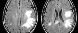
Meningiomas: large and small
This example clearly demonstrates that the manifestations of brain tumors largely depend not on their size and “benign quality,” but on their location. In the first case, a 58-year-old patient came to me with the only complaint of “forgetfulness.” Perhaps the patient would not have contacted a neurosurgeon if it had not been for the persistence of his relatives, who noticed how significantly the patient’s character and behavior had changed. Irritability, decreased criticism, and inappropriate actions served as a reason for examination. According to the results of the MRI study, a giant tumor was identified - a large bilateral parasagittal meningioma of the anterior third of the phalx, more on the left. It was this formation that was the cause of the above complaints. The patient underwent surgery to remove a brain tumor. Histological examination confirmed the presumptive diagnosis of meningioma. After the patient “came to his senses,” he was able to remember that eight years before the operation, he was diagnosed with a small meningioma of the indicated location, but the patient refused treatment and was not observed by a neurosurgeon. According to a control MRI study performed two years after surgery, no signs of tumor recurrence were detected.
In the second case, a 40-year-old patient, in perfect health, decided to be examined “just in case” and, on his own initiative, performed an MRI examination. , a small meningioma of the pineal region was identified without signs of a volumetric effect or impaired outflow of cerebrospinal fluid. Given the absence of clinical manifestations of the disease, the patient refrained from surgery. However, after three months, the patient noted the appearance of episodes of persistent headache with nausea and vomiting at the height of the attack, and deterioration of vision. Another MRI study confirmed a slight increase in tumor size, which led to impaired cerebrospinal fluid circulation and the occurrence of occlusive attacks. This was the reason for hospitalization and surgical intervention. The patient was discharged from the hospital in satisfactory condition with complete regression of existing complaints. According to the control contrast-enhanced MRI study, no signs of disease recurrence were detected.
The MRI images provided below clearly show the impressive size of the patient’s tumor (images of patient A) with “memory deterioration” and the seemingly insignificant size of the meningioma of the pineal region (images of patient B) in a patient with pronounced and rapidly emerging symptoms of the disease.
2.Causes and risk factors of meningioma
The causes of brain meningioma are not fully understood. But there are two known risk factors -
- Exposure to radiation;
- Neurofibromatosis type 2 is a genetic disease.
Previous injuries may also be a risk factor. Thus, brain meningiomas were found in patients in places where there had previously been a skull fracture and where the membranes surrounding the brain were injured.
Some studies have confirmed a connection between the growth of brain meningioma and the hormone progesterone.
Visit our Oncology page
A comment
Petroclival meningiomas are among the most difficult tumors to surgically remove. Considering the localization and involvement of the main blood vessels and cranial nerves in the tumor stroma, radical removal of petroclival meningioma is always associated with a high risk of profound disability for patients. Despite the extremely high relevance of the problem, it must be recognized that recently there have been few publications on this topic in the domestic literature. The work is a detailed review of the scientific literature on modern approaches to the treatment of petroclival meningiomas, their diagnosis, and biology. The results of surgical and radiation treatment of large series of patients with petroclival meningiomas are described in detail. The authors reasonably come to the conclusion that it is advisable for patients with radically inoperable petroclival meningiomas to undergo palliative surgery followed by radiation or symptomatic therapy. Of particular interest is the review's mention of decompression of the craniovertebral junction and structures of the posterior cranial fossa as a means of preparing the patient for radiation therapy.
Article by V.N. Szymanski et al. is relevant and modern, includes an analysis of recent works on the use of fluorescence navigation in meningioma surgery, new methods in the diagnosis of meningiomas, for example positron emission tomography. The work is of undoubted interest for a wide range of neurosurgeons, radiologists, and neurologists.
Of course, the article is worthy of publication in a neurosurgical journal, since it broadens the horizons of neurosurgeons and doctors of related specialties regarding the choice of treatment tactics for petroclival meningiomas, and demonstrates most of the modern options for their diagnosis and treatment. After reading the review, the conclusion arises that the decision to choose one or another tactic for treating petroclival meningiomas should be made collectively, together with a neurosurgeon and a radiologist.
M.A. Stepanyan (Moscow)
4. Diagnosis and treatment of meningioma
Diagnosis of meningiomas
It is rarely possible to diagnose brain meningioma before patients complain of specific symptoms. But if the doctor has reason to suspect a tumor, a brain scan may be prescribed - MRI and/or computed tomography
. This will help the doctor find the brain meningioma and determine its size.
a biopsy is done to diagnose meningioma.
. The surgeon will remove the tumor or part of it to determine whether it is benign or cancerous.
Treatment of meningioma
If the tumor does not cause any unpleasant symptoms, doctors often recommend observation and watchful waiting. Regular brain scans will help detect tumor growth if it occurs.
If the growing tumor begins to pose a health risk or symptoms of meningioma appear, surgery may be necessary to treat brain meningioma.
Surgical treatment of brain meningioma usually involves craniotomy. The doctor will remove part of the bone from the skull and thereby gain access to the affected part of the brain. The surgeon will then remove as much of the meningioma as possible, or as much of it as possible. After this, the integrity of the skull will be restored.
The possibility of surgery depends on where the tumor is located. If meningioma cannot be treated with surgery, radiation therapy may be prescribed. Radiation will help shrink the tumor or stop it from growing.
Radiation therapy is used to treat meningioma and if the tumor is malignant and the cancer cells need to be killed.
Treatment options
There are many methods of recognized medicine (medication, surgical intervention, radiosurgery, constant monitoring). But besides this, you can also help cope with brain meningioma using folk remedies. They will help not only eliminate unpleasant manifestations, but also help in the resorption of the formation.
Propolis
Combine propolis with alcohol in a ratio of 1 to 10. Let it brew and take a teaspoon three times a day before meals. It must be remembered that the use of royal jelly is prohibited for patients with tumors, as it can provoke their growth. But other bee products are not prohibited.
Chestnut
Take dried and fresh chestnut flowers in a ratio of 1:2. Pour 200 ml of boiling water. Let it sit for 8 hours. Throughout the day, drink 1.5 liters in small sips.
Simple ways to treat complex diseases:
Horseradish is the only plant that can draw salt through the pores of the skin. Do it - you won't regret it! Horseradish leaves will help get rid of all the salt that has accumulated in the body and can lead to painful salt deposits...Check... Read more
Never give an antibiotic BEFORE you get a blood test with a leukemia formula. Remember, write to yourself somewhere in a visible place!!! INCREASED leukocytes, ESR, lymphocytes - VIRUS. INCREASED leukocytes, ESR, segmented and rod neutrophils... Read more
What 1 glass of this drink will do to your liver can be called a real miracle! If the liver is overloaded or does not work well, we immediately feel it. Weakness, lack of energy, dizziness, nausea, pain in the right hypochondrium, problems with food... Read more
Dandelion is the elixir of life, and what a medicine!!! The medicinal dandelion is an unpretentious plant, but contains a good half of the chemical elements of the periodic table. Sodium, potassium, manganese, magnesium, and... Read more
Seeds that repair tendons and reduce joint pain. We treat osteoporosis and osteoarthritis. Osteoarthritis of the knee is a type of degenerative joint disease or arthritis that is localized in the knee and can cause pain and di... Read more
INTERESTING fact: Treatment of cancer with aspen
Calendula
Pour a glass of hot water over a tablespoon of calendula flowers. Let it sit for a while and then drink.
Potato
Among other folk remedies for treating brain meningioma, one can highlight the use of potato flower stalks. You should not exceed the recommended dose, as they are toxic and can cause negative effects. Combine a couple of tablespoons of the main ingredient with 0.25 ml of boiling water. You need to wait 3 hours and take 1⁄2 cups.
mistletoe
The use of mistletoe can very often be found in different variations. Before you start using it, you should consult your doctor about the dosage and advisability of use. 200 ml of goat's milk should cover 3 g of dried flowers.
Celandine
Pour 5 g of celandine herb into an enamel bowl. Add 200 ml hot liquid. Cover with a lid and let stand for a quarter of an hour. Then strain and bring with boiled water to the original volume. Drink 1/3 glass twice a day before meals. Store the finished product for no more than 2 days in a cool place.
Aloe
Find a three year old aloe plant. Finely chop 5 leaves and place in a jar. Cover with 0.2 liters of vodka. Place in a place protected from sunlight for 12 days. Shake the contents regularly. After the allotted time, drink a tablespoon three times a day 2 hours before meals.
There are some tips that you should listen to when dealing with this disease:
- To slightly normalize the state of the nervous system, steam a tablespoon of shiksha herb in a glass of water in a water bath and drink.
- Try to exclude foods that contain large amounts of salt.
- Add honey to your drinks and food instead of sugar.
- Regularly drink teas and decoctions of rose hips, mint, blueberries, and raspberries.
- Products such as wild garlic, onions, and garlic help saturate the body with vitamins and antioxidants that resist negative cells.
- Steamed herbs of sage, linden, and pine needles, which are placed between layers in gauze and applied as a bandage to the head for 6-8 hours, have a good effect on relieving swelling. You can cover the temporal and frontal lobes with clay diluted with acetic acid. Leave it for several hours.
- Control your stool. Many people advise sometimes taking a laxative, because it also removes excess fluid.
INTERESTING fact: Pancreatic cancer with liver metastases, how long do they live?
Remember that self-medication can lead to worsening of the condition. That is why alternative treatment of brain meningioma should be carried out under the supervision of an experienced specialist.
Health to you!
METHODS FOR DIAGNOSIS OF BRAIN HEMANGIOMA
Rambam Medical Clinic provides modern diagnostics at a high-quality level. In this case, innovative methods and laboratory procedures are used using improved equipment and research methods. At the first stage of diagnosis, complaints are identified and anamnesis is collected. Auxiliary tests and examinations are carried out:
- RT of the brain is carried out to determine the presence of neoplasms of a certain popcorn shape.
- CT scan using a contrast agent is used to identify the qualities of a hemangioma, namely its size, shape and effect on vital brain structures, etc.
Some diseases have the same symptoms as hemangioma. Doctors at the Rambam Medical Center recommend additional studies and diagnostics to make an accurate diagnosis, excluding other types of diseases.
OPTIONS FOR COMPLICATIONS IN BRAIN HEMANGIOMA
Speaking about complications with hemangioma, we can highlight bleeding from the tumor sites. In this case, convulsive seizures may occur. They can be controlled with the help of special medications. But to completely eliminate them, surgical removal of the hemangioma is necessary. If bleeding occurs, it can be fatal. Especially if the tumor is located close to the brain stem. This may cause a stroke.
TREATMENT OF HEMANGIOMA OF THE BRAIN IN RAMBAM
At the Rambam Medical Clinic, the tumor is removed through an opening in the skull. This is the most common surgical method. It is the most effective and successful in the fight against this disease. Tumors that are localized in hard-to-reach areas, including near the brain stem, are removed using focused radioactive radiation, namely stereotactic radiosurgery.
METHODS FOR PREVENTION OF BRAIN HEMANGIOMA
In the modern world, there are no specific ways to prevent the development of hemangioma in the brain. This disease can occur due to hereditary factors. If there have been cases of this disease in your family, then it is better to eliminate the risks when planning a pregnancy. If radiation therapy is replaced with other procedures, the risk of this disease is reduced significantly. This has been proven by experts. In Israel, numerous studies are currently being conducted to identify new methods for eliminating and preventing this disease.
ABOUT THE FORECAST
When talking about the prognosis for hemangioma, it is necessary to take into account the severity of the disease. Most often, this disease occurs without identified symptoms or with minor signs. They can be controlled with therapy. Hemangioma increases in size over time, but sometimes its growth stops at a certain stage. However, if the disease causes convulsions and bleeding, surgical removal of the formation is necessary.
Diagnosis of meningioma
Small meningioma can be discovered incidentally during an examination for other diseases. If it is already large enough, unfavorable symptoms occur, which force the person to seek medical help. Sometimes a complex of symptoms leads to an erroneous diagnosis, so it is advisable to use magnetic resonance imaging (MRI) - today this is the most informative method for examining the brain.
For meningioma, surgery is almost always prescribed. Antitumor drugs for this type of formation cannot provide positive dynamics, but drugs are used to eliminate symptoms: diuretics, antiemetics, anticonvulsants, etc.
If surgery is not possible (the tumor is inoperable), the patient is offered to undergo stereotactic radiation therapy for meningioma.