Cerebral leukoencephalopathy is a pathology in which white matter is damaged, leading to dementia. For various reasons, there are several nosological forms. The occurrence of leukoencephalopathy is common for them.
The disease can be caused by:
- viruses;
- vascular abnormalities;
- insufficient oxygen supply to the brain.
Other names for the disease: encephalopathy, Binswanger disease. This pathology was first described at the end of the 19th century by the German psychiatrist Otto Binswanger, who named it after himself. From this article you will learn what it is, what are the causes of the disease, how it manifests itself, how it is diagnosed and treated.
Leukoencephalopathy - clinical forms, diagnosis
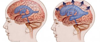
The medical term “leukoencephalopathy” is used to define a group of diseases accompanied by damage to the white matter and a number of deep structures of the brain.
Rapid progression leads to the formation of senile dementia. In children, there are vascular varieties, congenital forms with a long chronic course. The survival time for this type is longer compared to its multifocal counterpart.
MR image of vascular leukoencephalopathy
To distinguish the pathology from a number of other neurodegenerative diseases with similar clinical symptoms, a classification according to ICD 10 has been developed, which clearly distinguishes the forms of the nosology.
Therapy
Leukoencephalopathy is an incurable disease. But be sure to go to the hospital to choose drug addiction treatment. The goal of therapy is to slow the progression of the disease and activate brain function.
Treatment of leukoencephalopathy is complex, symptomatic and etiotropic. In each case it is selected individually.
Your doctor may prescribe the following medications:
- drugs that improve cerebral circulation (Vinpocetine, Actovegin, Trental);
- neurometabolic stimulants (Phesam, Pantocalcin, Lucetam, Cerebrolysin);
- angioprotectors (Stugeron, Curantil, Zilt);
- multivitamins, including B vitamins, retinol and tocopherol;
- adaptogens such as aloe extract, vit;
- glucocorticosteroids that help reduce inflammation (prednisolone, dexamethasone);
- antidepressants (fluoxetine);
- anticoagulants that reduce the risk of thrombosis (Heparin, Warfarin);
- if the disease is viral in nature, Zovirax, Cycloferon, Viferon are prescribed.
- physiotherapy;
- reflexology;
- acupuncture;
- breathing exercises;
- homeopathy;
- phytotherapy;
- massage of the collar area;
- manual therapy.
The difficulty of therapy is that many antiviral and anti-inflammatory drugs do not cross the BBB and, therefore, do not target pathological lesions.
What is cerebral leukoencephalopathy
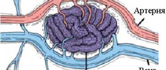
Damage to the white matter of the brain is caused in most cases by viruses. Vascular and discirculatory forms are caused by impaired blood supply to a certain area of the brain. Chronic ischemia causes irreversible changes.
Clinical symptoms of the disease more often occur when affected by papillomaviruses. The probability of nosology in patients with HIV is less than 6% according to brain MRI statistics in St. Petersburg.
Forms of vascular origin progress slowly. The chronic course of the disease is characterized by gradual irreversible tissue damage. Mild ischemia provokes the formation of small necrotic areas. Diffuse location leads to neurological disorders.
Progressive multifocal leukoencephalopathy (PML) is a demyelinating disease caused by an opportunistic infection, affecting mainly people with immunodeficiency states who have suppressed T-cell immunity (HIV-infected patients, hematological oncology patients undergoing chemotherapy, patients with autoimmune diseases, with renal and liver failure) [52]. The occurrence of PML is associated with the activation of one of the forms of the so-called JC virus, which has tropism for oligodendrocytes, which was isolated by B. Padgett in 1971 [48]. It has been established that for the development of the disease, a combination of the following factors is necessary: a mutation in the viral genome (in the capsid protein VP1), a decrease in the patient’s immune status, a mutation in the host genome [57, 58]. For two decades after its first description, PML was a rare disease, but as HIV incidence increased, the incidence of PML increased almost 50-fold [13, 25]. Before the advent of highly active antiretroviral therapy (HAART), PML occurred in 5% of cases in patients with HIV [57].
The currently increased interest in this pathology is associated with the development of the disease in patients receiving immunosuppressive therapy, in particular purine analogues (fludarabine, nelarabine), immunosuppressants (methotrexate, azathioprine, mitoxantrone, etc.), monoclonal antibodies such as natalizumab ( a monoclonal antibody that binds to the alpha-4 subunit of alpha-4-beta-1 and alpha-4-beta-7 integrins on the surface of all leukocytes except neutrophils and is used to treat MS), efalizumab (a monoclonal antibody that binds to CD11a and is used for the treatment of psoriasis) and rituximab (a monoclonal antibody to CD20, also used to treat a number of pathologies) [1, 3, 15, 16, 34, 36, 44, 61]. The main risk factors for the development of PML in patients receiving natalizumab are: previous immunosuppressive therapy, the presence of antibodies to the JC virus in the blood, and duration of use of natalizumab for more than 24 months [19]. Moreover, the course of PML associated with the use of natalizumab is more favorable than in patients with HIV [35].
Successful treatment of PML primarily depends on a timely diagnosis. The first stage of treatment is the abolition of immunosuppressants and plasmapheresis to accelerate their elimination from the body, which, in turn, can lead to reactivation of the immune system and the development of immune reconstitution inflammatory syndrome (IRIS, the English abbreviation IRIS - immune reconstitution inflammatory syndrome), which can manifest itself in the aggravation of previously identified symptoms of PML [11, 22, 31]. The development of IRIS has also been described in patients with HIV infection during HAART therapy [55, 50]. It should be noted that in most cases of IRIS development after plasmapheresis for PML, timely administration of corticosteroids led to improvement of the condition [31].
Clinical manifestations of PML are extremely varied and nonspecific, depend on the affected area of the brain, often occur subacutely and are manifested by focal or multifocal neurological deficits that increase from several days to several weeks. In patients without HIV, the cerebral hemispheres are predominantly affected; the frequency of involvement of the brainstem in the pathological process is approximately 1:10, in contrast to HIV-positive patients, where damage to brainstem structures occurs in approximately 25% of cases [2]. In patients with immunosuppression and subacute onset of progressive neurological symptoms, the presence of PML should be considered. The final diagnosis is made based on clinical, radiological and laboratory data [1].
MRI diagnosis of PML
Among the radiation research methods, the most sensitive is magnetic resonance imaging (MRI), which can detect pathology in the early stages, often even before clinical manifestations. In the presence of clear clinical signs of PML, changes are always detected on MRI, and conversely, the absence of changes on MRI when new symptoms appear is evidence against the development of PML [2].
Damage to the brain substance in advanced PML looks like pathological zones, often more than 3 cm in size, which are located mainly in the subcortical structures, involving arcuate fibers in the pathological process, sometimes spreading to the gray matter of the cortex. The lesion is often asymmetrical, with the periventricular white matter remaining relatively spared [52]. Involvement in the pathological process of the white matter of the occipital and parietal lobes, the corpus callosum, as well as the structures of the posterior cranial fossa, mainly the middle cerebellar peduncles, is often observed [5, 12, 40, 47, 50, 63, 64]. The distribution of the pathological area may be limited only to the cerebellum and/or brainstem, and may also selectively affect the corticospinal tract. Crescent-shaped zones in the cerebellum are characteristic almost exclusively of patients with PML [12, 33, 42, 54, 59]. Despite the predominant lesion of the white matter, in PML, as already indicated, the gray matter of the cortex, as well as the deep gray matter (basal ganglia), is sometimes involved in the pathological process. In addition, pinpoint hemorrhages may be observed [41, 53]. The boundaries of pathological zones are uneven, characterized by unclear contours in the white matter and a clear contour facing the gray matter [65]. When examining the dynamics, a rapidly progressing involvement of new zones in the pathological process, merging of existing foci, varying degrees of cortical atrophy and ventriculomegaly are noted, appearing within a few months or even weeks.
Traditional MRI sequences.
Pathological zones in PML have an increased intensity of the MR signal on T2-weighted and T2-FLAIR-weighted images (T2-WI and T2-FLAIR-WI), different shapes and sizes; as the disease progresses, the foci increase and merge, which, however , is not accompanied by volumetric effects.
Small-sized zones of diffuse increase in MR signal intensity in the subcortical white matter can best be seen on T2-FLAIR-WI (Fig. 1)
, they are considered the most sensitive for diagnosing newly appeared changes in the brain substance [26].
Figure 1. Progressive multifocal leukoencephalopathy in a patient with HIV infection. MRI of the brain, T2FLAIR-weighted images in the sagittal projection. Multifocal zones of diffusely increased MR signal intensity are visualized (indicated by arrows). One of the features of pathological zones in PML is the heterogeneity of increased signal intensity in T2-weighted images (Fig. 2)
, probably due to small cystic components [52].
Figure 2. Progressive multifocal leukoencephalopathy in a patient with HIV infection. MRI of the brain, T2-weighted images, axial projection. Multifocal zones of heterogeneously increased MR signal intensity are visualized (indicated by arrows) with clear contours on the gray matter side and unclear contours in the white matter. In addition, numerous point lesions are identified along the periphery of pathological zones in most patients [65]. On T1-weighted images (T1WI), the zones themselves initially appear weakly hypointense; as the disease progresses, the signal intensity continues to decrease (Fig. 3)
. Figure 3. Progressive multifocal leukoencephalopathy in a patient with HIV infection. MRI of the brain, T1-weighted images in the coronal projection. Zones of heterogeneously reduced MR signal intensity are visualized (indicated by arrows). Signal restoration on T1-weighted images does not occur without appropriate therapy [64].
The appearance of pathological changes in the brain substance in PML on diffusion-weighted images (DWI) depends on the stage of the disease. In the acute period, the pathological zone consists of a central component of reduced MR signal intensity on DWI with an increased measurable diffusion coefficient (MDI) on the ICD map and a peripheral zone of increased MR signal intensity on DWI, which decreases over time, thereby reflecting the resolution of the acute inflammatory process. The ICD of the peripheral zone is heterogeneous and can vary from slightly increased to decreased [10, 14, 19, 40, 47]. However, an increase in the intensity of the MR signal from the entire or part of the pathological zone in the DWI mode is characteristic of PML (Fig. 4)
[51].
Figure 4. Progressive multifocal leukoencephalopathy in a patient with HIV infection.
MRI of the brain, diffusion-weighted images, axial projection. Diffuse zones of increased MR signal intensity are visualized (indicated by arrows). MRI with intravenous contrast agent.
Histologically, PML is characterized by an insignificant inflammatory component or its absence, therefore, most often, after intravenous administration of a contrast agent, pathological areas of its accumulation are not detected, however, in the early stages, foci accumulating the contrast agent may be observed, probably due to damage to the blood-brain barrier [7, 55]. It may also accumulate slightly in the periphery, especially in patients with HIV receiving antiviral therapy [5, 12]. The typical patterns of contrast agent accumulation in patients with MS (diffuse, ring-like or open-ring) were not observed in patients with PML [65].
MTR.
Application of the magnetization transfer method allows one to calculate the magnetization transfer index (MTR), which reflects the efficiency of magnetization exchange between protons of macromolecules and free water before and after application of a suppressive pulse. In intact white matter, MTR is quite high due to myelin protons. Demyelination disrupts the structure of white matter, the exchange of magnetization increases, which leads to a decrease in MTR. Both in pathological areas in PML and in MS lesions, a decrease in MTR is observed, however, when studied over time, MTR in MS lesions often increases due to the processes of remyelination, while in PML its progressive decrease is observed [12].
M.R.S.
. With proton MR spectroscopy (MRS), in the center of the pathological zone there is a decrease in the N-acetylaspartate peak as a marker of neuronal death, an increase in the choline peak due to the breakdown of myelin and increased membrane metabolism, and an increase in the lactate peak due to increased activation of macrophages after membrane breakdown. Creatine content usually does not change and can be used as a reference value. Typically, there is an increase in the myoinositol peak as a marker of glial inflammation, which subsequently decreases. Changes in the content of metabolites, and, consequently, in the state of neurons along the periphery of the pathological zone are generally less pronounced than in the longer-existing central part [55].
Features of the MRI picture of PML in patients with MS
. To update the MRI criteria for PML in patients with MS published in 2006 and compare them with those in patients with HIV infection, a retrospective study was conducted of 22 MS patients treated with natalizumab for 2 years who were diagnosed with PML. , 10 of them also had IRIS (IRIS) [64, 65]. An MR pattern of PML similar to findings in HIV-infected patients was detected in the majority of MS patients treated with natalizumab. However, it can be noted that in patients with MS, involvement of the gray matter of the cerebral cortex in the pathological process is more common; pathological zones are most often localized in the frontal lobes of the cerebral hemispheres; much more often (in 59-67%) accumulation of contrast is observed in them substances, pointwise or along the periphery of the zone [17, 51]. In addition, numerous point lesions are also identified along the periphery of the lesions in most patients. Absolutely all patients had large lesions in the cortical-subcortical region, which may indicate the onset of the pathological process in the area with the most active blood flow. The same may be evidenced by the detected pinpoint subcortical foci of increased MR signal intensity in T2-WI and T2FLAIR-WI modes in pre-symptomatic patients, in the place of which a characteristic MR picture of PML was then discovered during a dynamic study [49]. These data suggest that clustered punctate lesions may be the first sign of the onset of the pathological process, and early diagnosis of PML, which is associated with higher patient survival [62], should be aimed primarily at detecting this pattern.
Immune reconstitution inflammatory syndrome
IRIS is an inflammatory response to clinically manifested or silent infectious agents during restoration of immune system function after a state of immunosuppression or immunodeficiency, first described in 1992 in a patient with HIV infection [21]. IRIS can also occur in the absence of any opportunistic disease in the form of a paradoxical deterioration of neurological symptoms, manifested by severe progressive encephalitis or demyelination in the central nervous system [18, 43]. Based on clinical manifestations, IRIS is divided into asymptomatic (absence of clinical signs), symptomatic (objective deterioration of neurological symptoms) and fatal (significant neurological deterioration, cerebral edema, coma, signs of brain herniation) [30, 43]. In HIV-infected patients, IRIS begins subacutely and most often manifests as worsening symptoms of PML; the time of onset of clinical manifestations varies from 1 to 104 weeks after the start of antiretroviral therapy, but most often symptoms are observed between the 4th and 8th weeks. [46, 60]. In patients with PML taking natalizumab, IRIS develops more often when the drug is discontinued, and symptoms are usually more severe, which can be explained by the intact immune system of patients with MS, which quickly releases large numbers of lymphocytes from the blood into the brain matter as soon as the drug is discontinued [17 , 52].
When conducting an MRI study in patients with MS, there is an increase in the size of existing pathological zones and the appearance of areas accumulating a contrast agent due to local inflammation and destruction of the blood-brain barrier [17, 43]. It has been shown that 86% of patients with PML-IRIS exhibit contrast agent accumulation, 23–57% higher than in patients with HIV, which, in part, confirms the presence of significant inflammatory response and demyelination [7, 60]. This inflammation can be accompanied by cerebral edema, significant volumetric effects, and in rare severe cases, the development of brain herniation and death [24]. It should be noted that IRIS can have a lightning-fast course.
In 86% of patients, pinpoint hyperintense foci were also found surrounding the main pathological zone on T2-weighted images, most of which accumulated contrast agent. A histological study showed that inflammation in patients with PML-IRIS is associated with a pronounced infiltration of CD8+ lymphocytes, mainly in the perivascular spaces, which can appear on T2-weighted images as pinpoint hyperintense foci that do not accumulate contrast material in the early stage and begin to accumulate it later [65 ]. With IRIS, linear areas of increased MR signal intensity can be visualized along the edge of pathological zones on T1-WI. With the exception of these slit-like areas, which can serve to differentiate the two pathologies, the MR features of PML and PML-IRIS are very similar [65].
Differential diagnosis
The cause of changes in the brain substance during MRI studies that are similar to the neuroimaging picture of PML can be various pathological conditions. Avoiding errors when performing MRI diagnostics is primarily possible through careful collection of anamnesis and data from other research methods. Below are the most, from our point of view, difficult conditions in terms of differential diagnosis, which is associated, among other things, with the possibility of developing PML against the background of these diseases.
RS
. The lesion in PML may be similar to the newly emerging demyelination lesion in MS. New lesions in MS can also most often have a hypointense signal on T1-weighted images, however, as a rule, about 80% of them eventually change it to isointense. Also, when conducting a magnetization transfer study in foci of demyelination in MS, over time, as a rule, remyelination and restoration of MTR are observed, while in PML the damage to the brain substance is irreversible [6, 32]. At the same time, new foci of demyelination in MS are accompanied by one or another pattern of contrast enhancement and often insignificant volumetric effects [52]. Large, confluent lesions with a heterogeneously increased MR signal in T2-WI mode, involvement of the deep gray matter in the pathological process, pathological zones in the cerebellum in the form of a crescent are generally more characteristic of PML, while periventricular lesions accumulating contrast agent are much less common [12].
HIV encephalitis.
Damage to the brain substance occurs quite often in HIV-infected patients, manifesting radiologically as damage to the white matter and a pronounced atrophic process, and clinically as progressive dementia [8]. The discovery in HIV-infected patients of bilateral, asymmetrically located, multifocal pathological zones affecting the subcortical white matter, as well as the absence of signs of pronounced atrophy of the brain substance, rather indicates the development of PML, while diffuse symmetrical bilateral lesions with signs of atrophy, but without involvement the pathological process of arcuate fibers rather indicates the presence of HIV encephalitis in the patient [55]. In addition, lesions in HIV encephalitis rarely have a pronounced hypointense signal on T1-weighted images, and are also reversible with timely antiretroviral therapy [8].
Vasculitis in systemic lupus erythematosus.
The brain substance is affected in various systemic autoimmune diseases, such as systemic lupus erythematosus (SLE), Wegener's granulomatosis, primary angiitis of the central nervous system, Behçet's syndrome. However, only SLE lesions are discussed in terms of differential diagnosis with PML due to the possibility of the development of this pathology during SLE therapy. Pathological areas in the white matter are usually bilateral, symmetrically located and may be indistinguishable from PML. Clinically, cerebral vascular lesions in SLE are characterized by mental disorders, which, along with signs of systemic pathology and the results of cerebrospinal fluid analysis, can help in differential diagnosis with PML [28, 29].
Posterior reversible encephalopathy syndrome (PRES)
is a radiological and clinical syndrome characterized by reversible damage to the brain substance and developing as a result of arterial hypertension (a sharp significant increase in blood pressure) [9, 37], eclampsia and preeclampsia [4], taking chemotherapy drugs [56], organ transplantation, especially bone brain, which also requires the use of immunosuppressants [9], septicemia leading to multiple organ failure [9], chronic renal failure and hemodialysis [23], autoimmune diseases (SLE, Wegener's granulomatosis, systemic scleroderma), even during periods between treatment with immunosuppressants [38 ]. When performing MRI of the brain, pathological areas are accompanied by a hyperintense MR signal on T2-WI, have an iso- or weakly hypointense MR signal on T1-WI, and are most often located in the gray, subcortical and deep white matter of the occipital and parietal lobes of the cerebral hemispheres , although the process may also involve the frontal, temporal lobes and cerebellar hemispheres, the most typical is bilateral symmetrical lesions [9]. Accumulation of contrast agent in pathological zones, as a rule, is not observed [27]. On DWI, an increase in the diffusion coefficient is noted due to the presence of vasogenic edema in these areas due to dysfunction of autoregulation mechanisms. One of the assumptions about the nature of the occurrence of SDOE is the theory of toxic endothelial dysfunction leading to vasculopathy. The location of pathological zones predominantly in the posterior parts of the cerebral hemispheres can be explained by the less pronounced influence on this area of the sympathetic nervous system, which is assumed to have a compensatory effect when the mechanisms of blood flow regulation are disrupted in response to endothelial damage [45]. Bilateral strictly symmetrical involvement of the gray and white matter of the occipital lobes in the process can help in the differential diagnosis of this fairly common complication of the above pathologies with PML [26].
Summarizing the above data, it should be noted that the observed heterogeneity of clinical signs of PML emphasizes the diagnostic value of regular MRI studies, especially in immunocompromised patients. Currently, an algorithm and protocol for conducting MRI studies have already been developed for patients with an increased risk of developing natalizumab-induced PML (taking natalizumab for more than 24 months, a history of immunosuppressant therapy and the presence of antibodies to the JC virus in the blood serum [20]). Every year, such patients are recommended to undergo scanning, which necessarily includes sequences to obtain T2-3D-FLAIR-WI, T2-WI, DWI, as well as T1-WI before and after intravenous administration of a contrast agent. In addition, for possible early detection of the pathological process, 3D-FLAIR scanning should be performed every 3 months, which is the most sensitive for detecting new white matter lesions in PML [39]. When stopping natalizumab for any reason other than PML, it is necessary to monitor patients for the risk of developing this pathology for several, presumably 6, months [20]. Considering the fact that successful treatment of PML primarily depends on the timely diagnosis, clinical alertness of neurologists and correct adherence to the above algorithm for diagnosing PML by radiologists are key provisions of the risk minimization plan for treatment-induced PML.
Types of leukoencephalopathy
The least dangerous form is focal. Formed by chronic inflammatory processes of vascular origin. Lack of microcirculation in a certain part of the brain provokes hypoxia, a lack of oxygen. The death of white matter zones takes several years to develop.
Morphological changes occur more aggressively in hypertension. An increase in intracranial pressure causes ruptures of small capillaries with areas of necrosis of the brain parenchyma. A type of medical language is called “discircular encephalopathy.” Appears in people over 55 years of age.
Progressive multifocal leukoencephalopathy has an aggressive course. People with pathology live no more than 5 years. Lethal outcomes are associated with extensive heart attacks and strokes.
Further actions
Although vascular disease cannot be completely cured, usually the doctor’s main efforts after diagnosing this disease are aimed at prolonging the patient’s life, as well as making symptomatic manifestations less obvious
This is why it is so important to see a doctor as soon as possible if symptoms occur. The prognosis in this case will depend solely on the prescribed therapy, as well as the type and stage of development of the disease
Treatment method
The goal of therapy in the presence of the disease is to stop the development of abnormalities in the functioning of the subcortical substance, delay death and alleviate symptoms.
To do this, it is important to identify the root cause and direct efforts to stop the development of this disease. This is a viral disease, so it is necessary to choose the right antibacterial therapy
The basis of treatment is fat-soluble drugs, since water-soluble ones cannot give the desired effect.
According to recent studies, the following were effective: dexamethasone; heparin. Cidofovir also helps improve brain function. This drug helps reduce depression of brain activity.
Drugs should be prescribed only in combination to ensure full results
It is important to pay attention to the presence of concomitant pathologies, as well as the characteristics of the body, in order to be able to choose the appropriate treatment in accordance with all these requirements. Otherwise, you may not only not achieve the desired result, but also provoke the development of additional other related problems and an even greater deterioration of the patient’s condition
Forecasts
With ideal treatment and a non-acute stage of the disease, the patient can live a maximum of 1 year after the first symptoms appear. If the pathology develops acutely, then death occurs within a month.
If present, you should carefully monitor your health to prevent deterioration of your immune system.
It is also important to pay attention to the fact that you need to treat these diseases as quickly and seriously as possible. If you put the underlying disease into remission, you will be able to prevent the development of this disease as well.
In any case, if the disease has already been diagnosed, then no treatment can help - therapy can only delay death and make the symptoms less obvious, but it is impossible to cure the patient.
Although the disease is incurable, with the right treatment, the patient will be able to live on. The main thing is to see a doctor as quickly as possible, since the disease usually develops rapidly
In addition, you need to pay attention to the background against which the disease can often develop. Therefore, in the presence of underlying pathologies, more attention should be paid to the state of health in order to initially prevent the occurrence of such a terrible disease
Classification of types of leukoencephalopathy according to ICD 10
A progressive type of vascular origin (Binswanger's disease) is coded with the symbols “I67.3”. Subcortical dementia with code “F01.2” has been excluded from the classification of diseases of the tenth revision.
Progressive multifocal (multifocal) leukoencephalopathy - “A81.2”. The group of the same name includes phenylketonuria, Alexander disease, and Canavan disease. Pathologies of category “IA” are distinguished for reasons, as they are of autoimmune origin - caused by tissue damage by immunoglobulins of the body’s own. Antibodies become aggressive when the structure of the membrane or the genetic information of the cell changes under the influence of viruses, chemical, and physical factors.
Let's look at the complete classification algorithm:
- Diseases of the circulatory system – “IX. 100-199";
- Cerebrovascular diseases “I60-69”;
- Other cerebrovascular diseases - “I67”;
- Progressive vascular leukoencephalopathy – “I67.3”;
- Other specified vascular lesions – “I67.8”.
The international classification of the tenth revision is valid. When encoding the diagnosis, discirculatory encephalopathy, acute cerebrovascular insufficiency, NOS, and cerebral ischemia (chronic) are often encountered.
Clinical symptoms of small focal leukoencephalopathy
Focal symptoms have a subacute course. The initial stages of the disease are identified by neurologists:
- Visual impairment, speech impairment;
- Pathology of the innervation of the muscles of one half of the body;
- Epilepsy attacks;
- Headaches, dizziness;
- Ataxia, anopsia.
Differential diagnosis of focal types is carried out to distinguish them from changes in white matter in HIV and dementia. Spinal lesions occur without impairment of mental functions. Damage to white matter is accompanied by cognitive impairment.
Progressive multifocal leukoencephalopathy
The cause of multifocal white matter damage is the JC virus, which leads to widespread damage to the nervous system. The disease develops against a background of reduced activity of the immune system. Antiretroviral treatment is expensive, so most people die.
Progressive encephalopathy quickly leads to the destruction of the myelin of most nerve cells. The changes are irreversible, the symptoms gradually increase.
About 80% of the country's population are carriers of human polyomavirus type 2, but encephalopathy does not occur. Only immunodeficiencies in AIDS create the possibility of rapid reproduction of the pathogen.
The immunity of older people cannot cope with the activity of polyomavirus (JC) after immunomodulatory or immunosuppressive therapy after treatment of cancer or organ transplant surgery.
In children, the appearance of pathology is observed after the start of therapy for chronic lymphocytic leukemia and Hodgkin's disease.
The 1C virus is transmitted by airborne droplets or fecal-oral routes. The majority of the population is asymptomatic. Provoking factors:
- HIV infection;
- Taking immunosuppressants;
- Lymphogranulomatosis;
- Leukemia.
Magnetic resonance imaging is the only way to identify pathological foci within the white matter. After the appearance of visual impairment, dysarthria, hemiparesis, and aphasia, neurologists will be able to suggest a diagnosis. Final verification is possible only after a microscopic examination of brain biopsies - tissue sections taken from the site of injury.
Treatment
Therapy should be comprehensive, aimed at eliminating or mitigating the manifestations of the underlying disease, protecting cerebral structures from damage due to ischemia and hypoxia, improving tissue microcirculation and blood flow in the brain. The list of events includes:
- Etiotropic treatment. A special diet is prescribed. Insulin therapy is adjusted or hypoglycemic agents are selected. Antihypertensive medications and cholesterol-lowering medications are used.
- Improved hemodynamics. Includes taking phosphodiesterase inhibitors, calcium channel blockers, antiplatelet agents, alpha-adrenergic receptor antagonists.
- Neuroprotective therapy. Derivatives of GABA and pyrrolidone, membrane-stabilizing drugs, vitamins, and drugs of animal origin (hydrolysates, hemodialysates) are effective.
In case of pronounced, rapidly increasing narrowing of the carotid artery, especially in combination with acute cerebrovascular accidents, surgery is indicated. It is possible to create an anastomosis with the formation of a bypass for blood flow or endarterectomy with expansion of the lumen of the vessel. In case of anomalies of the vertebral artery, reconstructive interventions are performed.
Discircular encephalopathy
The chronic progressive course of cerebrovascular pathology is accompanied by diffuse multifocal changes leading to hemiparesis, ischemic stroke, and multiple neuropsychological and neurological disorders.
The progression of dyscirculatory encephalopathy is associated with tissue degeneration and the accumulation of aggressive metabolites.
Before using neuroimaging methods, experts attributed most of the causes of cognitive disorders to dyscirculatory encephalopathy. Practice shows overdiagnosis of nosology cases. Nuclear magnetic resonance indicates only a 20% incidence of white matter lesions in elderly patients with vascular disease.
The main difference between the discircular type and a stroke is that it affects not large cerebral arteries, but small penetrating vessels, arterioles. Diffuse damage to small branches causes a number of morphological changes:
- Numerous heart attacks (lacunar);
- Diffuse destruction of white matter;
- Mixed form.
Early detection of any category prevents progression after proper supportive care is given.
Features of periventricular and residual leukoencephalopathy in children
Chronic lack of oxygen supply and prolonged ischemia of brain tissue leads to damage to subcortical structures, hemispheres, and the brain stem. Pathological foci are found deep in the gray matter and are accompanied by changes in subcortical fibers.
Periventricular encephalopathy is characterized by the predominant localization of pathological foci around the ventricles of the brain.
The residual appearance has congenital and acquired causes. The provoking factor in a child is traumatic injuries to the skull, inflammatory processes inside the skull. A separate type, encephalomyelopathy, occurs due to abnormalities in the structure of the vascular network of the brain.
Symptoms of residual encephalopathy in children:
- Cerebral paralysis;
- Oligophrenia;
- Epilepsy;
- Vegetative-vascular dystonia;
- Restless sleep.
Practice shows the presence of a latent course of nosology in newborns weighing about four kilograms. Clinical symptoms appear after the onset of active blood supply. Cases of dementia in preschoolers and schoolchildren are associated with injuries to the skull.
How long do people live with encephalopathy?
Life expectancy is determined by the clinical form of the disease, the rate of progression, and individual changes in the human body.
Progressive multifocal encephalopathy is accompanied by death 1-3 years after detection. Maintenance therapy increases survival.
Varieties of vascular origin have chronic progression. People with this variety, with proper treatment, live for decades. Reduces the duration of hypertension, pronounced ischemic foci of the brain structure, hemorrhages inside the brain.
Prevention
There is no specific prevention of leukoencephalopathy.
To reduce the risk of developing this pathology, the following rules should be followed:
- Strengthen your immune system by hardening yourself and taking vitamin and mineral complexes;
- normalize your weight;
- to live an active lifestyle;
- regularly go out into fresh air;
- give up drugs and alcohol;
- quit smoking;
- Avoid casual sex;
- Use a condom for casual sex;
- Follow a balanced diet, which should be dominated by fruits and vegetables;
- Learn to cope with stress;
- and get enough rest;
- Avoid excessive physical activity;
- For diabetes, atherosclerosis and hypertension, you should take medications prescribed by your doctor to compensate for the disease.
All these actions minimize the risk of developing leukoencephalopathy. If you become ill, you should seek medical attention as soon as possible and begin treatment that will help prolong your life.