prepared by the author of the site based on materials
Guidelines for the diagnosis and management of syncope (version 2009)
The Task Force for the Diagnosis and Management of Syncope
of the European Society of Cardiology (ESC)
Syncope (syncope, fainting) is an episode of temporary loss of consciousness due to general hypoperfusion of the brain. Syncope is characterized by an acute onset and rapid self-regression of symptoms.
The term “collapse” means a sharp decrease in blood pressure, but loss of consciousness is not necessary. In some cases, collapse can be the cause of fainting.
The ICD-10 category for both conditions is R55 Syncope and collapse.
ICD-10 identifies the following types of fainting conditions (“Emergency Therapy”, 2004, No. 1-2 (16-17), A.L. Vertkin, O.B. Talibov):
- psychogenic syncope (F48.8);
- sinocarotid syndrome (G90.0);
- heat syncope (T67.1);
- orthostatic hypotension (I95.1), incl. neurogenic (G90.3) and
- Stokes-Adams attack (I45.9).
The clinical picture of certain types of syncope includes a prodromal period with various symptoms: dizziness, general weakness, dizziness and visual disturbances, indicating the subsequent development of syncope. However, syncope often develops without warning.
Typically, syncope is short-lived, in 90% of cases the duration is 5-22 s. However, in some cases, attacks last longer - up to several minutes.
- Up to 90% of cases of syncope lasting more than half a minute are accompanied by clonic
- convulsions.
- The functions of the pelvic organs are usually monitored
- Pulse is weak,
- Blood pressure is reduced,
- Breathing is almost imperceptible.
(Algorithm for the diagnosis and treatment of syncope at the prehospital stage. Scientific and educational material. Moscow 2011)
After the end of syncope, a rapid and complete restoration of consciousness, orientation and adequate behavior is characteristic. Retrograde amnesia is rare and occurs mainly in older people. Some patients feel general weakness after an attack.
The adjective "presyncope" is used to describe the symptoms and signs that precede the loss of consciousness of syncope, being the prodrome of syncope. Thus, “presyncope” or “lipothymia” refers to the prodromal state of syncope without the subsequent development of loss of consciousness.
For the development of cerebral hypoperfusion, a decrease in systolic blood pressure below 60 mm Hg is sufficient. lasting 6-8 seconds. In addition, syncope can be caused by a rapid decrease in blood oxygenation.
Reflex (neurogenic) syncope
Vasovagal (neurocardiogenic) syncope
- mediated by emotional stress: fear, pain, the sight of blood;
- mediated by orthostatic stress
Situational syncope
- cough;
- sneezing;
- defecation;
- urination;
- swallowing (cold liquid);
- postprandial (after eating);
- manipulations in the hairdressing salon;
- playing the trumpet;
- strangulation;
- lifting weights;
- diving under water;
- stretching exercises.
Carotid sinus hypersensitivity
Atypical syncope (with/without obvious trigger)
Sources of development
In a healthy person, from 60 to 100 ml of blood moves through the cerebral arteries for every 100 g of GM substance every minute. A sharp drop in the reading to a level of up to 20 mg leads to short-term fainting. The list of factors influencing the occurrence of syncope is presented:
- decreased cardiac output (severe arrhythmia, heart attack, acute massive blood loss, ventricular tachycardia, slow heartbeat, hypovolemia arising from diarrhea of profuse etiology);
- decrease in the volume of the lumen of the cerebral arteries (atherosclerosis, spasm of blood vessels, occlusion of the carotid artery);
- dilatation of blood vessels;
- orthostatic collapse due to a sudden change in body position.
Neuroreflex changes in vascular tone are considered the main primary source of the formation of fainting. For their appearance, the following factors must be present:
- unstable psycho-emotional status;
- pain syndrome;
- irritation during swallowing, coughing or sneezing of the CS and the 10th pair of cranial nerves (Roemheld syndrome, examination of the eardrums);
- renal colic, exacerbation of inflammation of the gallbladder;
- neuralgia: trigeminal or glossopharyngeal nerve;
- VSD, excess of medication norms.
The second mechanism that causes syncoma is a drop in oxygen content in the blood. Fainting of this type occurs against the background of:
- blood pathologies - iron deficiency or sickle cell anemic condition;
- respiratory lesions, concentrated carbon monoxide poisoning.
A decrease in CO2 contained in the bloodstream during intense breathing also provokes fainting. About 41% of the causes of syncoma cannot be recognized.
Syncope due to orthostatic hypotension
Primary autonomic failure
- primary autonomic failure,
- multiple system atrophy,
- Parkinson's disease with autonomic failure,
- dementia with Lewy bodies
Secondary autonomic failure
- diabetes mellitus, amyloidosis, uremia, spinal cord lesions;
- drug-induced orthostatic hypotension;
- alcohol, vasodilators, diuretics, phenothiazines, antidepressants.
Hypovolemia
- bleeding, diarrhea, vomiting, etc.
Cardiogenic syncope
Arrhythmogenic
Bradycardia
- complete AV block (III degree AV block);
- Wolff-Parkinson-White syndrome;
- sick sinus syndrome.
Tachycardia
- supraventricular;
- ventricular
Long QT syndrome
Channelopathies
Structural heart lesions
- aortic stenosis;
- dissection and aortic aneurysm;
- acute myocardial infarction/myocardial ischemia;
- hypertrophic obstructive cardiomyopathy;
- pathology of the coronary arteries;
- primary pulmonary hypertension;
- pulmonary embolism;
- Eisenmenger syndrome;
- heart valve diseases;
- atrial myxoma;
- arrhythmogenic dysplasia of the right ventricle.
Conditions with partial or complete loss of consciousness, but without global cerebral hypoperfusion
- epilepsy;
- metabolic disorders (hypoglycemia, hypoxia, hyperventilation);
- intoxication;
- TIA in the vertebrobasilar region.
Conditions without loss of consciousness
- functional (psychogenic) syncope;
- drop attacks;
- transient ischemic attacks in the carotid region;
- cataplexy;
- paroxysmal dyskinesias;
- paroxysmal kinesiogenic choreoathetosis;
- paroxysmal non-kinesiogenic choreoathetosis;
- paroxysmal dyskinesias caused by physical activity;
- periodic syndromes of childhood;
- benign paroxysmal vertigo;
- benign paroxysmal torticollis;
- cyclical vomiting.
Other paroxysmal nonepileptic disorders
- levodopa-sensitive dystonia;
- episodic ataxia;
- Sandifer syndrome;
- nodule spasm;
- paroxysmal tonic upward abduction of the eyes in children;
- Benign nonepileptic myoclonus of infancy;
- congenital hyperekplexia;
- Seizures of shuddering;
- stereotypies, self-irritation and masturbation;
- Hyperventilation syndrome.
Syncope is an emergency and requires immediate patient evaluation. In the absence of indications of known medical conditions or medications taken by the patient, the clinical characteristics of the attack provide basic information about the nature of syncope.
Symptoms
Depending on the type of manifestation of syncope, the following characteristic symptoms are distinguished:
- Vasodepressor syncope or vasovagal state - manifested by weakness, nausea, cramping abdominal pain. The attack can last up to 30 minutes.
- Cardiogenic conditions - before them the patient feels weakness, rapid heartbeat, chest pain. They account for the bulk of syncope in older people.
- Cerebrovascular syncope - ischemic attack, rapid loss of consciousness, general weakness, dizziness, impaired visual acuity.
Presyncope
When fainting, the patient’s consciousness turns off suddenly, but sometimes it may be preceded by a prefainting state, in which the following are observed:
- severe weakness;
- dizziness;
- noise in ears;
- numbness of the limbs;
- darkening of the eyes;
- yawn;
- nausea;
- pale face;
- convulsions;
- sweating
History taking
Character of attacks
In order to determine whether an attack was syncope or another type of paroxysmal state, it is necessary to answer the following questions:
- Was the loss of consciousness complete?
- Was the loss of consciousness transient: short-term with rapid recovery?
- Has the patient recovered completely, spontaneously, and without sequelae?
- Was there a decrease in the tone of the muscles supporting the posture?
If the answer to each question is “yes,” the patient is likely experiencing syncope; if the answer is “no” to at least one of the questions, it is necessary to consider other causes of paroxysm.
Number of episodes
As a rule, benign causes are characterized by a small number of syncope and their rare development. In contrast, dangerous causes of syncope cause frequent syncope.
Associated symptoms
Dyspnea may indicate a connection between syncope and pulmonary embolism;
Angina pectoris - for heart disease; The sudden development of syncope (without a prodrome) is also characteristic of cardiogenic syncope, however, due to the fact that reflex syncope is more common, there are more cases of reflex syncope with a sudden onset than cardiogenic ones.
Focal neurological symptoms are associated with neurogenic syncope.
Blood in stool and vomit, melena are signs of gastrointestinal bleeding.
Involuntary urination and defecation , prolonged course - the epileptic (not syncope) nature of the attack.
However, none of the clinical signs is pathognomonistic; a clinical indication of a certain type of syncope does not exclude the need for a complete examination of the patient.
A more detailed overview of possible causes is given in the table below:
Table 1 diagnostic signs of various types of syncope
| Recommendations | Class | Level |
| VVS is diagnosed when syncope is provoked by emotional stress or orthostatic load and is combined with a typical prodrome. | I | C |
| Situational syncope is diagnosed if it occurs during or immediately after the action of special triggers (triggers) | I | C |
| Orthostatic syncope is diagnosed if it occurs after moving to an upright position and/or the patient has had orthostatic hypotension. | I | C |
| Arrhythmogenic syncope is diagnosed if the following changes are detected on the ECG: | I | C |
| - persistent sinus bradycardia <40 beats/min while awake or repeated episodes of sinoatrial block, or sinus pauses ≥3 s; - atrioventricular block 2nd degree (Mobitz II) or 3rd degree; - alternating blockade of the right or left bundle branches; - ventricular tachycardia or paroxysmal supraventricular tachycardia; - unstable episodes of polymorphic ventricular tachycardia and long or short QT interval syndrome; - malfunction of a pacemaker or artificial pacemaker | ||
| Syncope due to myocardial ischemia is diagnosed if the patient has ECG evidence of acute ischemia, with or without evidence of infarction. | I | C |
| Syncope caused by structural heart disease is diagnosed in the case of atrial myxoma causing valve dysfunction, severe aortic stenosis, pulmonary hypertension, pulmonary embolism, or acute aortic dissection. | I | C |
European Heart Rhythm Association (EHRA), Heart Failure Association (HFA), Heart Rhythm Society (HRS), et al.
Guidelines for the diagnosis and management of syncope (version 2009): the Task Force for the Diagnosis and Management of Syncope of the European Society of Cardiology (ESC).
Eur Heart J 2009; 30:2631.
Clinical characteristics that indicate the type of syncope at initial evaluation
Neurogenic syncope:
- no signs of heart disease;
- long-term course of recurrent syncope;
- development after an attack of pain, a sudden unpleasant sight, sound or smell;
- standing for long periods of time or being in a crowd or in hot rooms;
- a combination of nausea and vomiting with syncope;
- during or after meals;
- connection with head turns or pressure on the carotid sinus area (neck tumors, shaving, tight collar);
- development after load.
Syncope due to orthostatic hypotension:
- after moving to a vertical position;
- temporary association with the initiation or change in dose of vasodilator drugs that cause hypotension;
- standing for long periods of time, especially in a crowd or in hot rooms;
- presence of autonomic polyneuropathy or parkinsonism
- getting up after physical activity.
Cardiogenic syncope:
- established structural abnormalities of the heart;
- history of unexplained sudden death or channelopathy;
- during exercise or in a lying position;
- sudden palpitations just before syncope develops;
- ECG changes indicating arrhythmogenic syncope:
- Bifascicular block (defined as either a left or right bundle branch block combined with a left anterior or left posterior bundle branch block)
— Other intraventricular conduction disorders (QRS interval duration ≥0.12 s)
— 2nd degree AV block, Mobitz I
— Asymptomatic sinus bradycardia (<50 beats/min), sinoatrial block or sinus pauses ≥3 s without taking drugs with a negative chronotropic effect
— Unsustained ventricular tachycardia
— Premature ventricular complexes (QRS complexes)
— Long or shortened QT interval
— Early repolarization
— Signs of right bundle branch block with ST elevation in leads V1-V3 (Brugada syndrome)
- Negative T waves in the right precordial leads, ε waves and late ventricular potentials indicating arrhythmogenic right ventricular cardiomyopathy
- Q waves indicating myocardial infarction
Treatment of fainting
Syncopal paroxysm requires treatment to provide emergency care, prevent recurrent syncope, reduce the risk of injury, death, improve the quality of life of patients and treat pathology. Hospitalization of a patient may occur in the following situations:
- to clarify the diagnosis of syncope;
- if heart disease is suspected;
- when syncope occurs during exercise;
- if the consequence of fainting is a serious injury;
- there was a family history of sudden death;
- before syncope there was an arrhythmia or disruption of the heart;
- fainting occurred while lying down;
- this is a repeated condition.
Therapy for syncope syndromes differs depending on the stage of syncope and the methods used:
- At the moment of fainting, doctors bring the patient back to consciousness with ammonia or cold water. If there is no effect, mezatone, ephedrine, atropine sulfate are administered, chest compressions are performed, and lung hyperventilation is performed.
- Between syncope attacks - take prescribed medications, install a defibrillator.
- Non-drug therapy – changing the patient’s lifestyle. Includes avoidance of alcohol, diuretics, sudden changes in body position, and overheating. Patients are prescribed a diet, fluid balance, abdominal bandages, exercises for the legs and abs.
- Drug treatment is the treatment of diseases that cause syncope. Medicines to get rid of pathogenesis are Locacorten, Fluvet, Gutron. The following procedures are indicated: defibrillator implantation, cardiac pacing, antiarrhythmic therapy.
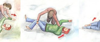
First aid
To quickly bring a patient out of a state of fainting on your own, without medical assistance, you should carry out the following manipulations:
- give a horizontal position, it is better to put the person on his side;
- loosen your tie, unbutton your shirt, provide fresh air;
- splash your face with cold water;
- bring ammonia to your nose.

