The most common complaint that doctors hear from their patients is headache. Both adults and children complain about it. It is impossible to ignore this. Especially if there are other symptoms. Parents should pay special attention to the child’s headaches and the baby’s behavior, because he cannot say that he is in pain. Perhaps these are the consequences of a difficult birth or congenital anomalies, which can be determined at an early age. Maybe these are liquorodynamic disturbances. What is it, what are the characteristic signs of this disease in children and adults and how to treat it, we will consider further.
What does liquorodynamic disturbances mean?
Liquor is cerebrospinal fluid that constantly circulates in the ventricles, cerebrospinal fluid ducts and in the subarachnoid space of the brain and spinal cord. Liquor plays an important role in metabolic processes in the central nervous system, in maintaining homeostasis in brain tissue, and also creates a certain mechanical protection for the brain.
Liquorodynamic disorders are conditions in which the circulation of cerebrospinal fluid, its secretion and reabsorption are impaired. These processes are regulated by glands located in the choroid plexuses of the brain ventricles that produce fluid.
In the normal state of the body, the composition of the cerebrospinal fluid and its pressure are stable.
Hydrocephalus in adults
Department of Emergency Neurosurgery > Diseases
This section is intended for people suffering from hydrocephalus, as well as for their relatives and friends. We hope that the information provided will help you better understand the disease and how to overcome it. Here we will talk about the causes of hydrocephalus in adults, methods for diagnosing it in the early stages, provide a list of necessary studies, reflect modern techniques used in our institute for the diagnosis and treatment of hydrocephalus, as well as prospects for the development of this field of neurosurgery.
Most people, including doctors, classify hydrocephalus as a childhood disease. Indeed, from 1 to 10 children out of every thousand newborns suffer from hydrocele. During a specialized examination of patients over 18 years of age in neurosurgical hospitals, hydrocephalic syndrome is detected in every fourth patient. Due to the lack of clear criteria for diagnosing hydrocephalus, only single operations for the disease in question are performed annually in non-core neurosurgical departments. Patients are discharged from such hospitals with diagnoses: “psycho-organic syndrome”, “dyscirculatory or post-traumatic encephalopathy”, “dementia of mixed origin”, “consequences of traumatic brain injury”, consequences of stroke.” This is not a complete list of diseases, under the guise of which patients are unsuccessfully treated in clinics, neurological hospitals and psychiatric hospitals. Timely and correct diagnosis of hydrocephalus and adequate surgical treatment allow patients to achieve recovery, work and social rehabilitation in almost 100% of cases. Thus, most of our patients return to their previous work, and some of the sick, despite incomplete work adaptation, can live without outside help, perform simpler activities, becoming full-fledged members of society.
A special group of patients in our department are patients with acute forms of hydrocephalus
, mainly with intraventricular hemorrhages and hemotamponade of the ventricles of the brain due to
non-traumatic subarachnoid hemorrhages
.
In cases where there is no specialized surgical care for such patients, they die within the first 12 to 48 hours from the onset of the disease. Modern methods of external drainage
with the introduction of thrombolytics into the ventricles of the brain, used in our department, can not only reduce mortality in this pathology, but also stabilize the condition of patients for a long time.
Below we present the basic concepts and terms necessary to understand the problem of hydrocephalus in adults and how to control it.
Functional anatomy of the cerebrospinal fluid-containing spaces of the brain and the definition of hydrocephalus.
Normally, each person's central nervous system contains about 120-150 ml of cerebrospinal fluid (CSF, cerebrospinal fluid). The physiological significance of CSF is as follows:
- is a kind of shock absorber for the brain, thus providing its mechanical protection during shocks and shocks
- performs nutritional functions
- maintains osmotic and oncotic balance at the tissue level
- has protective (bactericidal) properties, accumulating antibodies
- takes part in the mechanisms of regulation of blood circulation in the confined space of the cranial cavity and spinal canal.
CSF is formed in the cells of the choroid plexuses of the ventricles of the brain.
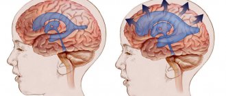
The largest amount of CSF is produced by the plexuses of the lateral ventricles of the brain (Fig. 1). The volume of CSF in the cranial cavity and in the spinal canal in an adult does not exceed 125–150 ml. About 500-600 ml of CSF is produced per day and the same amount is continuously absorbed. From the lateral ventricles of the brain, which contains about 25 ml of CSF, which enters the third ventricle through the foramen of Monroe, and from there, through the cerebral aqueduct (Aqueduct of Sylvius), the fluid enters the cavity of the fourth ventricle. The third and fourth ventricles of the brain contain approximately 5 ml of CSF. From the fourth ventricle, through the median foramen of Magendie and two lateral foramina of Luschka, located in the area of the lateral inversions of the fourth ventricle, CSF enters the subarachnoid space of the brain. At the base of the brain, the subarachnoid space expands and forms cavities filled with CSF (basal cisterns). The largest of them is located between the cerebellum and the medulla oblongata - the large cistern of the brain (cerebellar-medullary cistern). From it, CSF enters the premedullary and lateral cerebellar-medullary cisterns, located on the lower and lateral surfaces of the medulla oblongata, respectively. On the lower surface of the pons of the brain there is a rather large prepontine (prepontine) tank, which receives CSF from the above tanks. The prepontine cistern is separated from the midbrain and diencephalon cisterns (covering, interpeduncular, peduncular, chiasmatic, optic nerve) by a sheet of semipermeable membrane (Liliekvist membrane), which promotes one-way flow of CSF in the direction from back to front and from bottom to top. From the cisterns of the brain, CSF enters the convexital part of the subarachnoid space, washing the cerebral hemispheres, then is absorbed into the venous bed through the arachnoid cells and villi. The accumulation of such villi around the venous sinuses of the dura mater of the brain (there are especially many of them in the superior sagittal sinus) is called pachyon granulations. Liquid is partially absorbed into the lymphatic system, which occurs at the level of the nerve sheaths. The movement of CSF in different directions is also associated with vascular pulsation, breathing, and muscle contractions. (Fig. 1-2). If there is a violation of the relationship between the production and absorption of CSF at any of the listed levels (increased production of CSF by the choroid plexuses; closure of the ventricular openings by a tumor, adhesions, blood clots; obstruction of cells, villi and pachyonic granulations by erythrocytes, fibrosis of the membranes after hemorrhage or previous meningitis; occlusion of the sinuses) leads to a significant (maximum up to 12 liters in congenital hydrocephalus) accumulation of CSF, forming the development of hydrocephalus. The term “hydrocephalus” itself is formed by the merger of two Greek words “hydro” - water and “cephalus” - head (“dropsy of the brain”).
Below is the most complete definition of the concept of “adult hydrocephalus”.
Hydrocephalus in adults is an independent nosological form, or a complication of a number of brain diseases (tumor, hemorrhage, trauma, stroke, infectious process, etc.), characterized by an active progressive process of excessive accumulation of CSF in the cerebrospinal fluid spaces, caused by disturbances in its circulation (proximal and distal forms of occlusive hydrocephalus), absorption (aresorptive and dysresorptive forms), or production (hypersecretory form) and manifested morphologically by enlargement of the ventricles of the brain, periventricular leukareosis (decrease in the density of the medulla due to its saturation with CSF) and narrowing of the subarachnoid spaces. Clinical manifestations of hydrocephalus depend on its form.
Diseases that contribute to the formation of hydrocephalus in adults.
It has now been established that almost any pathology of the central nervous system can lead to such a complication as hydrocephalus.
Below we provide a list of only the main diseases in which hydrocephalus most often occurs:
- Brain tumors (usually stem, para-stem, or intraventricular localization).
- Inflammatory and infectious diseases of the central nervous system (meningitis, ventriculitis, encephalitis, tuberculosis, etc.).
- Subarachnoid and intraventricular hemorrhages (traumatic and non-traumatic), most often due to rupture of aneurysms and arteriovenous malformations of cerebral vessels.
- Acute cerebrovascular accidents of ischemic and hemorrhagic type.
- Encephalopathies of various origins (alcoholism, chronic hypoxic conditions, etc.).
In the department of emergency neurosurgery of the Research Institute of Emergency Medicine named after.
N.V. Sklifosovsky's priorities are the problems of diagnosis and treatment of acute and chronic hydrocephalus in non-traumatic subarachnoid hemorrhages due to rupture of arterial aneurysms of cerebral vessels or arteriovenous malformations, as well as post-traumatic hydrocephalus. Classification and pathogenesis of hydrocephalus.
- Based on its origin, hydrocephalus is divided into congenital and acquired.
Congenital hydrocephalus, as a rule, debuts in childhood. The causes of its occurrence are various intrauterine infections, hypoxia and, mainly, congenital developmental anomalies, leading either to disruption of the CSF circulation (stenosis and occlusion of the Sylvian aqueduct, Dandy-Walker anomaly, Arnold-Chiari anomaly, etc.), or accompanied by underdevelopment of structures, involved in CSF resorption (aresorptive hydrocephalus).Acquired hydrocephalus is further classified depending on the etiological factor (see causes of hydrocephalus).
- According to pathogenesis, there are three main forms of hydrocephalus.
- Occlusive (closed, non-communicating) hydrocephalus, in which the flow of cerebrospinal fluid is disrupted due to closure (occlusion) of the cerebrospinal fluid pathways by a tumor, a blood clot, or a post-inflammatory adhesive process. In the event that occlusion occurs at the level of the ventricular system (foramen of Monro, aqueduct of Sylvius, foramina of Magendie and Luschka), we are talking about proximal occlusive hydrocephalus. If the block in the path of the CSF flow is at the level of the basal cisterns, then they speak of a distal form of occlusive hydrocephalus.
- Communicating (open, dysresorptive) hydrocephalus, in which the processes of CSF resorption are disrupted due to damage to the structures involved in the absorption of CSF into the venous bed (arachnoid villi, cells, Pachionian granulations, venous sinuses).
- Hypersecretory hydrocephalus, which develops due to excess production of CSF (choroid plexus papilloma).
Previously, a fourth form of hydrocephalus was also identified, the so-called external (mixed, ex vacuo) hydrocephalus, which was characterized by an enlargement of the cerebral ventricles and subarachnoid space in conditions of progressive brain atrophy. However, this process should still be attributed to brain atrophy, and not to hydrocephalus, because the enlargement of the ventricles of the brain and the expansion of the subarachnoid space are not caused by excessive accumulation of CSF, due to disruption of the processes of its production, circulation and resorption, but by a decrease in the mass of brain tissue against the background of atrophy. - Based on current rates, they are distinguished:
- Acute hydrocephalus, when no more than 3 days pass from the first symptoms of the disease to severe decompensation.
- Subacute progressive hydrocephalus, developing within a month from the onset of the disease.
- Chronic hydrocephalus, which develops over a period of 3 weeks to 6 months or more.
- Based on the level of cerebrospinal fluid pressure, hydrocephalus is divided into the following groups:
- Hypertensive
- Normotensive
- Hypotensive
Clinical picture and diagnosis of hydrocephalus in adults.
With occlusive hydrocephalus, especially acutely developing, symptoms of increased intracranial pressure come first, which include:
- Headache;
- Nausea and/or vomiting;
- Drowsiness;
- Stagnation of the optic discs;
- Symptoms of axial dislocation of the brain.
Headache is most pronounced in the morning upon awakening, which is associated with an additional increase in intracranial pressure during sleep.
This is facilitated by vasodilation due to the accumulation of carbon dioxide, which is accompanied by blood flow, stretching of the walls of blood vessels and the dura mater of the brain at the base of the skull. Nausea and vomiting also worsen in the morning and sometimes lead to a decrease in headaches. Drowsiness is the most dangerous sign of increased intracranial pressure; its appearance precedes a period of sharp and rapid deterioration of neurological symptoms.
The development of stagnation of the optic discs is caused by an increase in pressure in the subarachnoid space surrounding the nerve and a disruption of the axoplasmic flow in it.
With the development of dislocation syndrome, there is a rapid depression of the patient's consciousness to a deep coma, oculomotor disorders appear (due to the expansion of the cerebral aqueduct), and sometimes a forced position of the head. Compression of the medulla oblongata manifests itself in rapid depression of breathing and cardiovascular activity, leading to the death of the patient.
The clinical picture is fundamentally different in the formation of chronic hydrocephalus
. The main manifestation of chronic dysresorptive hydrocephalus is a triad of symptoms:
- dementia;
- apraxia of walking or lower paraparesis;
- urinary incontinence.
The first symptoms of the disease usually appear 3 weeks after hemorrhage, trauma, meningitis or another disease leading to the development of hydrocephalus.
Disturbances in the sleep-wake cycle come first: patients become drowsy during the day with disturbances in night sleep. Subsequently, the general level of activity of the patients sharply decreases, they become spontaneous, lacking initiative, and inert. Among memory disorders, short-term memory disorders, especially numerical memory, come first. Thus, a patient with hydrocephalus cannot name the date, month, year, and incorrectly indicates his age. In the later stages of the disease, severe mnestic-intellectual impairments develop; patients can no longer care for themselves; they answer questions asked in monosyllables with long pauses, often inadequately. Apraxia of walking is that a patient with hydrocephalus can freely pretend to walk in a lying position or ride a bicycle, but as soon as he takes a vertical position, this ability is instantly lost, the patient walks with his legs wide apart, unsteadily, his gait becomes shuffling. In the later stages stages of the disease, lower paraparesis develops.
Urinary incontinence is the most late and variable symptom.
Stagnation of the optic discs is atypical for chronic hydrocephalus; as a rule, there are no changes in the fundus of the eye in such patients.
Diagnosis of hydrocephalus.
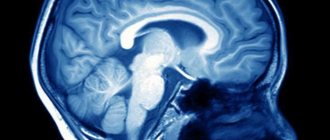
Computed tomography and magnetic resonance imaging play a leading role in the diagnosis of hydrocephalus (Fig. 1).
Rice. 1. Chronic dysresorptive hydrocephalus on CT: symmetrical expansion of the ventricular system with balloon-like enlargement of the anterior horns (one arrow), lack of visualization of subarachnoid fissures, foci of periventricular leukareosis (two arrows).
In the department of emergency neurosurgery of the Research Institute of Emergency Medicine named after. N.V. Sklifosovsky organized round-the-clock operation of the computed tomography department. We perform a CT scan on incoming patients with subarachnoid hemorrhage in the first hours after hospitalization to assess the nature and extent of the hemorrhage, then we conduct a CT scan as a control after surgery, or when the first signs of hydrocephalus appear.
To assess the stage of hydrocephalus and determine indications for surgical intervention, our department calculates ventriculo-cranial coefficients, which show the degree of expansion of the ventricular system and its decrease after the operation.
Computed tomography also makes it possible to clarify the presence and extent of concomitant ischemic brain damage in patients with subarachnoid hemorrhages.
To predict the outcome of surgical treatment of hydrocephalus, all patients undergo a tap-test
. The essence of the test is that when at least 40 ml of cerebrospinal fluid is removed during lumbar puncture, patients with chronic hydrocephalus experience a short-term improvement. In the case of a positive test, the patient's recovery after surgery can be more likely to be predicted. However, a negative result often does not indicate the impossibility of a good outcome in the late postoperative period.
Treatment of hydrocephalus.
Conservative treatment of hydrocephalus in adults with a full-blown clinical picture is ineffective.
- Treatment of acute hydrocephalus.
- Treatment of chronic hydrocephalus.
Acute hydrocephalus, which often occurs with intraventricular hemorrhages with the development of ventricular hemotamponade, is a serious complication requiring immediate neurosurgical intervention, the purpose of which is to “unload” the ventricular system, ensure normal cerebrospinal fluid flow, and reduce intracranial pressure
and
express sanitation of cerebrospinal fluid
.
In our department, similar operations are performed, which involve the application of external ventricular drains followed by the introduction of streptokinase into the ventricular cavity - a drug that dissolves blood clots and thereby ensures normal cerebrospinal fluid flow.
In addition, the department carries out direct measurement of intracranial pressure
such patients in order to select optimal infusion therapy and adequately monitor the dynamics of the patient’s condition.
The goal of the operation is to create an artificial pathway to drain excess CSF to an area where fluid can be easily absorbed. To achieve this goal, special liquor shunt systems are used.
Shunt system design. Each CSF shunt system consists of three components:
- Ventricular catheter - intended for installation in the lateral ventricles of the brain.
- A valve is a device that allows you to regulate the outflow of CSF. The valve is designed for certain values of cerebrospinal fluid pressure and when a certain pressure is reached, the valve opens and CSF begins to flow out of the ventricular system. When the pressure normalizes, the valve closes and the flow of cerebrospinal fluid from the ventricles stops.
- Peripheral catheter - designed for installation in various cavities of the body that have the ability to absorb fluid (abdominal cavity, pelvic cavity, atrium, etc.) (Fig. 2).
a) b)
Rice. 2. Scheme of the ventriculoperitoneal shunt operation: a) cranial stage; after puncture and drainage of the anterior horn of the right lateral ventricle from Kocher's point (one arrow), the ventricular catheter is passed into the postauricular region, where it is connected to the valve of the shunt system at the level of the projection of the foramen of Monroe (two arrows); b) peritoneal stage; additional incisions in the supraclavicular region and at the level of the xiphoid process are marked with an arrow.
Every year, our department performs about 50 surgeries for hydrocephalus in adults.
We use the most advanced technologies in the installation of cerebrospinal fluid shunt systems: we use programmable valves
that allow non-invasive regulation of CSF pressure;
we install valve systems with a built-in anti-siphon device
that prevents the reverse flow of cerebrospinal fluid when the body position changes;
To implant a catheter into the abdominal cavity, endovideolaparoscopic equipment
, which allows to minimize the trauma of the operation and achieve the best results (Fig. 3).
a) b)
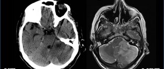
Rice.
3. Outcome of shunt surgery for chronic dysresorptive hydrocephalus. a) CT scan after non-traumatic subarachnoid hemorrhage. Severe internal hydrocephalus is determined (VKK2-26%). b) CT scan after implantation of a ventriculoperitoneal shunt. The normalization of the dimensions of the ventricular system is determined with the regression of foci of periventricular leukareosis and the appearance of visualization of subarachnoid fissures. Timely and correctly performed surgical intervention for hydrocephalus allows in almost 100% of cases to achieve recovery of patients, their labor and social rehabilitation.
What is the mechanism of violations
Let's consider how liquorodynamic disorders of the brain can develop:
- The rate of production and release of cerebrospinal fluid by the choroid plexuses increases.
- The rate of absorption of cerebrospinal fluid from the subarachnoid space slows down due to the blocking of the narrowing of the cerebrospinal fluid vessels due to previous subarachnoid hemorrhages or inflammatory diseases of the meninges.
- The rate of CSF production decreases during the normal absorption process.
The rate of absorption, production and release of cerebrospinal fluid is influenced by:
- On the state of cerebral hemodynamics.
- State of the blood-brain barrier.
The inflammatory process in the brain increases its volume and increases intracranial pressure. The result is poor circulation and blockage of the vessels through which the cerebrospinal fluid moves. Due to the accumulation of fluid in the cavities, partial death of intracranial tissue may begin, and this will lead to the development of hydrocephalus.
Discussion
Among the publications devoted to the study of cerebrospinal fluid dynamics according to FC-MRI data, studies in Chiari malformation [13-16] and hydrocephalus [22, 27] dominate. There are no works on the study of liquor dynamics in meningiomas of the GZ area in the available literature.
The closest pathology to BZ tumors in terms of the pathogenetic basis of liquorodynamic changes is Chiari malformation. An analysis of the literature demonstrates conflicting data on the parameters of the CSF flow at the upper cervical level both in healthy volunteers and in patients with Chiari malformation type I before and after surgery [13-16, 28, 29]. This may be due to the following reasons: different technical characteristics of MRI scanners; performing measurements at different levels; heterogeneity of the age composition of the study population; the study does not take into account degenerative-dystrophic changes that affect the processes of cerebrospinal fluid dynamics; Many authors do not present all parameters of liquor dynamics, which makes it difficult to compare results.
We used a technique previously developed at the institute and demonstrated in the article by N.V. Arutyunov when studying liquor dynamics in patients with type I Chiari malformations [26]. According to the conclusions made by N.V. Arutyunov et al. (2009), all parameters of CSF flow in patients with Chiari malformation type I before surgery are statistically significantly lower than normal, and after surgery there is a significant increase in all characteristics of CSF flow to normal levels. In our work, in patients with meningiomas of the GZ area before surgery, the opposite situation was noted: all parameters of cerebrospinal fluid dynamics are higher than normal. Our data on increasing the speed characteristics of the liquor flow correspond to the law of continuity in hydrodynamics (Castelli’s law), according to which the speed of fluid flow in pipes is inversely proportional to their cross-sectional area. An increase in the volumetric characteristics of the cerebrospinal fluid, in our opinion, occurs in connection with turbulent movements of the cerebrospinal fluid below the meningioma (at the C2-C3 level). After surgery, in patients with meningiomas of the GZ area, the velocity characteristics of the CSF flow (peak positive and negative velocities, the amplitude of the maximum linear velocity) significantly decrease, but do not reach normal values, the volumetric parameters (positive and negative volumes, stroke volume) do not change. Due to the fact that we carried out a repeat study 3-6 months after surgery, these liquorodynamic parameters can be explained by the development of scar postoperative changes.
Classification of violations
Liquorodynamic disorders are classified in the following areas:
- How does the pathological process proceed:
- Chronic course.
- Acute phase.
2. Stages of development:
- Progressive. Intracranial pressure increases and pathological processes progress.
- Compensated. Intracranial pressure is stable, but the ventricles of the brain remain dilated.
- Subcompensated. Great danger of crises. Unstable condition. Blood pressure can rise sharply at any moment.
3. In which cavity of the brain is the cerebrospinal fluid located:
- Intraventricular. Fluid accumulates in the ventricular system of the brain due to obstruction of the cerebrospinal fluid system.
- Subarachnoid. Liquorodynamic disturbances of the external type can lead to destructive lesions of brain tissue.
- Mixed.
4. Depending on the pressure of the cerebrospinal fluid:
- Hypertension. Characterized by high intracranial pressure. The outflow of cerebrospinal fluid is impaired.
- Normotensive stage. Intracranial pressure is normal, but the ventricular cavity is enlarged. This condition is most common in childhood.
- Hypotension. After surgery, excessive outflow of cerebrospinal fluid from the ventricular cavities.
Laboratory and instrumental diagnostics
Diagnosis of this type of pathology is based on a careful collection of clinical symptoms, possible causes and predisposing factors.
Laboratory diagnosis is based on the study of cerebrospinal fluid using a spinal puncture.
It is prohibited to perform a lumbar puncture if:
- hydrocephalus due to tumors;
- after surgery;
- ulcerative-necrotic changes in the skin at the puncture site (below the level of the 2nd lumbar vertebra);
- rupture of membranes with severe progressive bleeding;
- presence or signs of decreased intracranial pressure.
When examining the cerebrospinal fluid, an increase in protein levels is detected. The presence of lymphocytes is excluded. An increased number of lymphocytes is determined only in the presence of inflammatory changes - with tuberculosis, syphilis or encephalitis.
The most diagnostically valuable methods are computed tomography, magnetic resonance imaging and angiography. Computed tomography and magnetic resonance imaging determine the cause of irrational cerebrospinal fluid dynamics and possible methods of correction through surgery.
Angiography can reveal the presence of changes in the vascular walls, thrombotic and embolic obstacles that cause occlusion and increased ICP.
Causes congenital
There are congenital anomalies that can contribute to the development of liquorodynamic disorders:
- Genetic disorders in intrauterine development of the fetus.
- Agenesis of the corpus callosum.
- Dandy-Walker syndrome.
- Arnold-Chiari syndrome.
- Encephalocele.
- Stenosis of the cerebral aqueduct, primary or secondary.
- Porencephalic cysts.
Acquired reasons
Liquorodynamic disorders can begin to develop for acquired reasons:
- Spinal cord and brain injuries.
- Various infectious diseases and parasitic infections that affect the nervous system.
- Neoplasms inside the skull that block the cerebrospinal fluid pathways.
- Thrombosis.
- Intrauterine hypoxia in the first two days after birth.
- Papillomas of the choroid plexus.
Symptoms of disorders in infants
Liquorodynamic disorders in children under one year of age have the following symptoms:
- Frequent and profuse regurgitation.
- Unexpected crying for no apparent reason.
- Slow overgrowth of the fontanel.
- Monotonous crying.
- The child is lethargic and sleepy.
- Sleep is disturbed.
- Seams coming apart.
Over time, the disease progresses more and more, and signs of liquorodynamic disorders become more pronounced:
- Tremor of the chin.
- Twitching of limbs.
- Involuntary shudders.
- Life support functions are disrupted.
- Disturbances in the functioning of internal organs for no apparent reason.
- Possible squint.
Visually, you can notice the vascular network in the area of the nose, neck, and chest. When crying or tense muscles, it becomes more pronounced.
The neurologist may also note the following signs:
- Hemiplegia.
- Extensor hypertonicity.
- Meningeal signs.
- Paralysis and paresis.
- Paraplegia.
- Graefe's symptom.
- Nystagmus is horizontal.
- Delay in psychomotor development.
You should visit your pediatrician regularly. At the appointment, the doctor measures the volume of the head, and if pathology develops, changes will be noticeable. So, there may be such deviations in the development of the skull:
- The head grows quickly.
- It has an unnaturally elongated shape.
- The large and small fontanelles swell and pulsate.
- Sutures are coming apart due to high intracranial pressure.
All these are signs that a syndrome of liquorodynamic disorders is developing in an infant. Hydrocephalus progresses.
I would like to note that it is difficult to determine liquorodynamic crises in infants.
Vascular type of headache
The arteriodilatatory variant of the vascular type of pain is associated with a decrease in the tone of the craniocerebral arteries, which occurs when the blood vessels are excessively stretched by the pulse volume of blood. Excessive pulse stretching of the hypotonic arterial wall sometimes occurs at normal levels of systemic blood pressure, but more often when it increases. If the artery of the soft integument of the head (superficial, temporal artery) is subjected to excessive pulse stretching, then compression of the adductor trunk of the artery in the temporomygomatic region with a finger, as a rule, reduces pain.
Clinic: throbbing headache. When permeability is impaired and plasma impregnation of the arterial wall develops perivascular edema. In these cases, the amplitude of the pulsation decreases and the headache loses its pulsating character, becomes dull, then aching or bursting.
Therapy: anti-migraine drugs containing ergotamine, dihydroergotamine, sumatriptan are used to relieve pain. Ergotamine drugs relieve pain due to a powerful vasoconstrictor effect and prevent neurogenic inflammation. Ergotamine in combination with caffeine or with caffeine and dimenhydrinate is prescribed as monotherapy or in combination with analgesics, antiemetics and sedatives.
For vegetative-vascular dystonia, xanthine-type drugs are prescribed: aminophylline, pentoxifylline, xanthinol nicotinate, which are usually prescribed in a course 1-2 times a year. This group of drugs acts on smooth muscles, relaxing them, relieves spasm of blood vessels, reduces pressure in the pulmonary artery system, has a diuretic effect, and also inhibits platelet aggregation.
The arteriospastic variant occurs with spasm of the craniocerebral arteries and with an increase in arterial tone, which entails ischemic dyscirculation and ischemic hypoxia.
Clinic: the pain is aching and dull in nature, and is perceived as a feeling of compression. Pain, as a rule, is accompanied by nausea, unsystematic dizziness, darkening and flickering of “black spots” in the eyes.
Therapy: antispasmodic drugs are prescribed: phosphodiesterase inhibitors (papaverine, drotaverine), α-adrenergic blockers (dihydroergotoxin, nicergoline), calcium antagonists (nifedipine, nimodipine). For hypertension associated with impaired blood supply to the brain, drugs that improve cerebral circulation are used. Among the prescription drugs of this series, the most famous are the cerebrovasodilator vinpocetine, BMCA cinnarizine, piracetam, nicotinoyl gamma-aminobutyric acid. It must be remembered that these drugs are contraindicated in patients with acute cerebrovascular accidents, gastric and duodenal ulcers.
The venous variant is caused by excessive blood filling of the venous vessels and difficulty in venous outflow.
Clinic: a feeling of heaviness in the head and a feeling of dull distension. It occurs or worsens when lying down, as well as when working with your head low, as well as when straining or coughing. Morning headache is typical. Sometimes the pain is limited only to the back of the head.
Therapy: drugs of choice are also xanthine drugs, due to their venotonic effect and ability to reduce platelet aggregation, as well as mild venotonics, for example diosmin + hesperidin.
Signs of liquorodynamic disorders in children after one year
After one year, a child’s skull is already formed. The fontanelles have completely closed and the sutures have ossified. If there are liquorodynamic disturbances in a child, signs of increased intracranial pressure appear.
There may be such complaints:
- Headache.
- Apathy.
- Worry for no reason.
- Nausea.
- Vomiting, after which there is no relief.
The following signs are also characteristic:
- Gait and speech are impaired.
- There are disturbances in the coordination of movements.
- Vision decreases.
- Horizontal nystagmus.
- In advanced cases, “bobble doll head”.
And also, if liquorodynamic disorders of the brain progress, the following deviations will be noticeable:
- The child speaks poorly.
- They use standard, memorized phrases without understanding their meaning.
- Always in a good mood.
- Delayed sexual development.
- Convulsive syndrome develops.
- Obesity.
- Disturbances in the functioning of the endocrine system.
- Lag in the educational process.
Clinical symptoms
A pathognomonic clinical sign in adults associated with a disorder of cerebrospinal fluid dynamics is an unbearable headache in the morning, localized in the crown area. Pain can spread over the entire surface of the skull. The occurrence of a headache is associated with various reflex-protective acts - sneezing, loud talking, severe coughing. Often the pain syndrome is caused by a change in the position of the skull, a sharp turn of the torso, or an uncomfortable posture during sleep.
The pain syndrome is accompanied by dyspeptic disorders in the form of nausea and vomiting. Vomiting is caused by irritation of the chemoreceptor trigger zone located in the medulla oblongata. Vomiting is one-time, with a small amount of contents.
In addition to pain, ophthalmological disorders may be observed in the form of flickering spots, decreased visual acuity, frequent flashes of light, and nystagmus.
With pathological changes in liquor dynamics, dizziness often occurs, causing short-term loss of consciousness. With a severe and long-term course of the disease, cognitive disorders appear: memory impairment, the ability to transmit and analyze information.
With a persistent increase in intracranial pressure, a liquorodynamic crisis develops, which is characterized by a bursting paroxysmal headache. The pain syndrome is caused by irritation of the vessels of the dura mater.
A liquorodynamic crisis leads to hemodynamic disturbances, causing stagnant changes in the fundus.
During a crisis, tachycardia, convulsions, and stupor may occur.
Diagnosis of the disease in adults
If you experience headaches and the symptoms described above, you should consult a neurologist. To clarify the diagnosis and prescribe treatment, the following studies may be prescribed:
- Computed tomography.
- Angiography.
- Pneumoencephalography.
- ECHO of the brain.
- NMRI.
If there is a suspicion of a syndrome of cerebrospinal fluid dynamics disorders, a lumbar puncture may be prescribed with a change in cerebrospinal fluid pressure.
When diagnosing adults, much attention is paid to the underlying disease.
Consequences and residual effects
Late detection of pathology and lack of treatment in the early stages leads to the development of adverse residual effects.
In adults, memory loss, speech impairment, decreased performance, cardiac dysfunction, etc. occur.
Infants develop disorders of neuropsychic functions, lethargy, and lag in physical development from their peers in all respects. Most often, children with incorrect, late treatment of pathology become disabled.
Treatment of liquorodynamic disorders
The earlier the disease is detected, the greater the chance of restoring lost brain functions. The type of treatment is selected based on the presence of pathological changes in the course of the disease, as well as the age of the patient.
In the presence of increased intracranial pressure, diuretics are usually prescribed: Furosemide, Diacarb. Antibacterial agents are used in the treatment of infectious processes. Normalization of intracranial pressure and its treatment is the main task.
To relieve swelling and inflammation, glucocorticoid drugs are used: Prednisolone, Dexamethasone.
Steroid medications are also used to reduce cerebral edema. It is necessary to eliminate the cause of the disease.
As soon as liquorodynamic disturbances are detected, treatment should be prescribed immediately. After undergoing complex therapy, positive results are noticeable. This is especially important during the period of child development. Speech improves, progress in psychomotor development is noticeable.
Surgical treatment is also possible. It may be prescribed in the following cases:
- Drug treatment is ineffective.
- Liquorodynamic crisis.
- Occlusive hydrocephalus.
Surgical treatment is considered for each case of the disease separately, taking into account age, characteristics of the body and the course of the disease. In most cases, surgery on the brain is avoided so as not to damage healthy brain tissue, and complex drug treatment is used.
It is known that if the syndrome of liquorodynamic disorders in a child is not treated, the mortality rate is 50% up to 3 years, 20-30% of children survive to adulthood. After surgery, mortality is 5-15% of sick children.
Mortality increases due to late diagnosis.
Treatment of intracranial pressure
Intracranial pressure needs to be normalized and treated. An integrated approach to the problem, even after the first courses of treatment, even in severely ill patients, gives positive dynamics, speech and psychomotor development in children improves. The earlier treatment is started, the faster the impaired functions are restored and intracranial pressure is normalized. At the first consultation, the doctor will tell you about the main factors and symptoms of the disease: what is hypertension syndrome in newborns, infants, infants, children, children, adults, what is mild, moderate, severe cerebral hypertensive syndrome , hypertensive syndrome , cerebrospinal fluid hypertensive syndrome , how the army and residual encephalopathy are related to hypertensive hydrocephalic syndrome, what intracranial pressure should be normal, how to measure, determine, check, and how to reduce, lower, relieve, treat intracranial pressure. On the website sarclinic.ru you can see a doctor online for free.
Sign up for a consultation. There are contraindications. Specialist consultation is required. Photo: Sergiyn | Dreamstime.com\Dreamstock.ru. The people depicted in the photo are models, do not suffer from the diseases described and/or all similarities are excluded.
Related posts:
Delay, mental development disorder of the child, children with mental retardation
Enuresis, treatment of enuresis, how to treat nocturnal enuresis in Saratov, Russia
Bruxism, teeth grinding during sleep, odonterism: symptoms and treatment
Acalculia, dyscalculia, treatment of acalculia
General speech underdevelopment in children, phonemic onset: levels, correction, treatment
Comments ()
Prevention of liquorodynamic disorders
Preventive measures include:
- Observation of pregnancy in the antenatal clinic. It is very important to register as early as possible.
- Timely detection of intrauterine infections and their treatment.
At 18-20 weeks, an ultrasound shows the development of the fetal brain and the state of the unborn child’s cerebrospinal fluid. At this time, it is possible to determine the presence or absence of pathologies.
- The right choice of delivery.
- Regular monitoring by a pediatrician. Measuring the circumference of the skull, if there is a need to conduct a fundus examination.
- If the fontanel does not close in a timely manner, it is necessary to conduct neurosonography and consult a neurosurgeon.
- Timely removal of tumors that block the cerebrospinal fluid pathways.
- Regular observation by a doctor and carrying out the necessary studies after suffering injuries to the brain and spinal cord.
- Timely treatment of infectious diseases.
- Prevention and therapy of chronic diseases.
- Quit smoking and alcohol.
- It is recommended to play sports and lead an active lifestyle.
It is easier to prevent any disease or take all measures to reduce the risk of developing pathology. If liquorodynamic disorders are diagnosed, then the earlier therapy is started, the greater the chance that the child will develop normally.