hindbrain
The hindbrain includes the pons and cerebellum. It develops from the fourth cerebral vesicle (metencephalon).
The pons borders the medulla oblongata from below, passes into the cerebral peduncles from above, and its lateral sections form the middle cerebellar peduncles. In the anterior (basilar) part of the pons there are accumulations of gray matter - the nuclei of the pons; in the posterior part of the tegmentum of the pons there are nuclei of the superior olive, reticular formation and V - VIII pairs of cranial nerves. These nerves emerge from the base of the brain lateral to and behind the pons, at the border with the cerebellum and medulla oblongata. The white matter of the pons in its anterior part is represented by transversely running fibers going to the middle cerebellar peduncles. They are penetrated by powerful longitudinal bundles of fibers of the pyramidal tracts, which then form the pyramids of the medulla oblongata and go to the spinal cord. In the rear part of the bridge (pontineum) there are ascending and descending fiber systems (Fig. 113).
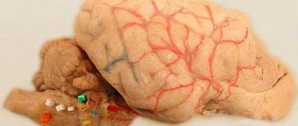
Rice. 113. Brain stem (front view). 1 - anterior median fissure; 2 - pyramids of the medulla oblongata; 3 - olive; 4 - cerebellum; 5 - decussation of the pyramids (the place of transition of the medulla oblongata into the spinal cord); 6 - middle cerebellar peduncle; 7 - bridge; 8 - interpeduncular fossa; 9 - cerebral peduncle; III - XII roots of cranial nerves; C - first spinal nerve
Physiology of the medulla oblongata and pons
The medulla oblongata and the pons perform two functions - reflex and conductive. Through the sensitive fibers of the roots of the cranial nerves, it receives impulses - information from the receptors of the scalp, mucous membranes of the eyes, nose, mouth (including taste buds), from the organ of hearing, the vestibular apparatus (organ of balance), from the receptors of the larynx, trachea, lungs, as well as from interoreceptors of the cardiovascular system and digestive apparatus.
Through the medulla oblongata, many simple and complex reflexes are carried out, covering not individual metameres of the body, but organ systems, for example, the digestive, respiratory, and circulatory systems. The reflex activity of the medulla oblongata can be observed in a bulbar cat, that is, a cat in which the brain stem above the medulla oblongata has been cut. The reflex activity of such a cat is complex and diverse.
The following reflexes occur through the medulla oblongata: 1) protective: coughing, sneezing, blinking, lacrimation, vomiting; 2) food: sucking, swallowing, secretion of juice from the digestive glands; 3) cardiovascular, regulating the activity of the heart and blood vessels; 4) in the medulla oblongata there is an automatically working respiratory center that provides ventilation to the lungs; 5) the vestibular nuclei are located in the medulla oblongata and the pons.
From the vestibular nuclei of the medulla oblongata begins the descending vestibulospinal tract, which is involved in the implementation of posture reflexes, namely in the redistribution of muscle tone. A bulbar cat can neither stand nor walk, but the medulla oblongata and cervical segments of the spinal cord provide those complex reflexes that are elements of standing and walking. All reflexes associated with the standing function are called positioning reflexes. Thanks to them, the animal holds its body, usually with the crown up.
The special importance of this part of the central nervous system is determined by the fact that the medulla oblongata contains vital centers: respiratory, cardiovascular. Therefore, not only removal, but even damage to the medulla oblongata ends in death.
In addition to the reflex function, the medulla oblongata performs a conductive function. Conducting pathways pass through it, connecting the cortex, diencephalon, midbrain, cerebellum and spinal cord with a two-way connection.
The cerebellum is located dorsal to the pons and medulla oblongata. It has two hemispheres and a middle part - the worm. The surface of the cerebellum is covered with a layer of gray matter (cerebellar cortex) and forms narrow convolutions separated by grooves. With their help, the surface of the cerebellum is divided into lobules. The central part of the cerebellum consists of white matter, which contains accumulations of gray matter - the cerebellar nuclei. The largest of them is the dentate nucleus. The cerebellum is connected to the brain stem by three pairs of peduncles: the upper ones connect it to the midbrain, the middle ones to the pons, and the lower ones to the medulla oblongata. The peduncles contain bundles of fibers connecting the cerebellum to various parts of the brain and spinal cord.
During development, the isthmus of the rhombencephalon forms the boundary between the hindbrain and midbrain. From it develop the superior cerebellar peduncles, the superior medullary velum located between them and the triangles of the loop, lying outward from the superior cerebellar peduncles.
During development, the fourth (IV) ventricle (ventriculus quartus) is a common cavity of the medulla oblongata and hindbrain. At the bottom, the IV ventricle communicates with the central canal of the spinal cord, at the top it passes into the cerebral aqueduct of the midbrain, and in the roof area it is connected by three openings to the subarachnoid space of the brain. Its anterior (ventral) wall—the bottom of the fourth ventricle—is called the rhomboid fossa. The lower part of the rhomboid fossa is formed by the medulla oblongata, and the upper part by the pons and isthmus. The posterior (dorsal) wall - the roof of the IV ventricle - is formed by the superior and inferior medullary sails and is supplemented at the back by a plate of the pia mater lined with ependyma. This area contains a large number of blood vessels that form the choroid plexus of the fourth ventricle. The convergence of the superior and inferior sails protrudes into the cerebellum and forms a tent. The rhomboid fossa is of vital importance, since most of the nuclei of the cranial nerves (V - XII pairs) are located in this area.
Physiology of the cerebellum
The cerebellum is a suprasegmental part of the central nervous system that does not have a direct connection with the receptors and effectors of the body. It is connected in numerous ways to all parts of the central nervous system. Afferent pathways are sent to it, carrying impulses from proprioceptors of muscles, tendons, vestibular nuclei of the medulla oblongata, subcortical nuclei and cerebral cortex. In turn, the cerebellum sends impulses to all parts of the central nervous system.
The functions of the cerebellum are studied by irritating it, partially or completely removing it, and studying bioelectrical phenomena. The Italian physiologist Luciani characterized the consequences of removal of the cerebellum and loss of its functions with the famous triad A: astasia, atony and asthenia. Subsequent researchers added another symptom: ataxia.
A dog without a cerebellum stands on widely spaced legs and makes continuous rocking movements (astasia). She has impaired proper distribution of flexor and extensor muscle tone (atony). Movements are poorly coordinated, sweeping, disproportionate, abrupt. When walking, the paws are thrown beyond the midline (ataxia), which is not observed in normal animals. Ataxia is explained by the fact that movement control is impaired. Analysis of signals from proprioceptors of muscles and tendons is missing. The dog cannot get its muzzle into the food bowl. Tilt of the head downwards or to the side causes a strong opposite movement.
The movements are very tiring: the animal, after walking a few steps, lies down and rests. This symptom is called asthenia.
Over time, movement disorders in a cerebellar dog smooth out. She eats on her own and her gait is almost normal. Only biased observation reveals some violations (compensation phase).
As E. A. Asratyan showed, compensation of functions occurs due to the cerebral cortex. If the bark of such a dog is removed, then all the violations are revealed again and are never compensated.
The cerebellum is involved in the regulation of movements, making them smooth, precise, proportionate. According to the figurative expression of L. A. Orbeli, the cerebellum is an assistant to the cerebral cortex in controlling skeletal muscles and the activity of autonomic organs. As studies by L.A. Orbeli have shown, cerebellar dogs have impaired autonomic functions. Blood constants, vascular tone, the functioning of the digestive tract and other autonomic functions become very unstable and easily shift under the influence of certain reasons (food intake, muscle work, temperature changes, etc.).
When half of the cerebellum is removed, motor functions on the side of the operation are impaired. This is explained by the fact that the cerebellar pathways either do not cross at all or cross twice.
Viscero-motor and motor-visceral reflexes - a review of the works of Mogendovich M.R.
The study of the presence of a reflex relationship between the visceral system and the human locomotor apparatus began with the question: “is there a response from the motor analyzer when an internal organ is irritated?”
The answer to this question was sought by many scientists of the late 19th and early 20th centuries, but the most important from the point of view of further development are the studies of Mikhail Romanovich, begun in the laboratory of Professor S.I. Galperin. The monograph he published in 1941 became a timely aid for understanding the connection between interoception and skeletal muscles: regulation occurs through viscero-motor reflexes [5]. In his studies of the influence of irritations of internal organs on the state of skeletal muscles, Mogendovich used the chronaximetry method, which determines the value of chronaxy, i.e., the shortest time during which a stimulus of twice the threshold strength will cause the excitation process. As it later turned out, this method made it possible to obtain the most accurate data on the state of skeletal muscle.
Thus, in the conditions of various acute and chronic experiments, it was found that normal processes occurring in the organs of the thoracic and abdominal cavities are reflected in the functional state in the form of changes in the chronaxy of not only nearby ones (as was shown in the works of A. A. Ukhtomsky [8]) , but also distant skeletal muscles. According to Mogendovich, the nature of the shifts in motor chronaxy is a more subtle indicator of visceral reception [5]. This assumption was later confirmed by E. Sh. Airapetyants in his article “Interoceptive conditioned reflex” (1949) [1].
It also became known that irritation of various internal organs and even individual membranes and sections of the same internal organ produces different patterns of shifts in motor chronaxy. Based on the research, Mikhail Romanovich made the following conclusion: “with any change in the state of various internal organs (heart, blood vessels, oral cavity, stomach, liver, spleen, lungs, bladder), complex reflex mechanisms come into play not only in the vegetative , but also in the animal system of the body. Apparently, these influences are immediate, direct reflexes of the unconditional type.”
The next step in the study of viscero-motor reflexes was the consideration of changes in muscle tone in pathology of internal organs. “When studying motor chronaxy, one should take into account the state of the internal organs,” the scientist wrote. Experimental data have shown that irritation of cardiac receptors under certain conditions causes a change in the chronaxy of the muscles of the limbs, regardless of the disturbance of blood circulation in them.
Based on these results, Mogendovich concluded that the decrease in the performance of heart patients has, at least in part, a reflex effect from an abnormally functioning heart, such as the viscero-motor reflex. This assumption was later confirmed, in particular, in the works of M. N. Tumanovsky, S. S. Sheinkman, L. A. Chakina (1949): in clinical conditions it was revealed that motor chronaxia in persons suffering from damage to the coronary vessels is prolonged [ 8].
These data made it possible to confirm the presence of the cortical section of the visceral analyzer. Indeed, a huge stream of visceral impulses continuously enters the central nervous system, sometimes having a profound impact on all functions of the cerebral hemispheres M. R. Mogendovich 2015 Final collection of works Applied kinesiology in sports Apostolova M. I. VISCERO-MOTOR AND MOTOR-VISCERAL REFLEXES. 30 31 brain. Signaling changes and dysfunctions of internal organs and the motor system, they cause special emotional states, “systemic feelings” (according to I.M. Sechenov). In this regard, S.I. Galperin wrote: “In our experiments, it was shown for the first time that interoceptive impulses, having risen to the cerebral cortex, can change its functional state (lability) and cause significant changes in higher nervous activity” [2]
Brain. Neural Factory
We pronounce the phrase “nerve cells do not recover” in dialogues, hinting to the interlocutor that there is no need to worry so much. But what is its origin? For more than 100 years, scientists believed that neurons were not capable of division. And, according to these views, when he died, an empty space remained in his brain forever. Stress is known to be detrimental to nerve cells. So what happens - the more nervous you are, the more “holes” there are in the nervous system?
Nursery for nerve cells
If nerve cells disappeared from the brain forever, then, probably, the Earth would not have seen the rise of civilization. A person would lose his cellular resources before acquiring any skills. Neurons are very “delicate” creatures and are easily destroyed by adverse influences. It is estimated that we lose 200,000 neurons every day. This is not much, but nevertheless, over the years, the shortage can affect health if the losses turn out to be irreparable. However, this does not happen.
Scientists' observation about the impossibility of dividing nerve cells was absolutely correct. But the fact is that nature has found another way to restore losses. Neurons can multiply, but only in three parts of the brain, one of the most active centers is the hippocampus
. And from there the cells slowly migrate to those areas of the brain where they are lacking. The rate of formation and death of neurons is almost the same, so no functions of the nervous system are impaired.
Who has more?
The amount of nerve cell loss varies greatly with age. It would probably be logical to assume that the older a person is, the more irreversible nervous losses he has. However, young children lose the most neurons. We are born with a significant supply of nerve cells, and in the first 3-4 years the brain gets rid of the excess. There are almost 70% fewer neurons. However, children do not become stupid at all, but, on the contrary, gain experience and knowledge. Such loss is a physiological process; the death of nerve cells is compensated by the formation of connections between them.
In older people, the loss of neurons is not fully compensated, even by the formation of new connections between nerve cells.
It's not just about quantity
In addition to restoring cell numbers, the brain has another amazing ability. If a neuron is lost and its place is not occupied for some reason, then neighbors can take over its functions by strengthening connections with each other. This ability of the brain is so developed that even after quite severe brain damage, a person can successfully recover. For example, after a stroke, when neurons in an entire area of the brain die, people begin to walk and talk.
Hit the hippocampus
With many adverse effects and diseases of the nervous system, the restorative function of the hippocampus is reduced, which leads to a decrease in neurons in the brain tissue. For example, regular drinking of alcohol slows down the proliferation of young nerve cells in this part of the brain. With a long “alcoholic experience,” the brain’s recovery abilities decrease, which affects the alcoholic’s state of mind. However, if you stop using it in time, the nervous tissue will recover.
But not all processes are reversible. For Alzheimer's disease
the hippocampus becomes depleted and ceases to perform its functions fully. With this disease, nerve cells not only die faster, but their losses become irreplaceable.
But acute stress is even beneficial because it mobilizes the brain. Another thing is chronic stress.
The nerve cells it kills can still be replaced by the hippocampus, but the recovery process is significantly slower. If stressful circumstances are strong and prolonged, the changes may become irreversible.