The concept of pathological reflex
When the main neuron of the brain or neural pathways are damaged, pathological reflexes occur. They are manifested by new connections between external stimuli and the body’s response to them, which cannot be called the norm. This means that the human body reacts inadequately to physical contact, compared to a normal person without pathologies.
Such reflexes indicate any mental or neurological diseases in a person. In children, many reflexes are considered normal (extensor-plantar, grasping, sucking), while in adults the same ones are considered a pathology. At the age of two years, all reflexes are caused by an immature nervous system. Both conditioned and unconditioned reflexes can be pathological. The former manifest themselves as an inadequate response to a stimulus, fixed in memory in the past. The latter are biologically unusual for a given age or situation.
Causes
Pathological reflexes can result from brain lesions and pathologies of the central nervous system, such as:
- damage to the cerebral cortex by infections, spinal cord diseases, tumors;
- hypoxia - brain functions are not performed due to lack of oxygen;
- stroke – damage to the blood vessels of the brain;
- Cerebral palsy (cerebral palsy) is a congenital pathology in which the reflexes of newborns do not fade over time, but develop;
- hypertension;
- paralysis;
- coma state;
- consequences of injuries.
Any diseases of the nervous system, damage to neural connections, or diseases of the brain can cause incorrect, unhealthy reflexes.
International Neurological Journal 5 (43) 2011
It is hardly necessary to explain what an important role the concept of reflexes plays in neurology. In the 70s of the 19th century, methods for studying reflexes appeared in neurology. In 1871–1872, Karl Westphal and Wilhelm Erb described tendon reflexes, and in particular the knee reflex. After the discovery of the knee reflex, a period of description of various cutaneous, tendon and periosteal reflexes began. In the second half of the 90s, the study of reflexes as an integral part of various symptom complexes began. French neurologist Joseph Frank Babinsky described a new type of reflexes, called pathological in neurology. On February 22, 1896, at a meeting of the Paris Biological Society, Babinsky reported the existence of a pathological foot reflex. By the end of the first quarter of the 20th century, almost all pathological reflexes currently known in neurological practice were discovered.
The study of pathological reflexes in the Russian Military Medical Academy was started by Vladimir Mikhailovich Bekhterev, a famous Russian neurologist, head of the department of mental and nervous diseases of the Military Medical Academy from 1873 to 1913. At a meeting of the scientific meeting of the clinic of mental and nervous diseases on February 22, 1901, the famous scientist demonstrated a special tarsal-digital reflex, expressed by slight bending of the fingers when tapping on the dorsal surface of the tarsal bones and the base of the metatarsals. In many manuals and reference books, this reflex is called the Bekhterev-Mendel reflex. The German neuropathologist Mendel, who described the same reflex, pointed out that it is also present in healthy people, expressed by dorsal flexion of fingers II–V. Arguing with Mendel, Bekhterev noted that extension movements were also observed by him, but they depend on the mechanical stimulus of the long and short extensors of the fingers, while the flexion reflex, named after him, is a completely special reflex, occurring due to the action of the muscles that flex the fingers, and It is observed precisely in pathological cases. In the scientist's circle of observations, there were cases with organic damage to the motor conductors, where the Babinski reflex was absent or was unclearly expressed, while the flexion reflex was evident. Thus, the flexion reflex of the toes, along with the Babinski reflex, has become a useful diagnostic sign of organic damage to the motor pyramidal tracts and is used to distinguish organic paralysis and determine the side of paralysis in coma, when the Babinski reflex is absent or unclearly expressed. The priority of Bekhterev in the discovery and description of the tarsal-digit reflex is beyond doubt, and its name as the Bekhterev-Mendel reflex is convenient to avoid confusion with others described by V.M. Bekhterev pathological reflexes. In 1902, in the Review of Psychiatry in the article “On the carpal-phalangeal reflex” V.M. Bekhterev described a pathological reflex in the hand. The carpophalangeal reflex is expressed by flexion of the finger phalanges when mechanically stimulated by a hammer on the back of the wrist. The reflex consists of transmitting irritation from the ligaments covering the back of the wrist and the beginning of the metacarpal bones of the upper limb to the flexors of the digital phalanges, including the interspinous muscles. Its arc passes through the level of the first thoracic and lower cervical roots. Therefore, detection of the carpophalangeal reflex indicates damage to the central motor neuron at levels lying above the cervical part of the spinal cord. According to the observations of V.M. Bekhterev, this reflex was noted more often in cases of capsular and cortical lesions [7, 9].
After a short break, the article “On the plantar-digital flexion reflex” by Mikhail Nikolaevich Zhukovsky, a student of V.M., appeared in the “Review of Psychiatry, Neurology and Experimental Psychology” for 1910. Bekhterev, later the first head of the department of nervous diseases of the Military Medical Academy. This is how Mikhail Nikolaevich described this reflex: “... if you strike with a percussion hammer in the middle of the sole of the foot, equally retreating from its front and rear edges, you will get a clearly expressed plantar flexion of all the fingers...”. Practical experience shows that Zhukovsky's symptom is superior to the symptoms of Bekhterev-Mendel and Oppenheim, second only to Babinsky's symptom. However, when the function of the pyramidal system is restored, Zhukovsky’s symptom persists longer than all others and in 6% of cases is observed in isolation from other foot pathological reflexes. Therefore, the plantar digital reflex is an important indicator of pyramidal tract lesions. For various reasons, this reflex is often called the Zhukovsky-Kornilov symptom. Professor Alexander Alekseevich Kornilov, speaking in 1920 with a report on his discovery of the plantar-digit reflex, responded to the remark of the staff of the Department of Nervous Diseases of the Military Medical Academy that he did not hold this magazine in his hands. Thus, the name of the Zhukovsky-Kornilov reflex seems completely unjustified. It remains a mystery what prevented M.N. Zhukovsky to describe a similar reflex on the upper limb. The similarity of these reflexes is so clear that the palmar flexion reflex is often called the Zhukovsky reflex, although Wartenberg was the first to describe this reflex [1, 5].
The study of pathological reflexes did not stop there. In 1923, student V.M. Bekhtereva, one of the founders of neurosurgery, organizer of a separate neurosurgical clinic at the University of Tartu L.M. Poussep described the abduction reflex of the fifth finger. It was initially discovered in patients with encephalitis without pyramidal lesions with cerebrospinal syphilis. In patients with the pyramidal symptom of extension of the first finger, the abduction reflex of the fifth finger was not observed. L.M. Poussep suggested that the reflex is an extrapyramidal symptom and indicates damage to the gray matter in the area of the Sylvian aqueduct. In 1924 L.M. Poussep described cases of striatal tumors in which the abduction reflex of the fifth finger was observed, which confirmed his opinion that the striatum and extrapyramidal system were damaged. However, explaining the significance of the Pussep reflex turned out to be difficult [6].
Babinsky in 1924 argued that abduction of the fifth finger is an integral part of the pyramidal fan reflex. Triumphov adhered to the same view. Some scientists argued that the abduction reflex of the fifth finger is observed with a joint lesion of the pyramidal and extrapyramidal systems. In 1935, Iprus presented an analysis of 3 cases: frontal tumors and one case of cerebral stroke with a pathological Poussep reflex on the side of the lesion, and came to the conclusion that the reflex is a sign of damage to the cortico-pontocerebellar pathways. Estonian scientists, neurologist Raudalhom and neurosurgeon Chevalier, proved that if the Poussep reflex is evoked contralaterally, it is usually combined with pyramidal symptoms, in particular with the Babinski reflex. In this case, the Pussep reflex is probably a component of the fan. But in most observed cases, this reflex manifested itself on the affected side without pyramidal reflexes or on the side opposite to these reflexes. Therefore, it should be recognized as an independent reflex, most often manifested in frontal and frontoparietal localization and on the side of the tumor. The mechanism of its occurrence in these cases is not clear enough, since it is not consistent with the anatomy of the cortico-ponto-cerebellar pathways. As is known, the main part of them undergoes crossover. It remains unclear whether the Pussep reflex is a consequence of the transfer of tumor pressure to the subcortical nodes and compression of the striatum, as L.M. believed. Pussep. Consequently, in cases where it is advisable to recognize the Pussep reflex as an independent symptom, it is observed more often on the side of the lesion and with various tumors of the frontal localization [8].
Of particular interest is a group of pathological reflexes in the facial area, the priority of discovery of which belongs to scientists from the Department of Nervous Diseases of the Military Medical Academy, professors M.I. Astvatsaturov and S.I. Karchikyan. According to Professor M.I. Astvatsaturov, the determining factor in reflex changes in cases of damage to the extrapyramidal tracts is the degree of their phylogenetic age. It is well known that phylogenetic ancient reflexes when the pyramidal fasciculus is damaged increase or come out of a latent state. They are recorded at previous stages of human development and are observed in newborns. When the pyramidal tract is affected, they are either weakened or lost. Based on these data, Professor M.I. Astvatsaturov assumed that with pseudobulbar palsy, reflexes should be detected on the part of the m. orbicularis oris, since the function of this muscle belongs to movements characteristic of all animals and present in children in infancy. M.I. Astvatsaturov described an oral phenomenon, which is obtained by lightly tapping the root of the nose with a hammer and is expressed in simultaneous bilateral contraction of m. orbicularis oris. With great vivacity of the reflex, a distinct protrusion of both lips is noted due to the raising of the chin upward and forward. The reflex is called nasolabial, and, in addition to the sucking movement of the lips, contraction of the masticatory muscles can simultaneously occur. More clearly, according to the author, the nasolabial reflex is obtained in the supine position. It is necessary to distract the patient, because the slightest volitional tension of the facial muscles delays the appearance of the reflex. According to the author, the nasolabial reflex, clearly expressed when the first blows of the hammer are applied to the back of the nose, is not subsequently evoked by repeated taps. The reflex was attributed by Professor M.I. Astvatsaturov to the category of periosteal reflexes and was interpreted by him as an unconditionally pathological reflex, occurring only in organic brain disease - bilateral damage to the pyramidal tracts, in particular in pseudobulbar palsy [2, 3].
Interest in reflex phenomena in the facial area increased significantly during the study of epidemic encephalitis, when a number of new phenomena were discovered in this area.
In 1938, at the Department of Nervous Diseases under the leadership of the head of the clinic, Professor B.S. Doynikova S.I. Karchikyan defended his dissertation for the degree of Doctor of Medical Sciences on the topic “Subcortical reflexes in the facial area, their biological essence and clinical significance.” In the dissertation, the author systematized the available data on this issue, gave their practical analysis based on personal long-term research, tried to clarify their mechanisms and distinguish these reflex reactions from purely muscular phenomena in the facial area. Motor reactions of muscles to mechanical stimulation in the facial area in a number of cases were incorrectly interpreted as reflexes, although they are local idiomuscular excitability. S.I. Karchikyan proved that the combined activity of human neurons immediately disappears from the moment of death of the organism. Idiomuscular excitability after death not only remains preserved, but even for some fairly long time - from 30 minutes to 2 hours - appears to be enhanced. The disappearance of reflexes during the period of agony begins with the head and ends with the limbs; reflexes on the face are one of the first to disappear. Using the method of natural experiment on corpses, S.I. Karchikyan and his students classified a number of phenomena described as reflexes as phenomena of idiomuscular excitability, including some of the subcortical reflexes in the facial area. These include: buccal reflex of Wurp, oral ankylosing spondylitis, medusenreflex of Raymista, reflex de la mane (grimace reflex) of Foix. The author identified 5 main phenomena of the maxillo-oral apparatus (phenomena of oral automatism) as true pathological reflexes: Oppenheim's fressreflex, Epstein's proboscis reflex, Marinesco-Radovici's palmomental, Astvatsaturov's nasolabial and the described new distance-oral reflex, which was later named after him. The author classified the distance-oral reflex as a subcortical reflex, or, to use Pavlov’s term, as an unconditioned reflex, which is a clear expression of the automatism of the maxillo-oral apparatus. According to S.I. Karchikyan, the arc of this reflex goes through the optic nerve, thalamus opticus, striopallidal system and the nucleus of the facial nerve. The participation of the cortex in the implementation of the distance-oral reflex is very insignificant, and in some cases it is completely excluded. The visual hillocks acquire the greatest importance here. It was not obtained from completely blind individuals, but the slightest visual stimulus is sufficient for the accompanying subcortical mechanisms to come into an active state and reveal this reflex. The distance-oral reflex is caused by bringing a hammer or finger closer to the mouth without touching it at all. In certain pathological conditions of the brain, the very sight of an object approaching the mouth causes a reflex reaction in patients. A stimulus in the form of an object approaching the face activates mainly the automatism of the maxillo-oral apparatus. The action of the facial muscles is expressed in the maximum protrusion forward of the lips gathered into a tube with the simultaneous closing of the eyelids and the approach of the head. According to S.I. Karchikyan, the presence of oral reflexes, in particular the distance-oral reflex, with only rare exceptions, always indicates a significant diffuse destructive change in both hemispheres with an increase in the automatism of the thalamostriopallidal system, dissociation of the functions of the cortex and large basal nodes due to disorders of the innervation mechanisms of the cortical and subcortical brain instances [4].
Thus, the pathological reflexes of Bekhterev, Zhukovsky, Pussep are characteristic of damage to the pyramidal system and represent forms of reactions of the underlying additional apparatus, disconnected from the cerebral cortex. Axial reflexes are rarely observed in healthy adults. Normally, they are present in newborns and infancy. Their appearance is characteristic of pathological processes in the brain, which are accompanied by the disconnection of reflex centers from the cerebral cortex, as a result of which the latter loses its inhibitory effect on the segmental apparatus of the brain stem. Reflexes of oral automatism can also be increased with extrapyramidal pathology. In practical neuropathology, subcortical reflexes in the facial area are often decisive in predicting the patient’s life. Thus, the appearance of the Karchikyan distance-oral reflex indicates severe brain damage and is often prognostically unfavorable.
Thus, the school of neurological scientists of the department and clinic of the Russian Military Medical Academy is one of the first neurological schools that had a great influence on the formation and development of domestic neurology.
Classification of pathological reflexes
Pathological reflexes are divided into the following groups:
- Reflexes of the upper limbs. This group includes pathological carpal reflexes, an unhealthy response to external stimuli of the upper extremities. May manifest as involuntary grasping and holding of an object. They occur when the skin of the palms at the base of the fingers is irritated.
- Reflexes of the lower extremities. These include pathological foot reflexes, reactions to tapping with a hammer in the form of flexion or extension of the phalanges of the toes, and flexion of the foot.
- Reflexes of the oral muscles are pathological contractions of the facial muscles.
Pathological reactions of unconditioned reflexes
In addition to pathological reflexes of the upper, lower extremities and oral muscles, pathological reactions of unconditioned reflexes are also distinguished:
- Reflexes are perverted . Such reflexes provoke the formation of a dominant focus in the area of the main center (for example, bending the arm). When the tendons are stretched, at the moment of irritation due to the dominant focus, the limb will not flex, but extend. This pathology can be triggered by intoxication with tetanus toxins, injury to nerve endings and pressure on the nerve fibers of scars.
- Reflex contractures . They appear in the area where the dominant focus has stagnated. Nerve impulses that will be transmitted through the joints from the area of injury will first create and later strengthen this focus in the spinal cord itself. As a result of this process, strong flexion of the injured limb occurs, which, if prolonged, causes severe pain and discomfort.
- Reflex paralysis . They appear due to the slowing down of motor neurons in the impulses of more sensitive neurons. An example is the formation of scars in the area of sensitive nerve endings. With strong pressure and pinching of the nerve, paralysis of the limbs and body can develop.
- Reflexes exhibiting nonspecific reflex projection . One of the striking examples of this type of reflex is Babinski’s symptom. It involves bending the toes when a stimulus is applied to the area from the end of the heel to the beginning of the toes.
Foot reflexes
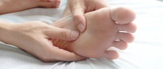
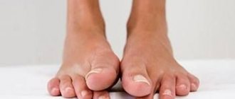
Extensor reflexes of the foot are an early manifestation of damage to the nervous system. The pathological Babinski reflex is most often tested in neurology. It is a sign of upper motor neuron syndrome. Belongs to the group of reflexes of the lower extremities. It manifests itself as follows: a stroking movement along the outer edge of the foot leads to extension of the big toe. May be accompanied by fanning out all the toes. In the absence of pathology, such irritation of the foot leads to involuntary flexion of the big toe or all the toes. Movements should be light and not cause pain. The reason for the formation of the Babinski reflex is the slow conduction of stimulation along the motor channels and impaired excitation of segments of the spinal cord. In children under one and a half years of age, the manifestation of the Babinski reflex is considered normal; then, with the formation of gait and vertical body position, it should disappear.
A similar effect can occur with other effects on receptors:
- Oppenheim reflex - extension of the finger occurs when pressing and moving from top to bottom with the thumb in the area of the tibia;
- Gordon's reflex - when the calf muscle is compressed;
- Schaeffer reflex - when the Achilles tendon is compressed.
Pathological flexion reflexes of the foot:
- Rossolimo reflex - when exposed to abrupt blows of the hammer or fingertips on the inner surface of the phalanges, rapid flexion of the II-V toes occurs;
- ankylosing spondylitis reflex - the same reaction occurs when lightly tapping the outer surface of the foot in the area of the metatarsal bones;
- Zhukovsky reflex - manifests itself when struck in the center of the foot, at the base of the toes.
Babinski reflex
The main extensor reflex is the Babinski foot extensor reflex. It is caused by streak irritation, carried out by pressing the blunt end of an injection needle on the outer edge of the plantar surface of the foot in the direction from the heel to the toes (Fig. 1.4.7).
Normally, in this case, the plantar reflex is evoked (flexion of all toes). When the central motor neuron (corticospinal part of the pyramidal tract, projection motor cortex) is damaged, extension of the thumb occurs. This reflex is of very great clinical importance. There are a number of modifications of the methods for inducing it: squeezing the Achilles tendon (Schaeffer reflex), squeezing the calf muscle in its distal section (Gordon reflex), pressing the thumb on the anterior inner surface of the shin with sliding down the entire shin (Oppenheim reflex), etc.
Oral automaticity reflexes
Oral automatism is the reaction of the oral muscles to a stimulus, manifested by their involuntary movement. Pathological reflexes of this kind are observed in the following manifestations:
- The nasolabial reflex, which occurs when the base of the nose is tapped with a hammer, is manifested by stretching the lips. The same effect can occur when approaching the mouth (distance-oral reflex) or when lightly hitting the lower or upper lip - oral reflex.
- Palmomental reflex, or Marinescu-Radovic reflex. The stroke movements in the area of the thumb from the side of the palm cause a reaction of the facial muscles and cause the chin to move.
Such reactions are considered normal only for infants; their presence in adults is pathological.
Synkinesis and defensive reflexes
Synkinesis are reflexes characterized by paired movements of the limbs. Pathological reflexes of this kind include:
- global synkinesia (when the arm is bent, the leg is extended or vice versa);
- imitation: involuntary repetition of movements of an unhealthy (paralyzed) limb after the movements of a healthy one;
- coordinator: spontaneous movements of an unhealthy limb.
Synkinesis automatically occurs during active movements. For example, when moving a healthy arm or leg, a spontaneous muscle contraction occurs in a paralyzed limb, a flexion movement of the arm occurs, and an extension movement of the legs occurs.
Protective reflexes arise when a paralyzed limb is irritated and are manifested by its involuntary movement. An irritant can be, for example, a needle prick. Such reactions are also called spinal automatisms. Protective reflexes include the Marie-Foy-Bekhtereva symptom - flexion of the toes leads to involuntary flexion of the leg at the knee and hip joint.
Synkinesis
Synkinesia is a reflex during which one reflex movement of the upper or lower limb is accompanied by a reflex reaction of the other.
Synkinesias are divided into:
- global (flexion of the paralyzed arm together with extension of the paralyzed leg);
- imitation (involuntary motor acts of paralyzed limbs, movements familiar to a healthy person);
- Coordinating (production of various movements by paralyzed parts of the body during the performance of other complex motor acts).
To avoid the development of pathological reflexes, both in childhood and in adulthood, it is very important to devote a lot of time to your health. Particular attention should be paid to the daily routine, healthy eating, alternating rest and physical activity.
If nonspecific signs of the disease appear, you will urgently need to consult a neurologist.
Tonic reflexes
Normally, tonic reflexes appear in children from birth to three months. Their continued manifestation even in the fifth month of life may indicate that the child has cerebral palsy. With cerebral palsy, congenital motor automatisms do not fade away, but continue to develop. These include pathological tonic reflexes:
- Labyrinthine tonic reflex. It is checked in two positions - on the back and on the stomach - and manifests itself depending on the location of the child’s head in space. In children with cerebral palsy, it is expressed in increased tone of the extensor muscles when lying on the back and flexor muscles when the child lies on the stomach.
- Symmetrical cervical tonic reflex. In cerebral palsy, it is manifested by the influence of head movements on the tone of the muscles of the limbs.
- Asymmetrical cervical tonic reflex. It manifests itself as increased muscle tone in the limbs when turning the head to the side. On the side where the face is turned, the extensor muscles are activated, and on the side of the back of the head, the flexor muscles.
With cerebral palsy, a combination of tonic reflexes is possible, which reflects the severity of the disease.
Tendon reflexes
Tendon reflexes are normally caused by hitting the tendon with a hammer. They are divided into several types:
- Biceps tendon reflex. In response to a hammer blow on it, the arm bends at the elbow joint.
- Triceps tendon reflex. The arm is bent at the elbow joint, and upon impact, extension occurs.
- Knee reflex. The blow falls on the quadriceps femoris muscle, under the kneecap. The result is extension of the leg at the knee joint.
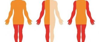
Pathological tendon reflexes manifest themselves in the absence of a reaction to hammer blows. They can occur with paralysis, coma, or spinal cord injuries.
Direct lesion of the pyramidal tract
Damage to the pyramidal tract has the following classification:
- Foot clonus . It appears when the foot is strongly compressed while a person is lying down. A positive reaction will consist of sharp clonic motor actions of the foot.
- Patellar clonus . To diagnose, you need to grab the upper part of the kneecap and pull it up a little, and then sharply release it. In the presence of a pathological disorder, contraction of the quadriceps femoris muscle will appear.
Is treatment possible?
Pathological reflexes in neurology themselves are not treated, since this is not a separate disease, but only a symptom of some mental disorder. They indicate problems with the functioning of the brain and nervous system. Therefore, it is necessary, first of all, to look for the reason for their appearance. Only after a doctor has made a diagnosis can we talk about specific treatment, because it is necessary to treat the cause itself, and not its manifestations. Pathological reflexes can only help in determining the disease and its severity.


