Stages of nervous system development
In evolution, the nervous system has undergone several stages of development, which became turning points in the qualitative organization of its activities.
These stages differ in the number and types of neuronal formations, synapses, signs of their functional specialization, and in the formation of groups of neurons interconnected by common functions. There are three main stages of the structural organization of the nervous system: diffuse, nodular, tubular. The diffuse nervous system is the most ancient, found in coelenterates (hydra). Such a nervous system is characterized by a multiplicity of connections between neighboring elements, which allows excitation to freely spread throughout the nervous network in all directions.
This type of nervous system provides wide interchangeability and thereby greater reliability of functioning, but these reactions are imprecise and vague.
The nodal type of nervous system is typical for worms, mollusks, and crustaceans.
It is characterized by the fact that the connections of nerve cells are organized in a certain way, excitation passes along strictly defined paths. This organization of the nervous system turns out to be more vulnerable. Damage to one node causes dysfunction of the entire organism as a whole, but its qualities are faster and more accurate.
The tubular nervous system is characteristic of chordates; it includes features of the diffuse and nodular types. The nervous system of higher animals took all the best: high reliability of the diffuse type, accuracy, locality, speed of organization of nodal type reactions.
The leading role of the nervous system
At the first stage of the development of the world of living beings, interaction between the simplest organisms was carried out through the aquatic environment of the primitive ocean, into which the chemical substances released by them entered. The first oldest form of interaction between the cells of a multicellular organism is chemical interaction through metabolic products entering the body fluids. Such metabolic products, or metabolites, are the breakdown products of proteins, carbon dioxide, etc. This is the humoral transmission of influences, the humoral mechanism of correlation, or connections between organs.
The humoral connection is characterized by the following features:
- lack of an exact address to which a chemical substance entering the blood or other body fluids is sent;
- the chemical spreads slowly;
- the chemical acts in minute quantities and is usually quickly broken down or eliminated from the body.
Humoral connections are common to both the animal and plant worlds. At a certain stage of development of the animal world, in connection with the appearance of the nervous system, a new, nervous form of connections and regulation is formed, which qualitatively distinguishes the animal world from the plant world. The higher the development of an animal’s organism, the greater the role played by the interaction of organs through the nervous system, which is designated as reflex. In higher living organisms, the nervous system regulates humoral connections. Unlike the humoral connection, the nervous connection has a precise direction to a specific organ and even a group of cells; communication is carried out hundreds of times faster than the speed of distribution of chemicals. The transition from a humoral connection to a nervous connection was not accompanied by the destruction of the humoral connection between the cells of the body, but by the subordination of nervous connections and the emergence of neurohumoral connections.
At the next stage of development of living beings, special organs appear - glands, in which hormones are produced, formed from food substances entering the body. The main function of the nervous system is both to regulate the activity of individual organs among themselves, and in the interaction of the body as a whole with its external environment. Any impact of the external environment on the body appears, first of all, on receptors (sensory organs) and is carried out through changes caused by the external environment and the nervous system. As the nervous system develops, its highest department—the cerebral hemispheres—becomes “the manager and distributor of all the activities of the body.”
Structure of the nervous system
The nervous system is formed by nervous tissue, which consists of a huge number of neurons - a nerve cell with processes.
The nervous system is conventionally divided into central and peripheral.
The central nervous system includes the brain and spinal cord, and the peripheral nervous system includes the nerves that arise from them.
The brain and spinal cord are a collection of neurons. In a cross section of the brain, white and gray matter are distinguished. Gray matter consists of nerve cells, and white matter consists of nerve fibers, which are processes of nerve cells. In different parts of the central nervous system, the location of white and gray matter is different. In the spinal cord, gray matter is located inside, and white matter is outside, but in the brain (cerebral hemispheres, cerebellum), on the contrary, gray matter is outside, white matter is inside. In various parts of the brain there are separate clusters of nerve cells (gray matter) located inside the white matter - the nuclei. Clusters of nerve cells are also located outside the central nervous system. They are called nodes and belong to the peripheral nervous system.
Cells of the brain and spinal cord
The brain and spinal cord are made up of cells whose names and characteristics are determined by their functions. Cells characteristic only of the nervous system are neurons and neuroglia .
Neurons –
These are the workhorses of the nervous system. They send and receive signals to and from the brain through a network of interconnections so numerous and complex that it is completely impossible to count them or chart them completely. At best, we can roughly say that the brain contains hundreds of billions of neurons and many times more connections between them.
Figure 1. Neurons
Brain tumors arising from neurons or their precursors include embryonal tumors (previously called primitive neuroectodermal tumors - PNETs)
, such as
medulloblastomas
and
pineoblastomas
.
The second type of brain cells are called neuroglia . In the literal sense, this word means “the glue that holds the nerves together” - thus, the auxiliary role of these cells is already clear from the name itself. Another part of neuroglia contributes to the work of neurons, surrounding them, nourishing them and removing their breakdown products. There are many more neuroglial cells in the brain than neurons, and more than half of brain tumors develop from neuroglia.
Tumors arising from neuroglial (glial) cells are generally called gliomas. However, depending on the specific type of glial cells involved in the tumor, it may have one specific name or another. The most common glial tumors in children are cerebellar and hemispheric astrocytomas, brainstem gliomas, optic pathway gliomas, ependymomas and gangliogliomas. The types of tumors are described in more detail in this article.
Reflex activity of the nervous system
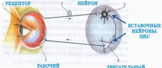
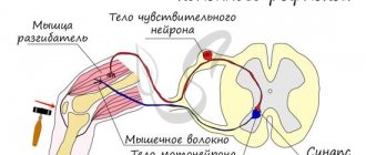
The main form of activity of the nervous system is the reflex. Reflex is the body’s reaction to changes in the internal or external environment, carried out with the participation of the central nervous system in response to irritation of receptors.
With any irritation, excitation from the receptors is transmitted along centripetal nerve fibers to the central nervous system, from where, through the interneuron along centrifugal fibers, it goes to the periphery to one or another organ, the activity of which changes. This entire path through the central nervous system to the working organ, called the reflex arc, is usually formed by three neurons: sensory, intercalary and motor. A reflex is a complex act in which a significantly larger number of neurons take part. Excitation, entering the central nervous system, spreads to many parts of the spinal cord and reaches the brain. As a result of the interaction of many neurons, the body responds to irritation.
Spinal cord
The spinal cord is a cord about 45 cm long, 1 cm in diameter, located in the spinal canal, covered with three meninges: dura, arachnoid and soft (vascular).
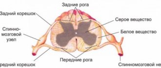
The spinal cord is located in the spinal canal and is a cord that at the top passes into the medulla oblongata and at the bottom ends at the level of the second lumbar vertebra. The spinal cord consists of gray matter containing nerve cells and white matter consisting of nerve fibers. Gray matter is located inside the spinal cord and is surrounded on all sides by white matter.
In a cross section, the gray matter resembles the letter H. It distinguishes the anterior and posterior horns, as well as the connecting crossbar, in the center of which there is a narrow canal of the spinal cord containing cerebrospinal fluid. In the thoracic region there are lateral horns. They contain the bodies of neurons that innervate internal organs. The white matter of the spinal cord is formed by nerve processes. Short processes connect sections of the spinal cord, and long ones make up the conductive apparatus of bilateral connections with the brain.
The spinal cord has two thickenings - cervical and lumbar, from which nerves extend to the upper and lower extremities. 31 pairs of spinal nerves arise from the spinal cord. Each nerve begins from the spinal cord with two roots - anterior and posterior. The dorsal roots are sensitive and consist of processes of centripetal neurons. Their bodies are located in the spinal ganglia. The anterior roots - motor - are processes of centrifugal neurons located in the gray matter of the spinal cord. As a result of the fusion of the anterior and posterior roots, a mixed spinal nerve is formed. The spinal cord contains centers that regulate the simplest reflex acts. The main functions of the spinal cord are reflex activity and conduction of excitation.
The human spinal cord contains reflex centers for the muscles of the upper and lower extremities, sweating and urination. The function of excitation is that impulses from the brain to all areas of the body and back pass through the spinal cord. Centrifugal impulses from organs (skin, muscles) are transmitted through ascending pathways to the brain. Along descending pathways, centrifugal impulses are transmitted from the brain to the spinal cord, then to the periphery, to the organs. When the pathways are damaged, there is a loss of sensitivity in various parts of the body, a violation of voluntary muscle contractions and the ability to move.
Human brain
August 25, 2014 Neurology
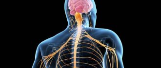
The human nervous system is represented by the brain, located in the cranial cavity; the spinal cord, located in the spinal cavity, and a branched system of nerves that extend from the brain (cranial nerves) and innervate the organs of the head; a system of nerves that branch from the spinal cord and innervate the arms, legs, torso, and internal organs. The brain and spinal cord represent the central nervous system, and the nerve system represents the peripheral nervous system.
All formations of the nervous system consist of many neurons (cells of the nervous system) and their processes, through which nerve impulses are transmitted in ascending and descending directions due to the diverse connections that exist between neurons.
Despite the fact that different neurons perform different functions and have differences in structure, they all have a body, a receptive structure, and a process, a dendrite, a conducting structure.
According to their functional characteristics, neurons are divided into motor - executive, and sensitive - perceiving, as well as interneurons that interact between them.
The nerve cell performs two main functions: 1) processing of incoming information, transmission of nerve impulses, and 2) biosynthetic, aimed at maintaining its vital functions.
This is the schematic diagram of the structure of a neuron.
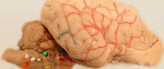
This is what the human brain looks like.
This is a complex structure, consisting of many different formations that are in close interaction; carrying out conducting, analyzing, regulating and coordinating functions. All body movements, human feelings, the work of internal organs, his mind, intellect, memory, consciousness, sleep, wakefulness, everything is controlled by the brain. The human brain can be compared to a complex computer with embedded programs that are constantly modified throughout a person’s life.
Schematically, the brain can be divided into lobes: frontal, occipital, temporal, parietal; cerebellum, brain stem. The lobes of the brain are covered with a cortex, which is a collection of highly differentiated neurons that carry out higher integrative activities.
In the frontal lobes there are centers for the regulation of voluntary movements, when damaged, weakness develops in the arms, legs on one side, or only the arms or legs. In the frontal lobes there are also rotations of the eyes and head, when damaged, deviation of the eyes and head occurs towards the pathological focus. The frontal lobes also contain centers for coordination of movements, which, when damaged, cause disturbances in standing and walking. And finally, when the frontal lobe cortex is damaged, behavioral and mental disorders develop.
The parietal lobes are responsible for a person’s ability to recognize objects by touch, the ability to perform complex goal-directed actions, the ability to decipher written characters and the ability to write.
The temporal lobes contain auditory, gustatory and olfactory centers, centers for understanding and reproducing speech, and centers for coordination of movements.
The visual lobes contain the centers for the perception of visual images and visual memory. The cerebellum is one of the main coordinating centers.
In the brain stem there are centers for the regulation of life-supporting organ systems, respiratory, cardiovascular, intermediate centers for the regulation of cranial nerves, the pathways of the motor and sensory systems.
In the brainstem, in its tegmentum, are located the nuclei of the cranial nerves, the bodies of nerve cells responsible for the innervation of the organs of the head and face, providing the functions of the gustatory, auditory, visual, vestibular and olfactory analyzers.
The cranial nerves of the caudal group are distinguished: 1) Accessory nerve, 11th pair, innervates the muscle that turns the head to the side. 2) Hypoglossal nerve, 12th pair, innervating the tongue. 3) Glossopharyngeal nerve, 9th pair, innervating the pharyngeal muscles, tongue, palate, middle ear, salivary glands. 4) Vagus nerve, 10th pair, innervating the muscles of the pharynx, soft palate, larynx, smooth muscles of the bronchi, trachea, esophagus, stomach, intestines.
Next, the cranial nerves of the cerebellopontine angle are distinguished: 1) Facial nerve, 7th pair, innervating the facial muscles. 2) Vestibulocochlear nerve, 8th pair, innervating the inner ear. 3) Trigeminal nerve, 3rd pair, innervating the skin of the face, jaws, and masticatory muscles. Next comes a group of oculomotor nerves: 3, 4, 6 pairs.
And finally, the optic nerve, 2nd pair, innervating the retina, and the olfactory nerve, 1st pair, innervating the nasal mucosa.
Evolution of the vertebrate brain
The formation of the central nervous system in the form of a neural tube first appears in chordates. In lower chordates, the neural tube is preserved throughout life; in higher chordates, vertebrates, a neural plate is formed on the dorsal side during the embryonic stage, which is immersed under the skin and folded into a tube. In the embryonic stage of development, the neural tube forms three swellings in the anterior part - three brain vesicles, from which parts of the brain develop: the anterior vesicle gives rise to the forebrain and diencephalon, the middle vesicle turns into the midbrain, the posterior vesicle forms the cerebellum and medulla oblongata. These five brain regions are characteristic of all vertebrates.
Lower vertebrates - fish and amphibians - are characterized by a predominance of the midbrain over other parts. In amphibians, the forebrain somewhat enlarges and a thin layer of nerve cells is formed in the roof of the hemispheres - the primary medullary vault, the ancient cortex. In reptiles, the forebrain increases significantly due to accumulations of nerve cells. Most of the roof of the hemispheres is occupied by the ancient cortex. For the first time in reptiles, the rudiment of a new cortex appears. The hemispheres of the forebrain creep onto other parts, as a result of which a bend is formed in the region of the diencephalon. Beginning with ancient reptiles, the cerebral hemispheres became the largest part of the brain.
The structure of the brain of birds and reptiles has much in common. On the roof of the brain is the primary cortex, the midbrain is well developed. However, in birds, compared to reptiles, the total brain mass and the relative size of the forebrain increase. The cerebellum is large and has a folded structure. In mammals, the forebrain reaches its greatest size and complexity. Most of the brain matter is made up of the neocortex, which serves as the center of higher nervous activity. The intermediate and middle parts of the brain in mammals are small. The expanding hemispheres of the forebrain cover them and crush them under themselves. Some mammals have a smooth brain without grooves or convolutions, but most mammals have grooves and convolutions in the cerebral cortex. The appearance of grooves and convolutions occurs due to the growth of the brain with limited dimensions of the skull. Further growth of the cortex leads to the appearance of folding in the form of grooves and convolutions.
STRUCTURE OF THE SPINAL CORD AND BRAIN
The structure of the spinal cord and brain. The nervous system is divided into central, located in the skull and spine, and peripheral, outside the skull and spine. The central nervous system consists of the spinal cord and the brain.
Rice. 105. Nervous system (diagram): 1 - cerebrum, 2 - cerebellum, 3 - cervical plexus, 4 - brachial plexus, 5 - spinal cord, 6 - sympathetic trunk, 7 - thoracic nerves, 8 - median nerve, 9 - solar plexus, 10 - radial nerve, 11 - ulnar nerve, 12 - lumbar plexus, 13 - sacral plexus, 14 - coccygeal plexus, 15 - femoral nerve, 16 - sciatic nerve, 17 - tibial nerve, 18 - peroneal nerve The spinal cord represents It is a long cord, approximately cylindrical in shape, located in the spinal canal. At the top it gradually passes into the medulla oblongata, at the bottom it ends at the level of the 1st-2nd lumbar vertebrae. At the site of the origin of the nerves to the upper and lower extremities there are 2 thickenings: the cervical - at the level of the 2nd cervical to the 2nd thoracic vertebrae and the lumbar - from the level of the 10th thoracic with the greatest thickness at the level of the 12th thoracic vertebra. The average length of the spinal cord in a man is 45 cm, in a woman it is 41–42 cm, the average weight is 34–38 g.
The spinal cord consists of two symmetrical halves connected by a narrow bridge, or commissure. A cross-section of the spinal cord shows that in the middle there is a gray matter consisting of neurons and their processes, in which two large, wide anterior horns and two narrower posterior horns are distinguished. In the thoracic and lumbar segments there are also lateral projections—lateral horns. In the anterior horns there are motor neurons, from which centrifugal nerve fibers depart, forming the anterior, or motor, roots, and through the dorsal roots, centripetal nerve fibers of the neurons of the spinal ganglia enter the dorsal horns. The gray matter also contains blood vessels. There are 3 main groups of neurons in the spinal cord: 1) large motor neurons with long, low-branching axons, 2) forming the intermediate zone of gray matter; their axons are divided into 2-3 long branches, and 3) sensitive, part of the spinal ganglia, with highly branching axons and dendrites. The gray matter is surrounded by white matter, which consists of longitudinally located pulpal and partly non-pulphate nerve fibers, neuroglia and blood vessels. In each half of the spinal cord, the white matter is divided into three columns by the horns of gray matter. The white matter located between the anterior groove and the anterior horn is called the anterior columns, between the anterior and posterior horn - the lateral columns, between the posterior bridge and the posterior horn - the posterior columns. Each column consists of separate bundles of nerve fibers. In addition to the thick pulpy fibers of the motor neurons, thin nerve fibers of the lateral horn neurons belonging to the autonomic nervous system emerge along the anterior roots. In the dorsal horns there are intercalary, or fascicle, neurons, the nerve fibers of which connect motor neurons of different segments and are part of the white matter bundles. Pulmonary nerve fibers are divided into short - local pathways of the spinal cord, and long - long pathways connecting the spinal cord to the brain.
Rice. 106. Cross section of the spinal cord. Diagram of pathways. On the left are the ascending paths, on the right are the descending paths. Ascending paths: / - delicate bundle; XI - wedge-shaped bundle; X - posterior spinocerebellar tract; VIII - anterior spinocerebellar tract; IX, VI - lateral and anterior spinothalamic tracts; XII - spinotectal tract. Descending tracts: II, V - lateral and anterior pyramidal tracts; III - rubrospinal tract; IV - vestibulo-spinal tract; VII - olivospinal tract. Circles (without numbering) indicate pathways connecting segments of the spinal cord. The ratio of gray and white matter in different segments of the spinal cord is not the same. The lumbar and sacral segments contain, due to a significant decrease in the content of nerve fibers in the descending tracts and the beginning of the formation of the ascending tracts, more gray matter than white matter. In the middle and especially upper thoracic segments there is relatively more white matter than gray matter. In the cervical segments, the amount of gray matter increases and white matter increases significantly. Thickening of the spinal cord in the cervical region depends on the development of innervation of the arm muscles, and thickening of the lumbar region depends on the development of innervation of the leg muscles. Consequently, the development of the spinal cord is determined by the activity of skeletal muscles. The supporting basis of the spinal cord is neuroglia and the connective tissue layer of the pia mater penetrating into the white matter. The surface of the spinal cord is covered with a thin neuroglial membrane, which contains blood vessels. Outside the soft tissue there is an arachnoid membrane connected to it, made of loose connective tissue, in which cerebrospinal fluid circulates. The arachnoid membrane adheres tightly to the outer hard shell of dense connective tissue with a large number of elastic fibers.
Rice. 107. Layout of spinal cord segments. The location of the spinal cord segments in relation to the corresponding vertebrae and the places where the roots exit the spinal canal are shown.
The human spinal cord consists of 31-33 segments, or segments: cervical - 8, thoracic - 12, lumbar - 5, sacral - 5, coccygeal - 1-3. Two pairs of roots emerge from each segment, connecting into two spinal nerves, consisting of centripetal - sensory and centrifugal - motor nerve fibers. Each nerve begins at a certain segment of the spinal cord with two roots: anterior and posterior, which end at the spinal ganglion and, joining together outward from the node, form a mixed nerve. Mixed spinal nerves exit the spinal canal through the intervertebral foramina, except for the first pair, passing between the edge of the occipital bone and the upper edge of the 1st cervical vertebra, and the coccygeal root, between the edges of the vertebrae of the coccyx. The spinal cord is shorter than the spine, so there is no correspondence between the spinal cord segments and the vertebrae. Spinal nerves innervate the skin and muscles of the trunk, arms and legs. They form: 1) the cervical plexus, consisting of 4 upper cervical nerves, innervating the skin of the neck, back of the head, auricle and skin on the collarbone, neck muscles and diaphragm; 2) brachial plexus of 4 lower cervical nerves and 1st thoracic nerve, innervating the skin and muscles of the shoulder girdle and arm; 3) thoracic nerves, which correspond to the 12 thoracic segments of the spinal cord and innervate the skin and muscles of the chest and abdomen (anterior branch) and the skin and muscles of the back (posterior branch), therefore, the thoracic spinal nerves have the correct segmental location and are clearly divided into anterior - abdominal part and back - dorsal part; 4) lumbar plexus, consisting of the 12th thoracic and 4 upper lumbar nerves, innervating the skin and part of the muscles of the pelvis, thigh, leg and foot; 5) sacral plexus, consisting of the lower lumbar, sacral and coccygeal nerves, innervating the skin and other muscles of the pelvis, thigh, leg and foot.
Rice. 108. Brain, median surface: I - frontal lobe of the cerebrum, 2 - parietal lobe, 3 - occipital lobe, 4 - corpus callosum, 5 - cerebellum, 6 - optic thalamus (diencephalon), 7 - pituitary gland, 8 - quadrigeminal (midbrain), 9 - pineal gland, 10 - pons, 11 - medulla oblongata The brain also consists of gray and white matter. The gray matter of the brain is represented by a variety of neurons, grouped into numerous clusters - nuclei and covering different parts of the brain on top. In total, there are approximately 14 billion neurons in the human brain. In addition, the gray matter contains neuroglial cells, which are approximately 10 times more numerous than neurons; they make up 60-90% of the total brain mass. Neuroglia are scaffolding tissues that support neurons. It is also involved in the metabolism of the brain and especially neurons; hormones and hormone-like substances are formed in it (neurosecretion).
The brain is divided into the medulla oblongata and the pons, the cerebellum, midbrain and diencephalon, which make up its trunk, and the telencephalon, or cerebral hemispheres, covering the brain stem from above (Fig. 108). In humans, unlike animals, the volume and weight of the brain sharply predominate over the spinal cord: approximately 40-45 times or more (in chimpanzees, the weight of the brain exceeds the weight of the spinal cord by only 15 times). The average weight of the adult human brain is approximately 1400 g in men and, due to the relatively lower average body weight, approximately 10% less in women. A person's mental development does not directly depend on the weight of his brain. Only in cases where the weight of a man’s brain is below 1000 g, and that of a woman is below 900 g, the structure of the brain is disrupted and mental abilities are reduced.
Rice. 109. Anterior surface of the brain stem. Origin of cranial nerves. The lower surface of the cerebellum: 1 - optic nerve, 2 - insula, 3 - pituitary gland, 4 - optic chiasm, 5 - infundibulum, 6 - gray tubercle, 7 - mamillary body, 8 - fossa between the peduncles, 9 - cerebral peduncle, 10 - semilunar node, 11 - minor root of the trigeminal nerve, 12 - major root of the trigeminal nerve, 13 - abducens nerve, 14 - glossopharyngeal nerve, 15 - choroid plexus of the IV ventricle, 16 - vagus nerve, 17 - accessory nerve, 18 - first cervical nerve, 19 - decussation of pyramids, 20 - pyramid, 21 - hypoglossal nerve, 22 - auditory nerve, 23 - intermediate nerve, 24 - facial nerve, 25 - trigeminal nerve, 26 - pons, 27 - trochlear nerve, 28 - external geniculate body, 29 - oculomotor nerve, 30 - optic tract, 31-32 - anterior perforated substance, 33 - external olfactory stripe, 34 - olfactory triangle, 35 - olfactory tract, 36 - olfactory bulb. 12 pairs of cranial nerves emerge from the nuclei of the brain stem, which in Unlike spinal ones, they do not have a correct segmental exit and a clear division into the abdominal and dorsal parts. Cranial nerves are divided into: 1) olfactory, 2) visual, 3) oculomotor, 4) trochlear, 5) trigeminal, 6) abducens, 7) facial, auditory, 9) glossopharyngeal, 10) vagus, 11) accessory, 12) sublingual .
Related materials:
Functions of the spinal cord
Reflex mechanism
Properties of nerve centers
Autonomic nervous system
Brain
If the spinal cord in all vertebrates is developed more or less equally, then the brain differs significantly in size and complexity of structure in different animals. The forebrain undergoes particularly dramatic changes during evolution. In lower vertebrates, the forebrain is poorly developed. In fish, it is represented by the olfactory lobes and nuclei of gray matter in the thickness of the brain. The intensive development of the forebrain is associated with the emergence of animals onto land. It differentiates into the diencephalon and two symmetrical hemispheres, which are called the telencephalon. Gray matter on the surface of the forebrain (cortex) first appears in reptiles, developing further in birds and especially in mammals. Truly large forebrain hemispheres become only in birds and mammals. In the latter, they cover almost all other parts of the brain.
The brain is located in the cranial cavity. It includes the brainstem and telencephalon (cerebral cortex).
The brainstem consists of the medulla oblongata, pons, midbrain and diencephalon.
The medulla oblongata is a direct continuation of the spinal cord and, expanding, passes into the hindbrain. It basically retains the shape and structure of the spinal cord. In the thickness of the medulla oblongata there are accumulations of gray matter - the nuclei of the cranial nerves. The posterior pons includes the cerebellum and the pons. The cerebellum is located above the medulla oblongata and has a complex structure. On the surface of the cerebellar hemispheres, gray matter forms the cortex, and inside the cerebellum - its nuclei. Like the spinal medulla oblongata, it performs two functions: reflex and conductive. However, the reflexes of the medulla oblongata are more complex. This is reflected in its importance in the regulation of cardiac activity, the condition of blood vessels, respiration, and sweating. The centers of all these functions are located in the medulla oblongata. Here are the centers for chewing, sucking, swallowing, saliva and gastric juice. Despite its small size (2.5–3 cm), the medulla oblongata is a vital part of the central nervous system. Damage to it can cause death due to cessation of breathing and heart activity. The conductor function of the medulla oblongata and the pons is to transmit impulses from the spinal cord to the brain and back.
In the midbrain there are primary (subcortical) centers of vision and hearing, which carry out reflexive orienting reactions to light and sound stimuli. These reactions are expressed in various movements of the torso, head and eyes towards the stimuli. The midbrain consists of the cerebral peduncles and quadrigeminalis. The midbrain regulates and distributes the tone (tension) of skeletal muscles.
The diencephalon consists of two sections - the thalamus and hypothalamus, each of which consists of a large number of nuclei of the visual thalamus and subthalamic region. Through the visual thalamus, centripetal impulses are transmitted to the cerebral cortex from all receptors of the body. Not a single centripetal impulse, no matter where it comes from, can pass to the cortex, bypassing the visual hillocks. Thus, through the diencephalon, all receptors communicate with the cerebral cortex. In the subtubercular region there are centers that influence metabolism, thermoregulation and endocrine glands.
The cerebellum is located behind the medulla oblongata. It consists of gray and white matter. However, unlike the spinal cord and brainstem, the gray matter - the cortex - is located on the surface of the cerebellum, and the white matter is located inside, under the cortex. The cerebellum coordinates movements, makes them clear and smooth, plays an important role in maintaining the balance of the body in space, and also influences muscle tone. When the cerebellum is damaged, a person experiences a decrease in muscle tone, movement disorders and changes in gait, speech slows down, etc. However, after some time, movement and muscle tone are restored due to the fact that the intact parts of the central nervous system take over the functions of the cerebellum.
The cerebral hemispheres are the largest and most developed part of the brain. In humans, they form the bulk of the brain and are covered with cortex over their entire surface. Gray matter covers the outside of the hemispheres and forms the cerebral cortex. The human cerebral cortex has a thickness of 2 to 4 mm and is composed of 6–8 layers formed by 14–16 billion cells, different in shape, size and functions. Under the cortex is a white substance. It consists of nerve fibers connecting the cortex with the lower parts of the central nervous system and the individual lobes of the hemispheres with each other.
The cerebral cortex has convolutions separated by grooves, which significantly increase its surface. The three deepest grooves divide the hemispheres into lobes. In each hemisphere there are four lobes: frontal, parietal, temporal, occipital. The excitation of different receptors enters the corresponding receptive areas of the cortex, called zones, and from here they are transmitted to a specific organ, prompting it to action. The following zones are distinguished in the cortex. The auditory zone is located in the temporal lobe and receives impulses from auditory receptors.
The visual zone lies in the occipital region. Impulses from the eye receptors arrive here.
The olfactory zone is located on the inner surface of the temporal lobe and is connected to the receptors of the nasal cavity.
The sensory-motor zone is located in the frontal and parietal lobes. This zone contains the main centers of movement of the legs, torso, arms, neck, tongue and lips. This is also where the center of speech lies.
The cerebral hemispheres are the highest division of the central nervous system, controlling the functioning of all organs in mammals. The importance of the cerebral hemispheres in humans also lies in the fact that they represent the material basis of mental activity. I.P. Pavlov showed that mental activity is based on physiological processes occurring in the cerebral cortex. Thinking is associated with the activity of the entire cerebral cortex, and not just with the function of its individual areas.
| Brain department | Functions | |
| Medulla | Conductor | Connection between the spinal and overlying parts of the brain. |
| Reflex | Regulation of the activity of the respiratory, cardiovascular, digestive systems:
| |
| Pons | Conductor | Connects the cerebellar hemispheres to each other and to the cerebral cortex. |
| Cerebellum | Coordination | Coordination of voluntary movements and maintaining body position in space. Regulation of muscle tone and balance |
| Midbrain | Conductor | Approximate reflexes to visual and sound stimuli (turns of the head and torso). |
| Reflex |
| |
| Diencephalon | thalamus
hypothalamus
| |
The structure of the nervous system. Spinal cord and brain
Nerve tissue is made up of cells called neurons, which are connected by processes. Communication between nerve cells occurs through the generation and transmission of nerve impulses.
Anatomically, that is, by location, the nervous system is divided into two parts : central
nervous system, abbreviated as (CNS)
and
peripheral .
Central
includes the brain and spinal cord. These sections are represented by clusters of nerve cells, forming nerve centers.
Peripheral
The nervous system consists of nerves, nerve ganglia and nerve endings.
Nerve
- this is a sheathed structure consisting of a bundle of nerve fibers (mainly axons) and the neuroglobula that supports
them
.
Nerves
There are three types:
sensitive
,
motor
and
mixed
.
Sensitive
, conduct nerve impulses from receptors to the central nervous system.
Motor
- conduct nerve impulses from the central nervous system to the executive organs.
Mixed ones
conduct nerve impulses in both directions.
Nerve nodes
(g
and
nglia) are clusters of neuron bodies located outside the central nervous system.
Physiologically, that is, according to the functions performed, the nervous system is also divided into two sections: somatic
and
vegetative (autonomous).
Somatic nervous system
controls the functioning of skeletal muscles. Thanks to it, voluntary movements are made.
Autonomic (autonomic) nervous system
regulates the functioning of internal organs. This part of the nervous system is not subject to our will (for example, the stomach, heart, kidneys function independently of a person’s desire) and works autonomously. This is where the name of this department comes from.
The human nervous system functions on the basis of reflexes.
Thoughts and actions are reflexive and fit into the scheme: perception of irritation - information processing and response.
Reflex
- the body’s response to influences from the external or internal environment with the participation of the nervous system.
In this case, the nerve impulse distributed through the neurons makes a certain path through the nervous system. This path is called the reflex arc
.
The reflex arc of the somatic nervous system consists of three neurons.
Let's trace the passage of the nerve impulse. For example, if a person pricks his finger, the skin receptors, in response to irritation, will generate a nerve impulse that will begin its journey along the sensitive neuron
.
Behind the sensory neuron is an interneuron
, from which irritation is transmitted to
the motor neuron
.
Thus, through sensitive neurons (the bodies of which are located in the nerve ganglia), the nerve impulse is transmitted to the central nervous system, where information is processed, and from there a signal is sent to the working organ to execute the command.
In reflex activity, the passage of a nerve impulse from the brain to the organs is an example of direct communication
.
And from organs to brain - the opposite
.
As we have already said, the autonomic (autonomic) nervous
the system regulates the functioning of internal organs.
Functionally, the autonomic (autonomic) nervous system consists of two sections: sympathetic
and
parasympathetic
. Thanks to this, information containing operating instructions reaches the organ in two different ways.
Sympathetic department
ensures the functioning of the body in stressful situations, and the parasympathetic - in a state of rest.
The sympathetic department ensures the functioning of organs during intense work, and the parasympathetic department - during normal physiological stress.
Motor part of the somatic nervous system
consists of one neuron. And in the autonomic (autonomic) nervous system it is represented by two neurons.
The connection of neurons occurs in the nerve ganglion, or g a
England.
,
the neuron that is located between the brain and the ganglion is called preganglionic
, and the neuron that connects the nerve ganglion with the executive organ
is
called
postganglionic .
The central part of the human nervous system, as we have already said, is represented by the spinal cord and brain.
The human spinal cord, like all vertebrates, is located in the spinal canal. It is a white strand 40-45 cm long, 1 to 1.5 cm wide and weighing about 35 grams.
It controls most of the reflexes and provides the ability to move.
At the top, the spinal cord passes into the lower part of the brain - the medulla oblongata
, and below it ends at the level of the first or second lumbar vertebrae.
The spinal cord does not occupy the entire cavity of the spinal canal: between the walls of the canal and the brain there remains a space filled with adipose tissue, blood vessels, meninges and cerebrospinal fluid, which washes the spinal cord and protects it from shocks.
Externally, the brain is covered with three membranes: dura
,
cobwebby
and
soft
.
On the surface of the spinal cord, two longitudinal grooves are clearly visible: the anterior
and
back
. They divide it into symmetrical halves - left and right.
31 pairs of spinal nerves arise from the spinal cord, dividing it into segments. The posterior roots extend from each nerve to the posterior surface, and the anterior roots extend from the anterior surface to each nerve, respectively.
In the internal structure of this section of the central nervous system, the central canal filled with cerebrospinal fluid and two parts that differ in color are clearly visible. In the middle, around the spinal canal, there is the so-called gray matter.
, which in cross section resembles the appearance of a butterfly, and is
white
.
Gray matter is represented by bodies
mi neurons and short branching processes - dendrites
and
tami.
And the white consists of long, non-branching axons that
form nerve fibers.
In the gray matter there are anterior
and
posterior horns
, and in the thoracic region and
lateral
.
Let's look at the direction of passage of the nerve impulse of the somatic reflex.
Along
the sensory neuron
, which enters the spinal cord as part of the dorsal roots, the nerve impulse reaches
the interneuron
, which is located entirely in the gray matter. From it, information is transmitted to the motor neuron, the body of which is located in the anterior horns of the gray matter, and its processes emerge from the spinal cord as part of the anterior roots.
Next, the nerve impulse enters the mixed spinal nerve
. It is called this because it contains both sensory and motor neurons. Along the mixed spinal nerve, the nerve impulse reaches the performing organ.
The nerve impulse enters the spinal cord through the dorsal roots and dorsal horns (through sensory neurons), and exits through the anterior horns and anterior roots (through motor neurons).
The spinal cord performs a reflex and conductive function.
That is, it conducts a nerve impulse to the brain and in the opposite direction.
The spinal cord provides the reflex. If a person unexpectedly pricks his finger, then thanks to a complex system of interactions in the body with the direct participation of the spinal cord, he will immediately withdraw his hand. This means that the reflex function of the spinal cord is that reflex arcs are closed on it.
However, when donating blood in a clinic during an injection, the person will not pull back his hand. Since information from the receptors is transmitted not only to the spinal cord, but also further to the brain, which processes it and in this situation sends instructions to slow down the spinal cord reflex to withdraw the hand.
Thus, the spinal cord conducts nerve impulses to the brain and in the opposite direction, performing a conductor function.
In addition to the spinal cord, the central nervous system also includes the brain.
Brain
located in the skull, the bones of which protect it from mechanical damage.
Externally, the brain resembles a yellowish jelly-like mass. The average weight of the human brain is 1300-1400 grams.
Like the spinal cord, the brain is covered with three membranes: dura
,
cobwebby
and
soft
.
The soft, or vascular, membrane of the brain is directly adjacent to the substance of the brain, extends into all grooves, and covers all convolutions. It consists of loose connective tissue, in which numerous blood vessels branch that feed the brain.
The arachnoid membrane of the brain is thin, translucent, and has no blood vessels.
The dura mater of the brain is the periosteum for the inner cerebral surface of the skull bones. This membrane contains the highest concentration of pain receptors in the human body, while the brain itself has no pain receptors.
Since metabolism is very active in the brain, it is richly supplied with blood vessels
providing it with oxygen and nutrients.
Inside the brain, like the spinal cord, one can distinguish gray
and
white matter
.
But their location is excellent. The cell bodies of neurons that form gray matter are found both on the surface of the brain and within it among the white matter, forming nuclei
.
The human brain is divided into five sections: medulla oblongata, posterior, middle, intermediate, terminal
.
Of these, three large sections are distinguished: posterior
,
middle
, and
front
.
The hindbrain is represented by the medulla oblongata
,
pons
and
cerebellum
.
And the forebrain consists of the intermediate
and
final brain
.
Medulla
is a continuation of the spinal cord, so their structure has a lot in common.
Only the gray matter of the medulla oblongata is located in separate clusters - idrama
.
The functions are also similar: reflex and conductive. Many reflex processes are carried out through the nuclei of the medulla oblongata, such as coughing, sneezing, lacrimation, etc.
The nuclei of the medulla oblongata contain nerve centers responsible for the acts of swallowing and the functioning of the digestive glands.
The medulla oblongata contains vital centers involved in regulating the activity of the heart and blood vessels, as well as breathing (respiratory center).
Over the bridge
All ascending and descending pathways pass through connecting
the forebrain
with the spinal cord, with
the cerebellum
and other structures.
Above the medulla oblongata is the cerebellum
. In principle, the structure of the cerebellum repeats the structure of the entire brain, which is where its name comes from. The surface of the cerebellum (cortex) is represented by gray matter and has folds, convolutions and grooves.
Inside the cerebellum there are also nuclei - accumulations of gray matter.
The cerebellum is a brain center that is extremely important for the coordination and regulation of motor activity. It works reflexively, maintaining the balance of the body and its orientation in space.
Midbrain
is a continuation of the bridge.
On its surface facing the cerebellum there are four tubercles - four about
the cerebellum. The superior colliculus carries out the primary processing of visual information. The lower ones are the centers for primary processing of information from the hearing organs.
Also in the midbrain are the most important motor centers that participate, together with the cerebellum, in coordinating body posture and maintaining muscle tone.
Diencephalon
consists of an upper part -
the thalamus
, or visual hillocks, and a lower part -
the hypothalamus
.
Thalamus
processes all types of information coming from the senses, except olfactory. It receives visual, tactile, gustatory and auditory information. Also in the thalamus are the highest centers of pain sensitivity.
Hypothalamus
, releases special neurohormones that affect the functioning of the
hypnophysis
. Therefore, it can be called the main link in the neurohumoral regulation of body functions.
The hypothalamus contains the hunger and thirst centers.
The large hemispheres of the human brain are divided into two halves by a deep longitudinal fissure - left and right.
The left and right hemispheres are connected by a plexus of nerve fibers - the corpus callosum
. Thanks to it, the collected information is transferred from one hemisphere to the cortical and subcortical structures of the other, which ensures an adequate and timely response.
The hemispheres of the brain are covered by a layer of gray matter called the cerebral cortex
. The cortex forms a large number of grooves of varying depth and length, between which convolutions are located.
The cerebral cortex consists of a huge number of neurons and provides higher nervous activity in humans. The number of neurons is between 10 and 11 billion, making up the majority of neurons in the entire human central nervous system.
The cortex of each hemisphere is divided into lobes
: frontal, parietal
,
occipital and temporal. The functions of the cortex are associated with different lobes.
The frontal lobes process information about all sensations. Here their summary analysis takes place and a holistic idea of the image is created. This zone of the cortex is called associative and it is with it that the ability to learn is associated. The centers that control muscle movements are also located here.
The parietal lobe is associated with musculocutaneous sensitivity.
The occipital region processes information from the visual organs.
Temporal - auditory information.
Cerebral cortex
The surface of the human cerebral cortex is about 1500 cm2, which is many times greater than the inner surface of the skull. This large surface of the cortex was formed due to the development of a large number of grooves and convolutions, as a result of which most of the cortex (about 70%) is concentrated in the grooves. The largest grooves of the cerebral hemispheres are the central one, which runs across both hemispheres, and the temporal one, which separates the temporal lobe from the rest. The cerebral cortex, despite its small thickness (1.5–3 mm), has a very complex structure. It has six main layers, which differ in the structure, shape and size of neurons and connections. The cortex contains the centers of all sensory (receptor) systems, representatives of all organs and parts of the body. In this regard, centripetal nerve impulses from all internal organs or parts of the body approach the cortex, and it can control their work. Through the cerebral cortex, conditioned reflexes are closed, through which the body constantly, throughout life, very accurately adapts to the changing conditions of existence, to the environment.