March 30, 2021
The brain is protected from external mechanical influences by the cranium. The most important sections are located at the bottom of the skull. The gray matter of the brain contains 25 billion neurons, which is almost 4 times the population of the globe (6.5 billion). The human brain is covered by three membranes:
- vascular (web-like),
- soft,
- hard.
In the area of the brain there are five ventricles - containers connected to each other by channels. Inside the cavities there is cerebrospinal fluid - a biological fluid that circulates both in the cisterns of the brain and in the spinal canal.
Finite brain
The terminal (telencephalon), or large, brain (cerebrum) develops from the anterior cerebral bladder and consists of highly developed paired parts - the right and left hemispheres of the cerebrum and the middle part connecting them. The hemispheres are separated by a longitudinal fissure, in the depth of which lies a plate of white matter - the corpus callosum. It consists of fibers connecting both hemispheres. Under the corpus callosum there is a vault, which consists of two curved fibrous cords, which are connected to each other in the middle part, and diverge in front and behind, forming the pillars and legs of the vault. Anterior to the columns of the arch is the anterior commissure. Between the anterior part of the corpus callosum and the fornix is a thin vertical plate of brain tissue - a transparent septum.
The cerebral hemisphere is formed by gray and white matter. It distinguishes the largest part, covered with grooves and convolutions, - a cloak formed by the gray matter lying on the surface - the cerebral cortex, the olfactory brain and accumulations of gray matter inside the hemispheres - the basal ganglia. The last two sections constitute the oldest part of the hemisphere in evolutionary development.
The cavities of the telencephalon are the lateral ventricles.
In each hemisphere, three surfaces are distinguished: the upper lateral - convex according to the cranial vault, the medial - flat, facing the same surface of the other hemisphere, and the lower - irregular in shape. The surfaces of the hemisphere have a complex pattern due to grooves running in different directions and folds between them - convolutions. The size and shape of the grooves and convolutions are subject to significant individual fluctuations. However, there are several permanent, clearly defined grooves that appear earlier than others during embryonic development. They are used to divide the hemispheres into large areas called lobes.
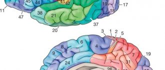
Each hemisphere consists of five lobes: frontal, parietal, occipital, temporal and insular, or insula, located deep in the lateral sulcus (Fig. 116). The boundary between the frontal and parietal lobes is the central sulcus, and between the parietal and occipital lobes is the parieto-occipital sulcus. The temporal lobe is separated from the rest by the lateral sulcus. On the superolateral surface of the hemisphere in the frontal lobe, there is a precentral sulcus, separating the precentral gyrus, and two frontal sulci: superior and inferior, dividing the rest of the frontal lobe into the superior, middle and inferior frontal gyri. The parietal lobe contains the postcentral sulcus, which separates the postcentral gyrus, and the intraparietal sulcus, which divides the rest of the parietal lobe into the superior and inferior parietal lobes. In the lower lobule, the marginal and angular gyri are distinguished. Two parallel sulci, the superior and inferior temporal, divide the temporal lobe into the superior, middle and inferior temporal gyri. In the occipital lobe, transverse occipital sulci and gyri are distinguished. On the medial surface of the hemisphere, the sulcus of the corpus callosum and the cingulate sulcus are clearly visible, between which the cingulate gyrus is located. Above it, surrounding the central sulcus, lies the paracentral lobule. The area between the parieto-occipital sulcus and the calcarine sulcus passing behind it is called the wedge, and the area in front of it is called the precuneus. At the point of transition to the lower surface of the hemisphere, the medial occipitotemporal, or lingual, gyrus stands out. On the lower surface, separating the hemisphere from the brain stem, there is a deep groove of the hippocampus, lateral to which is the parahippocampal gyrus. Laterally, it is separated by a collateral groove from the lateral occipitotemporal gyrus. The insula, located deep in the lateral sulcus, is also covered with grooves and convolutions.
Rice. 116. Convolutions and sulci of the cerebral hemispheres. I - central sulcus; II - lateral groove; III - transverse fissure of the cerebrum; 1 - precentral gyrus; 2 - superior frontal gyrus; 3 - middle frontal gyrus; 4 - inferior frontal gyrus; 5 - superior temporal gyrus; 6 - middle temporal gyrus; 7 - inferior temporal gyrus; 8 - cerebellum; 9 - occipital lobe; 10 - inferior parietal lobule; 11 - superior parietal lobule
The cerebral cortex (cortex cerebri) is a layer of gray matter up to 4 mm thick, covering the surface of the hemispheres and lying deep in the grooves. The cortex is formed by layers of nerve cells and fibers arranged in a certain order (Fig. 117). The most typically structured areas of the phylogenetically newer cortex consist of six layers of cells; the old and ancient cortex has fewer layers and is simpler in structure. Different areas of the cortex have different cellular and fibrous structures. In this regard, there is a doctrine about the cellular (cytoarchitectonics) and fibrous (myeloarchitectonics) structure of the cerebral cortex.
Rice. 117. Structure of the cerebral cortex (diagram), a - location of layers (1 - 6) of nerve cells; b - location of nerve fibers
The olfactory brain in humans is represented by rudimentary formations, well expressed in animals. It makes up the oldest parts of the cerebral cortex.
The basal ganglia are clusters of gray matter within the hemispheres. These include the striatum, consisting of the caudate and lenticular nuclei, interconnected. The lenticular nucleus is divided into two parts: the shell located on the outside and the globus pallidus lying inside. They are subcortical motor centers. Outside the lenticular nucleus there is a thin plate of gray matter - the fence; in the anterior part of the temporal lobe lies the amygdala. Between the basal ganglia and the thalamus there are layers of white matter, the internal, external and outermost capsules. Conducting pathways pass through the internal capsule.
The lateral ventricles (right and left) are cavities of the telencephalon, lie below the level of the corpus callosum in both hemispheres and communicate through the interventricular foramina with the third ventricle. They are irregular in shape and consist of anterior, posterior and lower horns and a central part connecting them. The anterior horn lies in the frontal lobe; posteriorly it continues into the central part, which corresponds to the parietal lobe. At the back, the central part passes into the posterior and inferior horns, located in the occipital and temporal lobes. In the lower horn there is a cushion - the hippocampus. From the medial side, the choroid plexus is invaginated into the central part of the lateral ventricles, continuing into the lower horn. The walls of the lateral ventricles are formed by the white matter of the hemispheres and the caudate nuclei. The thalamus is adjacent to the central part below.
The white matter of the hemispheres occupies the space between the cortex and the basal ganglia. It consists of a large number of nerve fibers running in different directions. There are three systems of hemispheric fibers: associative, connecting parts of the same hemisphere; commissural (commissural) connecting parts of the right and left hemispheres, which include the corpus callosum, anterior commissure and commissure of the fornix, and projection fibers, or pathways connecting the hemispheres with the underlying parts of the brain and spinal cord.
Functions of parts of the brain
The brain consists of five sections:
- Anterior - cerebral hemispheres. They are divided by deep grooves into the following lobes: frontal (motor center), parietal (sensitivity center), temporal (auditory zones) and occipital (visual zone). The areas of taste and smell are located in the temporal and frontal lobes. The centers of memory and the formation of unconditioned reflexes (work skills) are located in associative zones, which are located in all lobes of the gray matter of the brain.
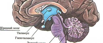
- Intermediate - thalamus (visual thalamus), hypothalamus, geniculate bodies, reticular formation.
- Middle - quadrigeminal (centers of visual and auditory analyzers), cerebral peduncles (place of passage of ascending and descending nerve fibers).
- Posterior - cerebellum (coordination of movements) and the pons (connection between parts of the central nervous system: spinal cord, brain, cerebellum. It contains the nuclei of cranial nerves - V, VI, VII, VIII).
- The medulla oblongata contains important regulatory centers:
- breathing;
- heart activity;
- body temperature;
- vascular tone (change in blood pressure);
- protective reflexes - sneezing, coughing, tearing, blinking.
Brain and spinal cord
Spinal cord
It is a nerve cord lying in the spinal canal formed by the vertebrae. Extends from the foramen magnum to the lumbar spine. At the top it passes into the medulla oblongata, at the bottom it ends with a conical point with a terminal filament.
The spinal cord is covered by several membranes: dura mater, arachnoid and pia mater. Between the arachnoid and soft membranes, cerebrospinal fluid circulates - cerebrospinal fluid, which surrounds the spinal cord and takes an active part in the metabolism of the spinal cord.
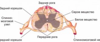
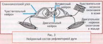
In cross section, the spinal cord (SC) resembles a butterfly. In the center is the gray matter, consisting of the cell bodies of neurons. At the periphery there is white matter, which is formed by the processes of neurons.
In the gray matter of the SC, there are two anterior projections (anterior horns), two lateral ones (lateral horns) and two posterior ones (posterior horns). In the next article we will study reflex arcs, so this knowledge will be very useful to us. The horns of the gray matter contain neurons that are part of the reflex arcs.
Numerous nerve fibers approach the posterior horns of the spinal cord, which unite to form bundles - the dorsal roots. Numerous nerve fibers emerge from the anterior horns of the spinal cord and form the anterior roots.
The white matter consists of numerous nerve fibers, bundles of which form cords. The spinal cord pathways are divided into ascending - from receptors to the brain, and descending - from the brain to effector organs. 31 pairs of spinal nerves arise from the spinal cord.
The spinal cord has two important functions:
- Reflex
- Conductor
Due to the bodies of neurons, which are located in the gray matter of the spinal cord and are part of the reflex arcs that provide reflexes.
Due to the presence of white matter in the spinal cord, which includes numerous nerve fibers that form bundles and cords around the gray matter.
Brain and its parts
We move on to the study of the human brain, the complex main organ of the central nervous system, located in a reliable bone container - the skull. The average brain weight ranges from 1300 to 1500 grams.
Let me note that the weight of the brain has nothing to do with intellectual abilities: for example, Albert Einstein’s brain weighed 1230 grams - less than that of the average person. Intelligence is rather determined by the complexity and ramification of the neural networks of the brain, but not by mass.
The human brain is divided into five sections: medulla oblongata, posterior (pons and cerebellum), middle, intermediate and terminal. The most ancient sections - the medulla oblongata, posterior and middle - form the brain stem, which resembles the structure of the spinal cord. Sometimes the intermediate section is also referred to as the brainstem. 12 pairs of cranial nerves arise from the brain stem.
The telencephalon differs from the structure of the brain stem; it is a huge accumulation (about 16 billion) of neurons that form the cerebral cortex (CCH). Neurons are arranged in several layers, their processes form thousands of synapses with other neurons and their processes. The centers of higher nervous activity - memory, thinking, speech - are located in the KBP.
We begin a fascinating journey through the parts of the brain. It is fundamentally important for you to separate and remember the functions of the various departments; for this, be sure to use your imagination!)
- Medulla
- Hindbrain (pons and cerebellum)
- Midbrain
- Diencephalon
- Finite brain
The most ancient part of the brain. Remember that it regulates vital functions: the cardiovascular system, respiratory and digestive processes. The centers of protective reflexes - vomiting, sneezing, coughing - are concentrated here.
The pons Varoliev performs a conductive function: all descending and ascending nerve pathways pass through the bridge. It also controls the work of the facial and chewing muscles of the face and the lacrimal gland.
The cerebellum has its own hemispheres connected to each other. The cerebellar cortex is formed by gray matter, the subcortical nuclei are surrounded by white matter.
The cerebellum takes part in the coordination of voluntary movements, helps maintain body position in space, regulates tone and balance. Thanks to the cerebellum, our movements are clear and smooth.
In the midbrain there are the superior (anterior) and inferior (posterior) tubercles of the quadrigeminal. The upper tubercles of the quadrigeminal are responsible for the visual orientation reflex, and the lower ones are responsible for the auditory orientation reflex.
What is the visual orientation reflex expressed in? Imagine walking into a dark room. In her corner the screen shines comfortably, the website (of course) Studarium is visible =) And then the visual orientation reflex begins: You move your eyes, turn your head in the direction of the source of intellectual light. At the same time, do not forget to regulate the size of the pupil and the accommodation of the eyes - all this is a visual orientation reflex.
The auditory orientation reflex is also necessary for us. It’s good if, while reading the textbook now, you are in silence. Suddenly your phone starts ringing: you immediately stop reading and head towards the source of the sound - the phone. Thanks to this orienting reflex, we can determine the location of the sound source relative to us (left, right, behind, in front).
The midbrain also performs a conductor function and is involved in the regulation of muscle tone and body posture.
Let me remind you that the hypothalamus we studied, the associated pituitary gland, pineal gland and thalamus belong to the diencephalon. You know that the hypothalamus controls the pituitary gland, the conductor of the endocrine glands, therefore the functions of the hypothalamus are: regulation of the metabolism of proteins, fats and carbohydrates, as well as water-salt metabolism.
In addition, the hypothalamus controls the sympathetic and parasympathetic systems, regulates body temperature, and is responsible for sleep-wake cycles. The hypothalamus contains the centers of hunger and satiety.
Consists of subcortical structures and cerebral cortex (CBC). The surface of the KBP reaches an average of 1.5-1.7 m2. Such a large area is due to the fact that the CBP forms convolutions - elevations of the brain matter, and grooves - depressions between the convolutions.
Cerebral cortex
The cortex has several layers of cells, between which numerous branched connections are formed. Despite the fact that the cortex functions as a single mechanism, its different parts analyze information from different peripheral receptors, which I.P. Pavlov called the cortical ends of analyzers.
The cortical representation of the visual analyzer is located in the occipital lobe of the CBP; it is in connection with this that, when falling on the back of the head, a person sees “sparks from the eyes” when the neurons of this lobe are excited mechanically, as a result of the impact.
The cortical representation of the auditory analyzer is located in the temporal lobe of the cerebral cortex.
Remember that the cortical representation of the motor analyzer - the motor zone - is located in the anterior central (precentral) gyrus, and the representation of the skin analyzer - the sensory zone - is in the posterior central (postcentral) gyrus.
Think about it! When making any voluntary (conscious) movement, a nerve impulse arises precisely in the neurons of the precentral gyrus, from where it begins its long journey through the brain stem, spinal cord and finally reaches the effector organ.
Impulses from skin receptors reach the neurons of the postcentral gyrus - the sensory department, thanks to which we receive information from them and are aware of our own sensations.
The number of neurons in these convolutions allocated for different organs is not the same. Thus, the projection area of the fingers of the hand takes up a lot of space, making fine movements of the fingers possible. The projection area of the torso muscles is much smaller than the area of the fingers, since the movements of the torso are more uniform and less complex.
The areas of the brain that we studied, in which the transformation and analysis of incoming information occurs, are called associative zones of the CBP. These zones connect different parts of the CBP, coordinate its work, and play a crucial role in the formation of conditioned reflexes.
Our conscious activity lies within the framework of the cerebral cortex: any conscious movement, any sensation (temperature, pain, tactile) - everything has representations in the CBP. The cortex is the basis for communication with the external environment and adaptation to it. QBP also lies at the foundation of the thinking process. In general, you understand how highly you should value it and how well you should know this topic 
You've probably heard that the right and left hemispheres are functionally different. The left hemisphere contains the mechanisms of abstract thinking (language abilities, analytical thinking, logic), and the right hemisphere contains the mechanisms of concrete figurative thinking (imagination, parallel processing of information). With injuries or damage to the left hemisphere, speech may be impaired.
Diseases
Depending on the level of damage to the spinal cord during trauma, the picture of neurological disorders manifests itself differently. The higher the level of damage, the more nerve pathways are “cut off” from the brain. So, for example, with a lumbar injury, arm movements are preserved, but with a cervical injury, arm movements are impossible.
Sometimes after a stroke (bleeding in brain tissue) or injury, paralysis (complete lack of movement) develops on one side of the body. Knowing the anatomy, you can conclude: if movements are lost in the right arm and leg, then the stroke occurred on the left.
Why does this pattern exist? The fact is that the nerve fibers running from the precentral gyrus to the working organs - the muscles - form the so-called physiological cross at the border of the medulla oblongata and the spinal cord. That is, to put it simply: part of the nerves that came from the left hemisphere pass to the right side and vice versa - nerves from the right hemisphere pass to the left side.
© Bellevich Yuri Sergeevich 2018-2021
This article was written by Yuri Sergeevich Bellevich and is his intellectual property. Copying, distribution (including by copying to other sites and resources on the Internet) or any other use of information and objects without the prior consent of the copyright holder is punishable by law. To obtain article materials and permission to use them, please contact Yuri Bellevich
.
END BRAIN
Lecture 8
The telencephalon, or cerebrum, in the process of evolution arose later than other parts of the brain. In its mass and size, it significantly exceeds all other parts of the brain and is directly related to the most complex manifestations of human mental and intellectual activity.
The telencephalon consists of two cerebral hemispheres connected to each other by the corpus callosum, the anterior and posterior commissures and the commissure of the fornix. The telencephalon cavities form the right and left lateral ventricles of the brain, each of which is located in the corresponding hemisphere. The medial wall of each lateral ventricle in the rostral region is formed by a transparent septum.
The cerebral hemispheres are covered on top by the cerebral cortex - a layer of gray matter formed by neurons of more than fifty varieties. Under the cerebral cortex in the cerebral hemispheres there is white matter, consisting of myelinated fibers, most of which connect the cortex with other parts and centers of the brain. In the thickness of the white matter of the hemispheres there are accumulations of gray matter - the basal ganglia.
The thalamus and cerebral peduncles are fused with the cerebral hemispheres. The layer of white matter that borders the hemispheres from the thalamus of the diencephalon is called the internal capsule.
The right and left hemispheres of the brain are separated from each other by a longitudinal fissure. In each hemisphere, there are three surfaces - lateral, medial and inferior, as well as three edges - superior, medial and inferior, and three poles - frontal, occipital and temporal.
The surface of the mantle of each hemisphere is divided by slits and grooves into lobes, lobules and convolutions. The fissures and primary sulci are deep and belong to the permanent formations of the brain. They appear in the 5th month of intrauterine development and divide the hemispheres into lobes. The largest fissures are the longitudinal fissure of the brain, which separates the hemispheres from each other, and the transverse fissure, which separates the cerebellum from the occipital lobes. Secondary and especially tertiary grooves determine the individual surface relief of the hemispheres. Their formation occurs from birth to 7-8 years.
In most people, the main relief - the location of deep permanent grooves and large convolutions - is of a similar nature. Large fissures and fissures divide each hemisphere into 6 lobes: frontal, parietal, occipital, temporal, insular and limbic.
On the lateral surface of the hemisphere, there is a central (Rolandian) fissure, which separates the frontal lobe from the parietal lobe, and a lateral (Sylvian) fissure, which separates the temporal lobe from the frontal and parietal lobes. The parietal lobe is bounded from the occipital parieto-occipital sulcus. The anteroinferior border of the occipital lobe is a conventional line drawn from the upper end of the parieto-occipital sulcus down to the lower edge of the hemisphere. Deep in the lateral sulcus is the insular lobe (or insula). This lobe is covered by parts of the frontal, parietal and temporal lobes. On the medial surface of the hemisphere, next to the corpus callosum, is its limbic lobe, separated from the other lobes by the cingulate groove.
THE FRONTAL LOBE contains the following sulci and gyri:
1 Precentral sulcus; between the precentral and central sulcus is the precentral gyrus;
2. The superior and inferior frontal sulci, between which the superior,
middle and inferior frontal gyri. The inferior frontal gyrus is divided into three parts: opercular (tegmental), triangular (triangular) and orbital (orbital).
3. Anterior horizontal sulcus and its ascending branch;
4. The medial frontal gyrus, separated from the limbic lobe by the cingulate sulcus;
5. Part of the cingulate cortex;
6. Olfactory and orbital grooves, located on the lower surface of the frontal lobe. The olfactory sulcus contains the olfactory bulb, olfactory tract and olfactory triangle.
7. Gyrus recta, located between the olfactory sulcus and the medial
edge of the hemisphere.
The frontal lobe corresponds to the anterior horn of the lateral ventricle.
Functional characteristics of the cortical zones of the frontal lobe. 1. In the area of the precentral gyrus of the frontal lobe there is the cortical nucleus of the motor analyzer - the kinesthetic center. This area is also called the sensorimotor cortex. Some of the afferent fibers from the thalamus come here, carrying proprioceptive information from the muscles and joints of the body. Descending pathways to the brain stem and spinal cord also begin here, providing the possibility of conscious regulation of movements (pyramidal tracts). Damage to this area of the cortex leads to paralysis of the opposite half of the body.
2. In the posterior third of the middle frontal gyrus lies the center of writing - the center of graphia, or the associative center of written characters. This zone of the cortex gives projections to the nuclei of the oculomotor cranial nerves, and also, through cortico-cortical connections, communicates with the center of vision in the occipital lobe and the control center for the muscles of the arms and neck in the precentral gyrus. Damage to this center leads to impaired writing skills under visual control (agraphia).
3. In the posterior third of the inferior frontal gyrus there is the speech motor center (Broca's center) - the center of speech articulation. It has pronounced functional asymmetry. When it is destroyed in the right hemisphere, the ability to regulate timbre and intonation is lost, speech becomes monotonous. When the speech motor center on the left is destroyed, speech articulation is irreversibly impaired, up to the loss of the ability to articulate speech (aphasia) and singing (amusia). With partial violations, agrammatism may be observed - the inability to form phrases correctly.
4. In the area of the anterior and middle third of the upper, middle and partially inferior frontal gyri there is an extensive anterior associative zone of the cortex, which programs complex forms of behavior (planning various forms of activity, making decisions, analyzing the results obtained, volitional reinforcement of activity, correction of the motivational hierarchy). The area of the frontal pole and medial frontal gyrus is associated with the regulation of the activity of emotiogenic areas of the brain included in the limbic system, and is related to the control of psycho-emotional states. Disturbances in this area of the brain can lead to changes in what is commonly called “personality structure” and will affect a person’s character, his value orientations, and intellectual activity.
The orbital region contains the centers of the olfactory analyzer and is closely connected anatomically and functionally with the limbic system of the brain.
5. In the anterior part of the middle frontal gyrus there is a center for combined rotation of the head and eyes.
PARIETAL LOBE. The structure of the parietal lobe includes the postcentral gyrus, postcentral sulcus, interparietal sulcus, superior and inferior parietal lobules; in the inferior parietal lobule - the supramarginal and angular gyri, the posterior part of the paracentral lobule; behind it lies the precuneus; parieto-occipital and subparietal sulci. The parietal lobe corresponds to the central part of the lateral ventricle.
Functional characteristics of the cortical zones of the parietal lobe. The cortical areas of the parietal lobe contain the following centers:
1. Projection center of general sensitivity - skin analyzer of general
sensitivity (tactile, pain, temperature and conscious proprioceptive) - the cortex of the postcentral gyrus.
2. The projection center of the body diagram is the edge of the intraparietal sulcus.
3. Associative - the core of the skin recognition analyzer
objects to the touch - the cortex of the superior parietal lobule.
4. Associative - analyzer of purposeful habitual
movements (playing the piano, working on a typewriter) - supramarginal cortex
convolutions.
5. Associative optical center of speech - visual analyzer of written
speech - center of lexia (Dejerine) - cortex of the angular gyrus.
TEMPORAL LOBE. In the region of the temporal lobe, on its lateral surface, the superior and inferior temporal sulci are distinguished. These grooves and the lateral sulcus are limited by the superior, middle, and inferior temporal gyri.
On the lower surface, the temporal lobe does not have clear boundaries with the occipital lobe. Next to the lingual gyrus is the lateral occipito-umoral gyrus of the temporal lobe, which is bounded superiorly by the collateral sulcus from the limbic lobe, and laterally by the occipital temporal sulcus. The temporal lobe corresponds to the inferior horn of the lateral ventricle.
Functional characteristics of the cortical zones of the temporal lobe.
1. In the region of the middle part of the superior temporal gyrus, on its upper surface, there is the cortical center of the auditory analyzer. Its damage leads to deafness. The auditory speech center (Wernicke's center) lies in the posterior third of the superior temporal gyrus. Injuries to this area lead to the inability to understand spoken language: it is perceived as noise.
2. In the area of the middle and inferior temporal gyri there is a cortical representation of the vestibular analyzer. Damage to this area leads to imbalance when standing and decreased sensitivity of the vestibular system.
INSALE LOBE (ISLANK). The insular lobe is located deep in the lateral sulcus. The islet is bounded by a circular groove.
Functional characteristics of the cortical zones of the insula. It is believed that the insula is related to the analysis of olfactory and taste sensations, as well as to the processing of somatosensory information and auditory perception of speech.
LIMBIC LOBE. This lobe is located on the medial surface of the hemisphere. It includes the cingulate gyrus, isthmus, dentate and parahippocampal gyri. One of the boundaries of this lobe is the groove of the corpus callosum. This groove, descending, continues into the hippocampal groove. Under the hippocampal sulcus in the cavity of the inferior horn of the lateral ventricle is the hippocampal gyrus, or hippocampus.
Above the sulcus of the corpus callosum there is another boundary of the limbic lobe - the cingulate sulcus, which separates the cingulate gyrus. The cingulate sulcus borders the limbic lobe from the frontal and parietal lobes. The cingulate gyrus passes through the isthmus into the parahippocampal gyrus, which ends in the uncus.
Functional characteristics of the cortical zones of the limbic lobe. The cingulate and parahippocampal gyri are directly related to the limbic system of the brain. This system controls a complex of vegetative and behavioral psycho-emotional reactions to external environmental influences. The cortical representation of the gustatory and olfactory analyzer is located in the uncus and parahippocampal gyrus. At the same time, the hippocampus plays an important role in learning: the mechanisms of short-term and long-term memory are associated with it.
OCCIPITAL LOBE. On the lateral surface of the occipital lobe there is a transverse occipital groove. On the medial surface there are: a wedge, bounded in front by the parieto-occipital groove, and behind by the calcarine groove; lingual gyrus, bounded above by the calcarine groove and below by the collateral groove. The occipital lobe corresponds to the posterior horn of the lateral ventricle.
Functional characteristics of the cortical zones of the occipital lobe. The occipital lobe has the following centers:
• The projection center of vision (nucleus of the visual analyzer) is located in the cortex bordering the calcarine sulcus.
• The associative center of vision (visual memory analyzer) is located in the cortex of the dorsal surface of the occipital lobe.
WHITE MATTER OF THE HEMISPHERES OF THE BRAIN. The white matter of the cerebral hemispheres is represented by numerous fibers, which are divided into three groups:
1. Projection fibers – bundles of afferent and efferent fibers that connect the projection centers of the cerebral cortex with the basal ganglia, nuclei of the brainstem and spinal cord. Projection fibers form the internal capsule and fornix of the brain.
2. Association fibers connect areas of the cortex within one hemisphere. They are divided into short and long.
3. Commissural fibers connect areas of the cortex of the opposite hemispheres of the brain. Commissural formations include the corpus callosum, anterior commissure of the brain, commissure of the fornix, and posterior commissure of the brain.
STRUCTURE OF THE CEREBRAL CORTEX. The cerebral cortex is
a huge accumulation of neurons and glial cells. The thickness of the cortex ranges from 1.2 to 4.5 mm, and the surface area in an adult is from 1700 to 2200 sq. cm. In the cortex
According to various sources, the large brain contains from 10 to 14 billion neurons.
The main part of the cerebral cortex (95.9% of the entire surface of the hemispheres) is the neocortex - the new cortex. Phylogenetically, this is the most recent formation of the brain. The remaining 4.1% of the area is covered
1. old cortex - archiocortex, located in the temporal lobe, called the hippocampus, or Amon's horn;
2. ancient cortex - paleocortex, which occupies a section of the frontal lobe cortex near the olfactory bulbs;
3. Small zones adjacent to the paleocortex are called mesocortex - interstitial cortex.
The ancient and old bark appears earlier in the phylogeny of vertebrates and carries features of a relatively primitive internal structure. Main feature
These cortical areas are weakly stratified (divided into layers). For example, the hippocampal cortex has five cortical layers, while the dentate gyrus cortex has only three layers. The neurons that form these layers also have a more primitive structure compared to the neurons of the neocortex.
The layer-by-layer arrangement of neurons in the cortex is called cytoarchitecture. In the neocortex of the cerebral hemispheres, neurons are grouped into six to seven cortical layers:
I - external molecular, or pleximorphic;
II - external granular, or external granular;
III - external pyramidal, or ganglionic;
IV - internal granular, or internal granular;
V - internal pyramidal, or internal ganglionic;
VI and VII are layers of polymorphic neurons.
In each of the layers of the cortex, neurons of certain sizes and shapes predominate.
Layer one is poor in cells and contains mainly branches of the apical dendrites of pyramidal neurons of the underlying layers, as well as branching of the axons of neurons. Thanks to the molecular layer, intra- and interhemispheric connections are made between different areas of the cortex.
Layer two includes small pyramidal and stellate (granular) neurons, which provide partial processing of information and its transmission from the structures of the molecular layer to the underlying cortical layers. These neurons are also called interneurons or interneurons.
Granular neurons are also located in layer IV, where they process and transmit information from the endings of afferent fibers coming to the cortex and branching within layer IV to pyramidal neurons of layers III and V.
Layers III and V contain a large number of large pyramidal neurons, the axons of which provide different types of intracortical, intercortical and cortical-subcortical connections. The fifth layer in the area of the precentral gyrus contains the largest pyramidal neurons, which are called Betz pyramidal cells. In layers III and V, interneurons of various sizes and shapes are also found in large numbers (double-fascicle cells, long-axonal and short-axonal basket neurons, chandelier cells). Interneurons provide selective intracortical interactions between neurons of different types. This is necessary for:
• transmission of information between afferent fibers arriving in the cortex and
pyramidal neurons;
• exchange of information between neurons located in different cortical layers;
• exchange of information between centers located in different convolutions, lobes and hemispheres;
• storage and reproduction of information.
Long-term circulation of excitation in the cortex and in associated parts and centers of the brain with the participation of interneurons accompanies cognitive operations and other higher forms of mental activity. Ultimately, all information processes occurring in brain structures are integrative, systemic in nature and are mediated by many interneurons.
The lowest cortical layers VI and VII differ mainly in the density of cells in the section: layer VI is more densely cellular and contains larger neurons than layer VII. The lower layers are older in origin than the rest, and therefore contain polymorphic cells that differ in shape from the pyramidal neurons and interneurons of the overlying layers. Neurons of layers VI and VII provide U-shaped connections between the cortex in adjacent gyri and projectional corticothalamic connections.
In addition to cellular elements (neurons and glia), the gray matter of the cortex contains branching fibers of various origins. Among them, associative, commissural and projection fibers are distinguished. The layer-by-layer arrangement of fibers in the cortex is called myeloarchitecture.
MODULAR ORGANIZATION OF THE LARGE HEMISPHERES CORTEX. The cortical module (neural ensemble) is a group of neurons, as well as glial cells and blood vessels, specially located in space and functionally interconnected. This module ensures the processing and storage of incoming information in the cerebral cortex. It has the appearance of a discrete columnar block of cells with a diameter of 300-600 microns, covering all cortical layers in the vertical direction. Associated with the module is a certain set of afferent fibers that bring information, which it subjects to standard discrete processing, as well as a set of efferent fibers that deliver it to certain areas of the brain. Various modules of the cortex are closely connected to each other via interneurons and intracortical fibers. The principle of modular structural and functional organization is valid for all departments of the central nervous system.