Vyacheslav Dubynin about the thalamus and hypothalamus
Vyacheslav Dubynin, Doctor of Biological Sciences, Professor of the Department of Human and Animal Physiology of the Faculty of Biology of Moscow State University, a specialist in the field of brain physiology, speaks about the similarities between the thalamus and the cerebral hemispheres, hyperactivity in children and the functions of the hypothalamus.
Hypothalamus
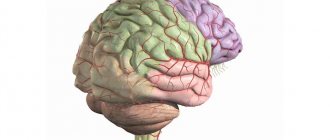
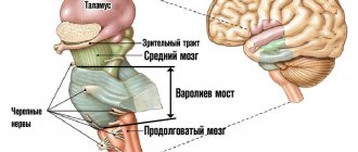
The most important part of our brain is the diencephalon, which is so named because it is located between the cerebral hemispheres. During evolution, the cerebral hemispheres and diencephalon are formed from a structure called the forebrain. The central part of the forebrain gives rise to two processes that turn into the cerebral hemispheres, while the center remains the diencephalon. Inside the diencephalon there is a small, narrow, slit-like cavity called the third ventricle.
The diencephalon consists of two main sections: the upper half is called the thalamus, and the lower half is the hypothalamus. Their actual size is 3–4 centimeters. In addition to the thalamus and hypothalamus, there is the epithalamus, to which the pineal gland is adjacent (this is our endocrine gland, it is located in the upper rear part of the thalamus) and the pituitary gland (this is another endocrine gland, adjacent to the hypothalamus below). If we go along the stem structures of the brain, we will first come across the medulla oblongata, the pons, then the midbrain, and then we will find ourselves in the zone of the thalamus and hypothalamus. Connected to the diencephalon is the optic nerve, the second cranial nerve that enters the brain at the border of the thalamus and hypothalamus.
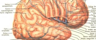
The thalamus is a key structure located at the entrance to the cerebral cortex. The cerebral cortex is the highest and most remarkable centers that deal with the most complex functions. In order for them to work effectively, they need to receive the right information flows in the right quantities. These functions are performed by the thalamus, which is why it is also called the “secretary” of the cerebral cortex.
The cerebral cortex contains visual, auditory, motor centers, as well as centers associated with emotions. The thalamus has the same set of centers, but only in a reduced size. There is a group of “secretaries” who help the cerebral cortex function correctly and efficiently. The thalamus can be compared to an information funnel, transmitting some signals to the cerebral cortex, and either blocking the remaining signals altogether or transmitting them in a weakened form. The problem is that the cerebral cortex cannot process the huge amount of information flows that constantly moves through our brain.
Visual centers supply visual information, auditory centers supply auditory information, memory centers remember last night, emotional centers experience emotions, motor centers want to move. The cerebellum constantly suggests to the cerebral cortex: “Let's do this! Let's do it! Why do we sit and not move, we can do so many things?” In order to really sit and not move, so that, for example, a schoolboy sits quietly in class, the thalamus must constantly block these information flows so that the cerebral cortex does not receive unnecessary exciting signals. That is, this is really an information funnel that should cut off a lot of things. The cutting occurs due to the work of inhibitory neurons, that is, in the thalamus, as well as in the cerebellum and basal ganglia, the function of gamma-aminobutyric acid (GABA) and inhibitory reactions are very important.
If the thalamus does not work well, then, for example, younger schoolchildren experience a fairly typical change in behavior called ADHD (attention deficit hyperactivity disorder). Analyze the name: attention deficit - the brain cannot hold the information channel for a long time, that is, the thalamus cannot block signals from the body, movement occurring outside the window for a long time. Therefore, the student cannot listen to the teacher for a long time, and his attention quickly dissipates. Hyperactivity is the inability to suppress for a long time those motor proposals that come from the cerebellum and basal ganglia. The student was just listening to you, but now he is spinning around, reaching into his briefcase, grabbing a textbook and throwing it at his neighbor - it’s difficult to control all this. Therefore, a truly mature thalamus is formed by the age of 8–10. And just when you are happy that everything is fine with the child and you are managing him, puberty begins, sex hormones again disrupt the functioning of the thalamus, and problems arise again.
If we go along the thalamus, we will see in it a lot of structures that correspond to different centers of the cerebral cortex. The anterior nuclei of the thalamus are nuclei associated with the transfer of information to memory centers and centers that work with emotions. Behind the anterior nuclei of the thalamus are the so-called ventral lateral, ventral lateral nuclei of the thalamus, which are associated with motor control, the anterior part of these nuclei works with the basal ganglia, and the posterior part with the cerebellum.
Next is the ventrobasal complex, which mainly carries information about the sensitivity of the body. This information is supplied to the thalamus by the spinal cord. As you know, there are neurons of the spinal ganglia, sensory neurons that collect skin and muscle sensitivity. Neurons of the spinal ganglia form bundles of axons, which, as part of the white matter of the spinal cord, without entering the gray matter, first rise to the medulla oblongata, and then go to the thalamus. These collections of fibers are called the dorsal columns, or the gracilis and cuneate fasciculi, or the gracilis and cuneate fasciculi of the spinal cord, and are very important for the conduction of cutaneous and muscular sensation. Muscle sensitivity from the spinal cord to the brain rises along two parallel pathways - to the thalamus and the cerebellum, because movement control occurs both through automated cerebellar programs and through voluntary programs generated by the cerebral cortex. The cerebral cortex, of course, needs these information flows.
Above the ventrobasal nucleus complex are the visual and auditory centers of the thalamus. The visual areas of the thalamus are very extensive, there is a pillow and the lateral geniculate body, into which the optic nerve arrives. The auditory nuclei of the thalamus are the medial geniculate bodies, they are smaller than the visual nuclei, and the main information flows come to them from the auditory nuclei of the medulla oblongata and the pons, from the nuclei of the eighth nerve.
In addition to what has already been listed, there are many other structures in the thalamus, associated, for example, with the associative zones of the cerebral cortex, and there are very well-known medial (innermost) nuclei of the thalamus, bordering the third ventricle. The medial nuclei contain clusters of nerve cells that process and transmit taste, pain signals, and vestibular sensitivity. In addition, the medial nuclei are associated with the centers of sleep and wakefulness.
There is a spinothalamic tract that comes directly from the spinal cord and ends in the medial nuclei of the thalamus. This is a specific tract, a path for pain signals. If some kind of malfunction occurs in the medial nuclei, then a pathology called chronic pain may occur, when a person constantly has pain, for example, in the thumb of his right hand. Moreover, everything is fine with the finger itself, but somewhere in the thalamus a microstroke occurred, and now a pathological pain signal arises there, preventing the person from living. This kind of pathology is not blocked by any analgesics, and in severe cases, people undergo an operation called thalamotomy, when the pinpoint zone of the medial thalamus is carefully destroyed, and then the transmission of the pathological pain signal stops.
The lower part of the diencephalon - the hypothalamus - deals with completely different tasks. The hypothalamus is oriented mainly towards the internal environment of our body. There we find nerve cells that are involved, firstly, in neuroendocrine regulation (the hypothalamus is the main endocrine center of our body). Secondly, in the hypothalamus there are neurons that are involved in autonomic regulation, that is, with the help of the sympathetic and parasympathetic systems, they control the activities of various internal organs. Thirdly, in the hypothalamus we find a number of important centers of biological needs. These three groups of hypothalamic functions are extremely important.
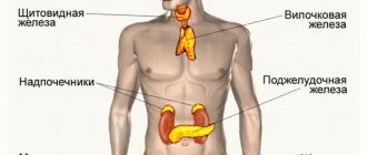
From the point of view of neuroendocrine regulation, it is important that the nerve cells of the hypothalamus constantly evaluate the concentration of the main hormones that are in our blood. Hormones of the thyroid gland, gonads, adrenal glands - all of these hormones are monitored by the hypothalamus. The hypothalamus innately knows how much there should be, and it has ways of conveying a signal to specific endocrine glands to secrete more or less hormones. In this case, the hypothalamus mainly influences the pituitary gland.
The endocrine system is structured on three floors. There is a specific endocrine gland, the thyroid. It secretes thyroxines - important hormones on which the overall level of activity of each cell in our body depends. In order for the thyroid gland to secrete the correct amount of thyroxine, there is a pituitary gland that secretes thyroid-stimulating hormone, and this hormone tells the thyroid gland how active it should be. But above the pituitary gland is the hypothalamus, which, with the help of its hormones called releasing hormones, tells the pituitary gland how much to secrete thyroid-stimulating hormones and ultimately change the activity of the thyroid gland. If there is too little thyroxine, the hypothalamus senses this and secretes thyroxin-releasing hormone, which causes the pituitary gland to secrete more thyroid-stimulating hormone, and the thyroid gland begins to secrete more thyroxine. Regulatory circuits of this kind are characteristic not only of the thyroid gland, but of the adrenal cortex and gonads; the release of growth hormones is controlled in a similar way.
In addition to these functions, hypothalamic neurons themselves are capable of releasing hormones directly into the blood - hormones such as oxytocin and vasopressin. Axons of nerve cells in the central zone of the hypothalamus (gray hillock of the hypothalamus) go to the posterior lobe of the pituitary gland, where oxytocin and vasopressin are released directly into the blood from these axons. Oxytocin is a known hormone that affects the contraction of the uterus during childbirth and the mammary glands when feeding a child. In addition, oxytocin is now known as a mediator of attachment. Vasopressin is a hormone that affects the functioning of the kidneys and thirst centers. Our current need for fluid depends on the concentration of vasopressin.
From the point of view of autonomic regulation, the anterior part of the hypothalamus is very important. There are thermoreceptor neurons that constantly assess the temperature of the blood flowing through the hypothalamus. If the blood is too warm, it is from the hypothalamus that reactions are triggered that reduce our body temperature. Skin blood vessels dilate and sweating begins. If the blood flowing through the hypothalamus is too cold, contraction reactions of the skin vessels are triggered, and trembling or goosebumps occur on the skin. These are all autonomic reactions that are controlled by the hypothalamus. The posterior part of the hypothalamus provides autonomic support for stress, which is also very important. Finally, the hypothalamus contains the centers of our six most important biological needs: the centers of hunger and thirst, the centers of sexual and parental behavior, and the centers of fear and aggression.
Link to source
Views: 812
Specific kernels
This group of nuclei of the thalamus has a number of distinctive features that unite them. First, they receive impulses from long nerve pathways that transmit information from somatosensory, visual and auditory receptors to the cerebral cortex. Through these nuclei, the impulse is transmitted further to the corresponding areas of the cortex: somatosensory, auditory and visual. In addition, information from them enters the premotor and motor areas of the cortex.
Also, specific nuclei receive feedback from the cortex. Experiments have proven that when a section of the cortex corresponding to a specific nucleus is removed, this nucleus is also destroyed. And when certain nuclei are stimulated, the nerve cells of the corresponding cortex are activated.
This group receives information from the cortex, reticular formation, and brain stem. It is precisely because of the presence of these connections that the cerebral cortex has the ability, among all incoming information, to select the most important at the moment.
In addition, the structure of the thalamus includes nuclei that receive information from the red and basal nuclei, the limbic system, and the dentate nucleus (located in the cerebellum). Next, the signal goes to the motor areas of the cortex.
Main functions
The thalamus is a key formation in the transmission of nerve impulses to the cerebral cortex. If the cortex is damaged, it is thanks to the work of the thalamus that partial restoration of functions such as touch, sensation of pain and temperature is possible.
Another important function of the thalamus is the integration of motor and sensory activities. This is possible due to the receipt of information into the thalamus from both the motor and sensory centers of the nervous system.
In addition, the thalamus is essential for attention and consciousness. He also takes part in the formation of behavioral reactions.
Thanks to its connection with the hypothalamus, which will be discussed later in the article, the functions of the thalamus also cover memorization and emotional behavior.
Associative kernels
A peculiarity of this group of nuclei is that they receive already processed signals from other parts of the thalamus.
Thanks to their work, it is possible to carry out integrative processes in which generalized signals are formed. They are then transmitted to the associative areas of the cerebral cortex (frontal, parietal and temporal lobes). It is thanks to the presence of this area of the cortex and associative nuclei that processes such as recognition of objects, coordination of speech with motor activity, understanding of the three-dimensionality of space and awareness of oneself in this space are possible.
Nonspecific nuclei
These nuclei consist of small nerve cells that receive information from neurons of other thalamic nuclei, limbic system, basal ganglia, hypothalamus, and brain stem. Through the ascending pathways, the nuclei receive signals from pain and temperature receptors, and through the reticular formation - from almost all other structures of the central nervous system.