| Postcentral gyrus | |
| Postcentral gyrus of the human brain | |
| Brodmann areas 3, 1 and 2 of the human brain. Brodmann area 3 is in red, area 1 is in green, and area 2 is in yellow. | |
| Details | |
| System | Somatosensory system |
| Location | Parietal lobe |
| Artery | Middle cerebral artery |
| Function | Primary somatosensory cortex |
| Identifiers | |
| Latin | Gyrus postcentralis |
| NeuroNames | 105 |
| NeuroLex ID | birnlex_1070 |
| TA98 | A14.1.09.128 |
| TA2 | 5469 |
| F.M.A. | 61896 |
| Anatomical terms of neuroanatomy [edit in Wikidata] | |
Postcentral gyrus
is a prominent gyrus in the lateral parietal lobe of the human brain.
It is the location of the primary somatosensory cortex, the main sensory receptive area for touch. Like other sensory areas, this location has a map of sensory space called the sensory homunculus
.
The primary somatosensory cortex was originally defined based on surface stimulation studies by Wilder Penfield and parallel surface potential studies by Bard, Woolsey, and Marshall. Initially identified as roughly the same as areas , and Brodmann, later work by Kaas suggested that for homogeneity with other sensory fields, only area 3 should be called "primary somatosensory cortex", since it receives the bulk of thalamocortical projections from sensory hollow input.
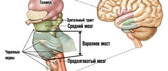
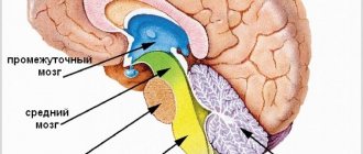
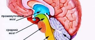
Parts of the brain
During intrauterine development, a complex brain was formed from an ordinary neural tube. This happened due to the bulging of five brain vesicles, which gave rise to the corresponding parts of the brain:
- telencephalon, or forebrain, from which the cerebral cortex, basal ganglia, and anterior part of the hypothalamus were formed;
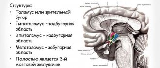
- diencephalon, or diencephalon, which gave rise to the thalamus, epithalamus, and posterior part of the hypothalamus;
- mesencephalon, or midbrain, from which the quadrigeminal peduncle and cerebral peduncles subsequently formed;
- the metencephalon, or hindbrain, which gave rise to the cerebellum and pons;
- myelencephalon, or medulla oblongata.
You may be interested in:How to treat allergies: causes, treatment methods, medications, therapeutic diet, reviews
Later in the article we will talk in more detail about the telencephalon, or forebrain. After all, the relief of the cerebral cortex belongs specifically to this part of the central nervous system.
You may be interested in: Is it possible to apply mustard plasters for a dry cough?
Structure of the cortex
Thanks to the presence of the cortex, a person is able to experience emotions, navigate himself and the surrounding space. What is noteworthy is that the structure of the bark is unique. The grooves and convolutions of the cerebral cortex of one person have a different shape and size than that of another. But the general plan of the building is the same.
What is the difference between the sulci and convolutions of the brain? Fissures are depressions in the cerebral cortex that look like slits. They are the ones who divide the bark into shares. There are four lobes of the cerebral hemispheres:
- frontal;
- parietal;
- temporal;
- occipital
Gyri are convex areas of the cortex that are located between the furrows.
How to teach the anatomy of the cerebral hemispheres?
Of course, in stages. First of all, I recommend making sure that you have studied and memorized the structure of the spinal cord, then the brain stem, diencephalon (not forgetting about physiology), and only then proceed to the anatomy of the cerebral hemispheres.
It is necessary to maintain this order because studying the anatomy, for example, of the telencephalon cortex separately is completely pointless. The cerebral hemispheres are the most developed part of our body, and they are interesting, first of all, because they can control and unite all other parts of the central nervous system, adding higher cortical functions, which we will talk about a little later. Only by first studying all the other underlying parts of the central nervous system will you be able to fully understand the structure and functions of the cerebral hemispheres themselves.
When you have passed these stages, you can begin to build the cerebral hemispheres themselves. Proceed step by step, step by step, strictly observing the order of the grooves and convolutions I drew. In this case, all the material will fit without problems and you will avoid confusion. By following these recommendations, you will understand that, in fact, the anatomy of the central nervous system is very simple and very interesting.
Formation of the cortex in embryogenesis
Embryogenesis is the intrauterine development of the fetus from conception to birth. First, uneven depressions form on the cerebral cortex, which give rise to furrows. The primary grooves are formed first. This occurs around the 10th week of intrauterine development. After this, secondary and tertiary depressions are formed.
You may be interested in: Patient's outpatient card: what is it for, form, sample filling
The deepest groove is the lateral one; it is one of the first to form. It is followed in depth by the central one, which separates the motor (motor) and sensory (sensitive) zones of the cerebral cortex.
Most of the cortical relief develops from 24 to 38 weeks of gestation, and some of it continues to develop after the baby is born.
Main grooves
Although the shape and size of some of the sulci and convolutions of the cerebral hemispheres differ from individual to individual, their number is normally unchanged. Every person, regardless of age and gender, has the following grooves:
- Sylvian fissure - separates the frontal lobe from the temporal lobe;
- lateral sulcus - separates the temporal, parietal and frontal lobes, and is also one of the deepest in the brain;
- Roland's fissure - separates the frontal lobe of the brain from the parietal lobe;
- parieto-occipital sulcus - separates the occipital region from the parietal;
- cingulate sulcus - located on the medial surface of the brain;
- circular - is the boundary for the insular part on the basal surface of the cerebral hemispheres;
- The hippocampal sulcus is a continuation of the cingulate sulcus.
Hemispheres
The longitudinal fissure divides the cerebral hemispheres into the right and left hemispheres. Each hemisphere controls processes on the opposite side of the body. This means that the right side of the brain receives and controls signals from the left side of the body, and the left side of the brain receives and controls signals from the right side of the body.
Although both hemispheres control many functions, some functions occur predominantly in one or the other. For example, the left hemisphere controls functions such as speech, writing, and mathematics. The right hemisphere controls aspects of creativity such as art and musical skills.
Main convolutions
The relief of the cerebral cortex is very complex. It consists of numerous convolutions of different shapes and sizes. But we can highlight the most important of them, which perform the most important functions. The main convolutions of the brain are presented below:
- angular gyrus - located in the parietal lobe, is involved in recognizing objects through vision and hearing;
- Broca's center - the posterior part of the inferior frontal gyrus on the left (in right-handers) or on the right (in left-handers), which is necessary for correct speech reproduction;
- Wernicke's center - located in the posterior part of the superior temporal gyrus on the left or right (similar to Broca's area), is involved in the understanding of oral and written speech;
- cingulate gyrus - located on the medial part of the brain, takes part in the formation of emotions;
- hippocampal gyrus - located in the temporal region of the brain, on its inner surface, necessary for normal memorization;
- fusiform gyrus - located in the temporal and occipital regions of the cerebral cortex, is involved in face recognition;
- lingual gyrus - located in the occipital lobe, plays an important role in processing information coming from the retina;
- precentral gyrus - located in the frontal lobe in front of the central sulcus, necessary for processing sensitive information entering the brain;
- postcentral gyrus - located in the parietal lobe behind the central sulcus, necessary for voluntary movements.
Postcentral gyrus
Brain structure
4. Postcentral gyrus
The postcentral gyrus (lat. gyrus postcentralis) is a section of the parietal lobe of the cerebral cortex. The paths of superficial and deep sensitivity end in the postcentral gyrus.
The postcentral gyrus is located in the parietal lobe, behind the central sulcus. Its boundaries are:
anteriorly - central groove
posterior - postcentral sulcus
medially - longitudinal fissure of the brain
Laterally - lateral groove
Consider the convolution function:
The afferent pathways of superficial and deep sensitivity end in the postcentral gyrus. In this case, closer to the longitudinal fissure of the brain are located the sections that receive information from the lower extremities and lower parts of the torso, and the fields of the upper parts of the body and head are projected lowest at the lateral sulcus. This pattern was noted by the Canadian neurosurgeon Penfield, and the image he obtained is called the “sensitive homunculus.”
Rice. 2. Penfield's Sensitive Homunculus
Semiotics of defeat:
When damaged, anesthesia or hypoesthesia of all types of sensitivity occurs in the corresponding (depending on the location of the lesion) parts of the body on the opposite side. When irritated, paresthesia occurs in areas of the body corresponding to the irritated zones of the cortex. Such paresthesias (sensitive attacks - epilepsy) can be an aura of a general epileptic attack.[5]
5. Parietal lobe analyzers
In addition to the cortex, which forms the superficial layers of the telencephalon, accumulations of gray matter in the cerebral hemispheres are present in the form of individual nuclei, or nodes. These nodes are located in the thickness of the white matter, closer to the base of the brain. Clusters of gray matter are called basal (subcortical, central) nuclei, or nodes, due to their special position.
The nucleus is also the site of concentration of cortical nerve cells, representing the exact projection of all the constituent elements of the corresponding peripheral receptor. The kernel carries out analysis, synthesis and integration of functions.
Scattered elements can be located in close proximity to the nucleus or at a great distance from it. Analysis and synthesis take place in these elements.
The core of the cortical analyzer of general (temperature, pain, tactile) and proprioceptive sensitivity is formed by neurons located in the cortex of the postcentral gyrus and superior parietal lobule. Sensory pathways leading to the cerebral cortex intersect either at the level of different segments of the spinal cord (pathways of pain, temperature sensitivity, touch and pressure), or at the level of the medulla oblongata - these are the pathways of proprioceptive sensitivity. As a result, the postcentral gyri of each hemisphere are connected to the opposite half of the body.
If this analyzer is damaged, general sensitivity is impaired, for example, loss of the sense of pain, or lack of a sense of temperature.
The core of the skin analyzer belongs to the general sensitivity analyzer - one of the particular types of sensitivity, which has the function of recognizing objects by touch (streognosia), located in the cortex of the superior parietal lobule. The cortical part of this analyzer is localized in the right hemisphere and is represented by the projection of the receptive fields of the left hand. Thus, the core of this analyzer for the right upper limb is located in the left hemisphere. Damage to the superficial layers of the cortex, i.e. damage to the analyzer, in this part of the brain is expressed in the loss of the function of recognizing objects by touch, but other types of general sensitivity are preserved with this lesion.
The core of the motor analyzer is located in the motor area of the cerebral cortex, at the location of the precentral gyrus and paracentral lobule on the inner surface of the hemisphere.
The precentral gyrus and paracentral lobule are located on the medial surface of the hemisphere. In layer V (plate) of the cortex of the precentral gyrus, giant pyramidal neurons (Betz cells) lie. These cells, through processes, are connected with the subcortical nuclei, motor cells of the nuclei of the cranial and spinal nerves. In the upper parts of the precentral gyrus and in the paracentral lobule there are neurons, impulses from which go to the muscles of the lower parts of the trunk and limbs. In the lower part of the precentral gyrus there are motor centers that regulate the facial muscles. We can say that all parts of the body are projected in the precentral gyrus, as if upside down. The pyramidal tracts, starting from the giganopyramidal neurons, intersect either at the level of the brain stem (corticonuclear fibers), or at the border with the bulb (lateral corticospinal tract), or in segments of the spinal cord (anterior corticospinal tract). , the motor areas of each hemisphere are connected to the skeletal muscles of the opposite side of the body. The muscles of the limbs have an isolated connection with one of the hemispheres, and the muscles of the trunk, larynx and pharynx have connections with the motor areas of both hemispheres.
The analyzer nucleus, which is responsible for the functions of turning the head and eyes in the opposite direction, is located in the posterior parts of the middle frontal gyrus, in the so-called premotor zone. The rotation of the eyes and head is also regulated by the receipt of impulses from the retina of the eye into the field of the occipital lobe, and not only by proprioceptive impulses from the muscles of the eyeball. In the field of the occipital lobe there is the nucleus of the visual analyzer.
The nucleus of the motor analyzer is located in the inferior parietal lobule, in the supramarginal gyrus (deep layer of the cytoarchitectonic field). The functional significance of this core is the synthesis of all purposeful, complex, combined movements. This is an asymmetrical core. For right-handers it is located in the left hemisphere, and for left-handers it is located in the right hemisphere. The ability to coordinate complex and precise movements is acquired by a person throughout life as a result of practice and accumulation of experience. Purposeful movements are carried out as a result of the emergence of temporary connections between cells located in the precentral and supramarginal gyri.
If this analyzer is damaged, purposeful movements are impaired, as well as the ability to coordinate complex and precise movements.[6]
Conclusion
The nervous system is the main control apparatus of the body, functioning as a highly complex cybernetic device. It continuously receives information encoded in the form of nerve impulses: through the receptors of internal organs, heart, blood vessels, muscles and joints - about the processes occurring in them and the state of the internal environment of the body (body temperature, sugar content, etc.), and through the senses , including skin receptors, - about all the effects of the external environment on the body. External information for a person includes not only direct stimuli (light, sound, etc.), but also oral and written speech, the meaning of perceived words.[7]
As a result of specific processing (analysis and synthesis) of incoming information in the brain centers, efferent impulses are transmitted from them to various organs, changing the vital functions of the body and its behavior. In other words, the nervous system regulates the work of all organs, coordinates the activities of different systems, adapting it to the constantly changing conditions in which the body finds itself.
Between the centers of the brain and spinal cord and the organs they regulate (between regulators and regulated objects) there is not only direct, but also feedback (reverse afferentation). From the organs whose activity changes under the influence of efferent impulses sent by the centers, information about the nature of these changes comes back to the brain.[8]
The human brain (its cortex) has achieved special development in connection with human labor activity and has become an organ of thinking and speech. According to F. Engels’ definition, “first work, and then, along with it, articulate speech were the two main stimuli, under the influence of which the monkey’s brain gradually turned into the human brain, which, despite all its similarities with the monkey’s, far surpasses it in size and perfection.” Physiological processes occurring in the cerebral cortex form the basis of human mental activity.
This work is based on the analysis of information about the telencephalon.
In the control work, the sulci, gyri and analyzers of the parietal lobe of the telencephalon were examined.
Based on this work, we can conclude that the telencephalon is the area where perception, evaluation and processing of sensory and motor impulses occur. This area is of great importance for a person, since it is responsible for certain and significant life processes.
Bibliography
1. Gavrilov L.F.; Tatarinov V.G. Anatomy: textbook, 2nd ed., revised. and additional – M.: Medicine, 1986. – 368 pp.: ill.
2. Pierce E. Anatomy for nurses trans. from English S. L. Kabak, V. V. Rudenok. – Mn.: BelADI (“Turtle”), 1996-416 p.: ill.
3. Priviz M.R. Human anatomy: textbook, - M.: Medicine, 1972. - 417 pp.: ill.
4. Sadikov O.N. Anatomy: Textbook. Volume I / ed. Doctor of Medical Sciences - “Contract”: “INFRA-M”, 2006. – 518 p.
5. Sinelnikov V.S. Atlas of Human Anatomy: Textbook, 1968. – 389 p.
6. Sumskoy D.A. Anatomy: Textbook for universities - JSC "anatominform", 2006. - 340 p.
7. Fedyukovich N.I. Anatomy and physiology: textbook. - Rostov-n/D.: Phoenix Publishing House, 2000. - 416 pp.: ill.
[1] Gavrilov L. F.; Tatarinov V.G. Anatomy: textbook, 2nd ed., revised. and additional – M.: Medicine, 1986. – 238s
[2] Pierce E. Anatomy for nurses_trans. from English S. L. Kabak, V. V. Rudenok. – Mn.: BelADI (“Turtle”), 1996-246s
[3] Priviz M.R. Human anatomy: textbook, - M.: Medicine, 1972. - 347s
[4] Sinelnikov V.S. Atlas of human anatomy: Textbook, 1968. – 39 p.
[5] Sadikov O.N. Anatomy: Textbook. Volume I / ed. Doctor of Medical Sciences - “Contract”: “INFRA-M”, 2006. – 318 p.
[6] Sumskoy D.A. Anatomy: Textbook for universities - JSC "anatominform", 2006. - 240 p.
[7] Gavrilov L. F.; Tatarinov V.G. Anatomy: textbook, 2nd ed., revised. and additional – M.: Medicine, 1986. – 345 p.
[8] Fedyukovich N.I. Anatomy and physiology: textbook. - Rostov-on-Don: publishing house “Phoenix”, 2000. – 413 p.
Brain structure
Information about the work “Structure of the Brain”
Section: Biology Number of characters with spaces: 17036 Number of tables: 0 Number of images: 2
Similar works
Animal brain
52940
0
0
... - with its dorsal section forms the bottom of the lateral ventricle of the brain, behind the caudate nucleus, from which the lateral ventricle is separated by the choroid plexus. The hippocampus is a fold of the cerebral cortex in the region of the hippocampal fissure and piriformis lobe. It curves crescent-shaped laterally-caudally and ventrally and is lost in the wall of the pyriform lobe. Ammon's horns lie dorsally on the optic...
Nervous system and brain
28009
1
2
... since the main receptor organs are located at the head end of the body, then, accordingly, the ganglia in the head part of the body develop more strongly, subordinate the activities of the others and form the brain. The nervous system of flatworms includes interneurons, which complicate the relationships and connections of nerve elements with each other. Centralization and cephalization are significantly expressed in round and...
Functions and structure of the brain
17648
0
0
... departments, other departments after some time can compensate for its function. Brain plasticity also plays a role in learning new skills. Embryonic development of the brain is one of the keys to understanding its structure and functions. The brain develops from the rostral part of the neural tube. Most of the brain (95%) is a derivative of the pterygoid plate. Brain embryogenesis takes place...
Structure and functions of the midbrain
16013
0
0
..., midges. The sequence of nerve impulses for the implementation of this kind of unconscious stereotypes of movements is contained in the “memory” of the lower levels of the brain. The purpose of this study is to study the structure and function of the midbrain. Objectives: 1. Give a brief description of the general structure of the human brain. 2. Explore the structure of the midbrain. ...
Outside surface
You may be interested in: APTT - what is it in a blood test?
The anatomy of the cerebral convolutions and sulci is best studied in sections. Let's start with the outer surface. It is on the outer surface of the brain that the deepest groove is located - the lateral one. It begins in the basal (lower) part of the cerebral hemispheres and moves to the outer surface. Here it branches into three more recesses: the ascending and anterior horizontal, which are shorter, and the posterior horizontal, which is much longer. The last branch has an upward direction. It is further divided into two parts: descending and ascending.
The bottom of the lateral groove is called the insula. It then continues as the transverse gyrus. The insula is divided into anterior and posterior lobes. These two formations are separated from each other by a central groove.
Parietal lobe
The boundaries of this part of the brain are outlined by the following grooves:
- central;
- parieto-occipital;
- transverse occipital;
- central.
Behind the central sulcus is the postcentral gyrus of the brain. At the back it is bounded by a groove with the appropriate name - postcentral. In some literary publications, the latter is further divided into two parts: upper and lower.
The parietal lobe, using the interparietal sulcus, is divided into two regions, or lobules: superior and inferior. The latter contains the supramarginal and angular gyri of the cerebral hemispheres.
In the postcentral, or posterior central, gyrus there are centers that receive sensory (sensitive) information. It is worth noting that the projection of different parts of the body in the posterior central gyrus is unevenly located. So, most of this formation is occupied by the face and hand - the lower and middle third, respectively. The last third is occupied by projections of the torso and legs.
Praxis centers are located in the lower part of the parietal lobe. It implies the development of automatic movements throughout life. This includes, for example, walking, writing, tying shoelaces, etc.
Function and semiotics of lesion
The afferent pathways of superficial and deep sensitivity end in the postcentral gyrus. In this case, closer to the longitudinal fissure of the brain are located the sections that receive information from the lower extremities and lower parts of the torso, and the fields of the upper parts of the body and head are projected lowest at the lateral sulcus[1]. This pattern was noted by the Canadian neurosurgeon Penfield, and the image he obtained is called the “sensitive homunculus.”
When damaged, anesthesia or hypoesthesia of all types of sensitivity occurs in the corresponding (depending on the location of the lesion) parts of the body on the opposite side. When irritated, paresthesia occurs in areas of the body corresponding to the irritated zones of the cortex. Such paresthesias (sensitive Jacksonian seizures) can be an aura of a general epileptic seizure [2].
Frontal lobe
The frontal part of the cerebral hemispheres is located in front of all other structures of the brain. Posteriorly, this area is limited from the parietal lobe by the central sulcus, and laterally, by the lateral sulcus, from the temporal region.
In front of the central sulcus is the precentral gyrus of the brain. The latter, in turn, is limited from other formations of the frontal lobe cortex by means of the precentral recess.
The precentral gyrus, together with the adjacent posterior parts of the frontal lobe, plays an important role. These structures are necessary for the implementation of voluntary movements, that is, those that are under the control of consciousness. In the fifth layer of the cortex of the precentral gyrus there are giant motor neurons, which are called pyramidal cells, or Betz cells. These neurons have a very long process (axon), the endings of which reach the corresponding segment of the spinal cord. This pathway is called the corticospinal pathway.
The relief of the frontal region of the brain is formed by three large convolutions:
- superior frontal;
- average;
- bottom.
These formations are delimited from one another by the superior and inferior frontal grooves.
In the posterior part of the superior frontal gyrus there is an extrapyramidal center, which is also involved in movements. This system is historically more ancient than the pyramidal one. It is necessary for accuracy and smoothness of movements, for automatic correction of motor acts that are already normal for humans.
In the posterior part of the inferior frontal gyrus there is Broca's motor center, which was already mentioned earlier in the article.
Cerebral cortex. Lobes of the cerebral hemispheres
The cerebral cortex is a unique evolutionary acquisition that allows people to do incredible things that are inaccessible to any other living beings. The cerebral cortex is a collection of neuron bodies covering the hemispheres of the telencephalon. Physiologists divide the cortex into three levels of development in accordance with evolutionary age:
- Ancient bark. This type of cortex is formed by structures that analyze olfactory signals. It is the sense of smell that is the most ancient sense, which even prehistoric fish possessed; accordingly, modern fish also have an ancient cortex;
- Old bark. This type of cortex is responsible for primitive emotions and primitive memory (for example, topographical). If you have ever seen how quickly a lizard runs to its hole over a considerable distance, you have observed the work of old bark. The neurons of the old cortex have formed a topographic memory, thanks to which the lizard instantly builds an escape route to its hole in its head. The old cortex is located in a structure called the hippocampus and other parts of the limbic system, which we will get to know shortly;
- New bark. This type of cortex is present in mammals and is especially developed in humans. More than 95% of our entire cortex is formed by new cortex. It is the new cortex that allows us to master speech, writing, use logic, imagination and other higher functions of the nervous system.
The cortex does not cover the hemispheres smoothly. Different areas of the cortex deepen, forming folds called fissures. This significantly increases the area of the cortex. The large grooves are the same in all people, so we can study them and learn to recognize them. The largest grooves divide the hemisphere into lobes. Slightly smaller grooves divide the lobes into convolutions.
The main lobes of the cerebral hemispheres on the superolateral surface
To study the lobes, sulci and convolutions of the cerebral hemispheres, I will use a step-by-step drawing method. Now I will draw a rather ugly schematic outline of the telencephalon, with the help of which we will disassemble and remember all these anatomical structures.
Already in this illustration we see one large groove, which looks like a large fold. This groove is called the lateral groove (sulcus lateralis), or Sylvian groove. The part of the hemisphere that is located below this groove is called the temporal lobe (lobus temporalis).
Next, we will draw another large groove, which starts from the longitudinal fissure and is located almost perpendicular to the lateral groove. This is the central sulcus, which according to the author is called Roland’s sulcus. Pay particular attention to the fact that the Sylvian fissure and Rolandic fissure do not touch.
Roland's fissure separates the frontal lobe (lobus frontalis) from the parietal lobe. By the way, let's label the formations we have already studied so as not to get confused (number 1 corresponds to the lateral groove, and number 2 to the central one):
Now we, in theory, should separate the parietal lobe (lobus parietalis) from the occipital lobe (lobus occipitalis). And here not everything is so obvious, because on the superolateral surface there is no single large groove that would delimit the occipital lobe. But on the medial surface we can see a pronounced parieto-occipital groove (number 3), which we can project onto the superolateral surface. As a result, both the parietal and occipital lobes received approximate boundaries in our diagram:
If we expand the substance of the hemisphere along the lateral sulcus, we will see another lobe, which is called the insular lobe (lobus insularis). Many English-language atlases do not recognize this anatomical formation as a full-fledged lobe and use the term “insular cortex” (cortex insularis). I can't draw a hemisphere with the brain tissue spread apart, however, I found an excellent illustration from Gray's atlas:
Just pay attention - in this illustration we see the cerebral hemispheres of a person looking to the right, and in our diagram we looked at the hemispheres of a person looking to the left. Also notable in this figure is the very large amount of brain tissue removed—to see parts of the insula, only small portions of the parietal and temporal lobes could be removed. Even the frontal part is affected here.
While editing this article, I found an illustration from Sapin’s textbook, where on a horizontal section of the cerebral hemispheres we can see both insular gyri:
Gyri and sulci of the superolateral surface
So, we divided the brain into lobes using large grooves. However, within the lobes there are folds of gray matter called gyri. The convolutions are also limited by grooves, only smaller in size. It is very important for clinicians to know the anatomy of these gyri because each of them is responsible for specific functions.
A patient after a stroke whose half of the face sag; an educated young man who suddenly no longer understands words in his native language; an elderly woman suddenly feeling the presence of a third hand on her back - this all sounds like something very strange and frightening, doesn’t it? In fact, behind each of these cases there is a violation of very specific functions of the cortex, which we will now study.
Precentral gyrus
First of all, let's draw a groove that is located within the frontal lobe just in front of the central gyrus. This groove is called the precentral sulcus (sulcus precentralis), I designated it as number 4. The gyrus that forms between the precentral and central sulcus is called the precentral gyrus (gyrus precentralis). Pay attention to the peculiarity of the topography - the precentral sulcus, unlike the central one, reaches the lateral sulcus.
Postcentral gyrus
From the side of the parietal lobe we can see another sulcus, which is located very close to the central sulcus. This is the postcentral sulcus (sulcus postcentralis). It, like the precentral sulcus, extends to the lateral sulcus. The postcentral and central sulci limit the postcentral gyrus (gyrus postcentralis) - the most anterior part of the parietal lobe.
Occipital lobe
The boundaries of the occipital region of the brain are outlined by the following formations: it is separated from the parietal lobe by the parieto-occipital recess, from below the occipital part smoothly flows into the basal surface of the brain.
It is in this area of the brain that the most unstable structures are located. But the posterior occipital gyrus of the brain is present in almost all individuals. Moving closer to the parietal region, transitional gyri are formed from it.
On the inner surface of this area there is a calcarine groove. It separates three convolutions from each other:
- wedge;
- lingular gyrus;
- occipitotemporal gyrus.
There are also polar grooves that have a vertical direction.
The function of the most posterior lobe of the brain is the perception and processing of visual information. It is noteworthy that the projection of the upper half of the retina of the eyeball is in the wedge, but it perceives the lower part of the visual field. And the lower half of the retina, which receives light from the upper visual field, is projected in the region of the lingual gyrus.