| Striatum | |
| lat. corpus striatum | |
| Striatum. | |
| Catalogs | |
| |
| Media files on Wikimedia Commons |
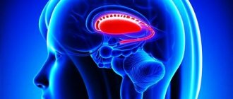
Striped body
(lat. corpus striatum) - anatomical structure of the telencephalon, belonging to the basal ganglia of the cerebral hemispheres. In horizontal and frontal sections of the brain, the striatum appears as alternating stripes of gray matter and white matter. The striatum includes the caudate nucleus and the lentiform nucleus.
Functions[ | ]
The striatum regulates muscle tone, reducing it; participates in the regulation of the work of internal organs; in the implementation of various behavioral reactions (food-procuring behavior); participates in the formation of conditioned reflexes. When the striatum is destroyed, the following occurs:
- hypertonicity of skeletal muscles,
- disturbance of complex motor reactions and food-procuring behavior;
- the formation of conditioned reflexes is inhibited.
The ventral striatum and nucleus accumbens regulate reward and reinforcement in the brain. The dorsal part of the striatum is more involved in regulating motor functions. The dorsal part is also associated with the presence of impulsive behavior.[2][3][4][5][6]
The striatum helped diagnose schizophrenia automatically
Ang Li et al.
/ Nature Medicine, 2020 Scientists from China have developed an algorithm that, based on the characteristics of striatal activity, diagnoses schizophrenia with an accuracy of more than 80 percent. This analysis also allows one to predict the patient’s susceptibility to antipsychotic drug therapy. Abnormalities in the functioning of the striatum correlate with the functioning of the dopaminergic system and the expression of genes that are associated with the risk of developing schizophrenia. The article was published in the journal Nature Medicine
.
Diagnosing schizophrenia is complicated by the very wide range of symptoms and the fact that the causes of this disease are poorly understood. The main hypothesis about the mechanism of schizophrenia is an imbalance of dopamine balance. Dopamine is a neurotransmitter with a wide range of functions and is closely linked to the reward system in the brain. For schizophrenia, antipsychotics are mainly prescribed, which block dopamine receptors and reduce the activity of this neurotransmitter.
Scientists believe that the structure that plays a central role in the development of schizophrenia is the striatum, a group of basal ganglia of the cerebral hemispheres. In patients with this structure, dopaminergic activity is often increased. An Li and his colleagues from the Chinese Academy of Sciences decided to use abnormalities in the functioning of the striatum as a marker for diagnosing schizophrenia. To do this, fMRI of the brain was performed on 560 patients with schizophrenia and 540 healthy people.
Then the scientists trained a classifier based on the support vector machine to determine the diagnosis (healthy or sick with schizophrenia) based on three characteristics of the striatum: the amplitude of low-frequency oscillations, the activity of connections within the striatum and with external structures of the brain (more than 12 thousand elements in total).
For each volunteer, the striatal abnormality coefficient was calculated from the position of the characteristics in support vector space: the coefficient was negative in patients and positive in control participants. In addition, the scientists cross-validated the algorithm, testing it on data from different medical centers and different MRI machines. The striatal abnormality coefficient was also calculated for patients with other mental illnesses.
The scientists tested whether abnormalities in the striatum were associated with increased activity of the dopaminergic system and the expression of genes that are associated with schizophrenia. For this purpose, we used the results of positron emission tomography and single-photon emission computed tomography of healthy volunteers. Data on the activity of 43 genes that are associated with schizophrenia were also analyzed. The spatial expression of dopaminergic system markers and selected genes was compared with the distribution of amplitudes of low-frequency oscillations in the striatum.
Researchers have found a number of differences in striatal function between people with schizophrenia and healthy volunteers. For example, the amplitude of low-frequency oscillations was increased in patients, and the connections of the striatum with other areas also differed (compared to the control group). The algorithm, which was trained on this data, distinguished patients with schizophrenia with an accuracy of more than 80 percent. The rate did not differ from control groups for all mental illnesses studied except for bipolar disorder—people with this diagnosis had a lower rate than controls.
Average amplitude of low-frequency oscillations in the striatum in schizophrenic patients (red) and healthy (blue) participants. On the horizontal axis are various medical centers in China, on whose MRI machines the data was obtained
Ang Li et al. / Nature Medicine, 2020
Share
Differences in external projections of the striatum of patients with schizophrenia and healthy people. Blue - projections in patients were less pronounced, red - stronger
Ang Li et al. / Nature Medicine, 2020
Share
The rate of striatal abnormality varied significantly among patients. It turned out that this indicator is associated with susceptibility to treatment with antipsychotic drugs. This observation may help tailor therapy individually, since often antipsychotic resistance is discovered only after failure of several drugs and their concentrations.
Both markers of the dopaminergic system and genes that are associated with the risk of schizophrenia were active in the same areas of the striatum in which the amplitude of low-frequency oscillations was high. This means that both of these mechanisms may underlie changes in striatal activity in schizophrenia.
Scientists have long been searching for the structural and molecular features of the brain that are characteristic of schizophrenia. A total of 413 genes have been identified that are associated with this disease. In addition to the striatum, the corpus callosum is suspected of participating in the development of schizophrenia, and this disease also causes changes in a number of other parts of the brain. You can read in detail about schizophrenia, its causes, symptoms and treatment in the material “Inheritance of Madness” (here is its continuation).
Alisa Bakhareva
Notes[ | ]
- ↑ 1234
Striatum // Fundamental Model of Anatomy - L. M. Yager, A. F. Garcia, A. M. Wunsch, S. M. Ferguson.
The ins and outs of the striatum: Role in drug addiction (English) // Neuroscience. — 2015-08. - Vol. 301. - P. 529–541. - doi:10.1016/j.neuroscience.2015.06.033. - M. Foster Olive, Taylor, Lewis.
The neurocircuitry of illicit psychostimulant addiction: acute and chronic effects in humans (English) // Substance Abuse and Rehabilitation. — 2013-02. - P. 29. - ISSN 1179-8467. - doi:10.2147/SAR.S39684. - Sergi Ferré, Carme Lluís, Zuzana Justinova, César Quiroz, Marco Orru.
Adenosine-cannabinoid receptor interactions. Implications for striatal function: Adenosine-cannabinoid receptor interactions // British Journal of Pharmacology. — 2010-06. - Vol. 160, iss. 3. - P. 443–453. - doi:10.1111/j.1476-5381.2010.00723.x. - Nestler, Eric J. (Eric Jonathan), 1954-.
Molecular neuropharmacology: a foundation for clinical neuroscience. — 2nd ed. — New York: McGraw-Hill Medical, 2009. — 1 online resource p. — ISBN 978-0-07-164119-7, 0-07-164119-X, 0-07-148127-3, 978-0-07-148127-4, 978-1-281-79174-0, 1- 281-79174-1. - BaekSun Kim, Heh‐In Im.
The role of the dorsal striatum in choice impulsivity // Annals of the New York Academy of Sciences. — 2019-09. - Vol. 1451, iss. 1. - P. 92–111. — ISSN 1749-6632 0077-8923, 1749-6632. - doi:10.1111/nyas.13961.
Structure
The striatum consists of:
- Caudate nucleus.
- Lenticular nucleus.
- Fences.
If we examine the body under a microscope, it consists of large neurons with long tails extending beyond the boundaries of the striopallidal system.
The parts of the caudate are the head, body and tail. The head forms the lateral wall of the anterior horn of the lateral ventricle; the body of the nucleus is extended along the central part of the ventricle; the tail is located on the upper wall of the inferior horn of the ventricle and ends at the level of the lateral geniculate body.
The posterior wall of the nuclear head is located on the border with the thalamus, separated by a strip of white matter.
The lenticular kernel, as the name implies, is shaped like a lentil.
It is located sideways to the caudate nucleus and thalamus. If you cut the kernel in half, it has a wedge-shaped shape, with the top facing the middle and the base facing the side.
And small layers of white matter divide the nucleus into several parts:
- The shell.
- Lateral globus pallidus.
- Medial globus pallidus.
The globus pallidus is a specific ancient formation (ancient body), which differs from other parts of the striatum both in macroscopic and histological appearance.
The fence is located outside the lenticular core. Externally, it is a thin, up to two millimeters, plate of a gray substance. The middle of the plate is smooth, and on the lateral edge there are small bulges of gray matter.
Does this mean that psychopaths are not to blame?
American judges are prone to The Double-Edged Sword: Does Biomechanism Increase or Decrease Judges' Sentencing of Psychopaths? make more lenient sentences when a biochemical cause of psychopathy is present. In such a situation, it seems that the person bears less personal responsibility for his actions. However, this is little consolation for people who have suffered from the actions of a psychopath and his future victims.
In Russia the term “psychopathy” is not used. In the international classification of diseases Specific Personality Disorders (F60), this disorder is numbered F60.2 - dissocial personality disorder - and currently has no effective treatments.
Probably the only thing that can be done is to recognize a psychopath in time and stay away from him.
Damage to the striatum and consequences
When the striatum stops functioning, a person experiences the following disorders:
- Athetosis. Banal alternating movements of the limbs.
- Chorea. Incorrect movements that are performed without any sequence or order, involving the entire musculature of the body.
- Strengthening unconditioned reflexes (defensive, indicative, etc.).
- Hyperkinesis. Significant strengthening of auxiliary movements that accompany each main movement.
- Hypotonicity. Upset muscle tone, its decrease.
- The appearance of Tourette's syndrome.
- The onset of Parkinson's disease contributes to the death of neurons in the body, which is why domafin, which is responsible for the motor system of the human body, is not produced.
- The appearance of Huntington's disease.
In addition, damage to the striatum and caudal nucleus in particular:
- Completely or partially prevents the perception of painful, visual, auditory and other types of stimulation.
- Reduces or increases salivation.
- Makes it difficult to orient in space.
- Impairs memory.
- Slows down the growth of the body.
- Promotes the disappearance of conditioned reflexes for a long time. Human behavior can be inert and stagnant.
Shells
This is a continuation of the spinal membranes, forming together with them a single system.
Hard
The outer one is in contact with the skull on one side, and on the other it is covered with a layer of endothelium. Between the arachnoid mater and it there is a subdural space with a small volume of fluid. Dividing in two, in some places it creates venous sinuses, of which there are 8 types.
This tissue creates seals. Those, penetrating between the hemispheres, separate them from each other, as well as from the skull.
Arachnoid
Separated from the previous one by a subdural space with fluid. Below it is the subarachnoid space with cerebrospinal fluid.
Soft
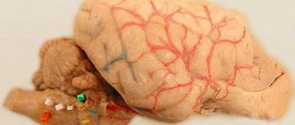
It fits tightly to the brain tissue, repeating its shape and tightly enveloping it. They contain vessels that provide blood supply.
Circulation
For restoration and normal functioning of the brain, it needs good blood supply. Such a small organ consumes from 10 to 20% of the body’s energy, and with a very high efficiency, weighing about 2% of the entire body, both in men and women. At the same time, it requires half the glucose that is available in the body.
The brain is supplied with arterial blood from 3 vessels: the main and a pair of carotid arteries. The main one is formed by the connection of the left and right vertebral vessels. Its branches form the posterior arteries of the brain. All three types of vessels form the so-called circle of Willis. The absence of certain connections between vessels increases the likelihood of strokes and is especially noticeable after they occur due to the enlarged area of the lesion.
When the activity of neurons in a certain area of the brain increases, blood flow to it increases, which does not affect breathing in any way. This became known thanks to MRI. There is a barrier between the vessels and brain tissues that prevents the penetration of certain substances and microorganisms into nerve cells thanks to special receptors.