The cerebral cortex is represented by a uniform layer of gray matter 1.3-4.5 mm thick, consisting of more than 14 billion nerve cells. Due to the folding of the bark, its surface reaches large sizes - about 2200 cm2.
The thickness of the cortex consists of six layers of cells, which are distinguished by special staining and examination under a microscope. The cells of the layers vary in shape and size. From them, processes extend deep into the brain.
Structure of the cerebral cortex
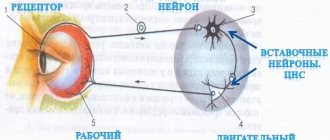
It was found that different areas - fields of the cerebral cortex differ in structure and function. There are from 50 to 200 such fields (also called zones, or centers). There are no strict boundaries between the zones of the cerebral cortex. They constitute an apparatus that provides reception, processing of incoming signals and response to incoming signals.
Areas of the cerebral cortex
In the posterior central gyrus, behind the central sulcus, there is an area of cutaneous and joint-muscular sensitivity . Here the signals that arise when touching our body, when it is exposed to cold or heat, or when it is painful are perceived and analyzed.
Areas of the cerebral cortex
In contrast to this zone, in the anterior central gyrus, in front of the central sulcus, the motor zone . It identifies areas that provide movement of the lower extremities, muscles of the trunk, arms, and head. When this area is irritated by electric current, contractions of the corresponding muscle groups occur. Injuries or other damage to the motor cortex lead to paralysis of the body muscles.
The temporal lobe contains the auditory area . The impulses arising in the receptors of the cochlea of the inner ear are received here and analyzed. Irritation of areas of the auditory zone causes sensations of sounds, and when they are affected by the disease, hearing is lost.
The visual zone is located in the cortex of the occipital lobes of the hemispheres. When it is irritated by electric current during brain surgery, a person experiences sensations of flashes of light and darkness. When it is affected by any disease, vision deteriorates and is lost.
Near the lateral sulcus there is a gustatory zone , where taste sensations are analyzed and formed based on signals arising in the receptors of the tongue. The olfactory zone is located in the so-called olfactory brain, at the base of the hemispheres. When these areas are irritated during surgery or during inflammation, people smell or taste something.
purely speech zone .
It is represented in the cortex of the temporal lobe, the inferior frontal gyrus on the left, and parts of the parietal lobe. Their diseases are accompanied by speech disorders.
The human brain, its structure and functions, the cerebral cortex (Table)
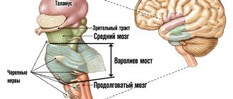
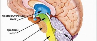
The brain is located in the cerebral part of the skull. Its average weight is 1360 g. There are three large sections of the brain: the brainstem, the subcortical section and the cerebral hemisphere. 12 pairs of cranial nerves emerge from the base of the brain.
| Longitudinal section of the brain (right half) | Base of the brain |
1 - upper section of the spinal cord; 2 - medulla oblongata, 3 - pons, 4 - cerebellum; 5 - midbrain; 6 - quadrigeminal; 7 - diencephalon; 8 - cerebral cortex; 9 - corpus callosum, connecting the right hemisphere with the new one; 10 - optic chiasm; 11 - olfactory bulbs.
Sections of the brain and their functions
| Brain parts | Department structures | Functions | |
| BRAINSTEM | hindbrain | Medulla Here are the nuclei with departing pairs of cranial nerves: XII - sublingual; XI - additional; X - wandering; IX - glossopharyngeal nerves | Conductor - connection between the spinal and overlying parts of the brain. Reflex: 1) regulation of the activity of the respiratory, cardiovascular and digestive systems; 2) food reflexes of salivation, chewing, swallowing; 3) protective reflexes: sneezing, blinking, coughing, vomiting; |
| Pons contains nuclei: VIII - auditory; VII - facial; VI - abducent; V - trigeminal nerves. | Conductor - contains ascending and descending nerve pathways and nerve fibers connecting the cerebellar hemispheres to each other and to the cerebral cortex. Reflex - responsible for vestibular and cervical reflexes that regulate muscle tone, incl. facial muscles. | ||
| Cerebellum The cerebellar hemispheres are connected to each other and are formed by gray and white matter. | Coordination of voluntary movements and maintaining body position in space. Regulation of muscle tone and balance. | ||
| The reticular formation is a network of nerve fibers intertwining the brain stem and diencephalon. Provides interaction between the ascending and descending pathways of the brain, coordination of various body functions and regulation of the excitability of all parts of the central nervous system. | |||
| Midbrain | Four Hills With the nuclei of the primary visual and auditory centers. Brain stems With the nuclei of the IV - oculomotor III - trochlear nerves. | Conductor. Reflexive: 1) indicative reflexes to visual and sound stimuli, which manifest themselves in turning the head and body; 2) regulation of muscle tone and body posture. | |
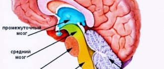
| SUBCORTEX | Forebrain | Diencephalon: a) thalamus (optic thalamus) with the nuclei of the 2nd pair of optic nerves; | Collection and evaluation of all incoming information from the senses. Isolation and transmission of the most important information to the cerebral cortex. Regulation of emotional behavior. |
| b) hypothalamus. | The highest subcortical center of the autonomic nervous system and all vital functions of the body. Ensuring the constancy of the internal environment and metabolic processes of the body. Regulation of motivated behavior and provision of defensive reactions (thirst, hunger, satiety, fear, rage, pleasure and displeasure). Participation in the transition between sleep and wakefulness. | ||
| Basal ganglia (subcortical nuclei) | Role in the regulation and coordination of motor activity (together with the thalamus and cerebellum). Participation in the creation and memorization of programs for purposeful movements, learning and memory. | ||
| CORTEX OF THE LARGE HEMISPHERES | Ancient and old cortex (olfactory and visceral brain) Contains the nuclei of the 1st pair of olfactory nerves. | The ancient and old cortex, together with some subcortical structures, forms the limbic system, which: 1) is responsible for innate behavioral acts and the formation of emotions; 2) provides homeostasis and control of reactions aimed at self-preservation and preservation of the species: 3 affects the regulation of autonomic functions. | |
| New crust | 1) Carries out higher nervous activity, is responsible for complex conscious behavior and thinking. The development of morality, will, and intelligence are associated with the activity of the cortex. 2) Performs perception, evaluation and processing of all incoming information from the senses. 3) Coordinates the activities of all body systems. 4) Provides interaction of the body with the external environment. | ||
Cerebral cortex
The cerebral cortex is phylogenetically the youngest formation of the brain. Due to the grooves, the total surface area of the adult human cortex is 1700–2000 cm2. The cortex contains from 12 to 18 billion nerve cells, which are located in several layers. The cortex is a layer of gray matter 1.5-4 mm thick.
The figure below shows the functional areas and lobes of the cerebral cortex
| Location of gray and white matter | Hemisphere shares | Hemisphere zones | Function |
| Cortex - gray matter, white matter is located under the cortex, in the white matter there are accumulations of gray matter in the form of nuclei | Frontal | Speech centers | |
| Parietal | Skin-muscular zone | Control of movements, ability to distinguish between irritations | |
| Temporal | Auditory zone | Arcs of reflexes that distinguish between sound stimuli | |
| Gustatory and olfactory zones | Reflexes for distinguishing tastes and smells | ||
| Occipital | Visual area | Discrimination of visual stimuli |
Sensory and motor areas of the cerebral cortex
| Left hemisphere of the brain | Right hemisphere of the brain |
| The left hemisphere (“mental”, logical) is responsible for the regulation of speech activity, oral speech, writing, counting and logical thinking. Dominant in right-handers. | The right hemisphere (“artistic”, emotional) is involved in the recognition of visual, musical images, the shape and structure of objects, and in conscious orientation in space. |
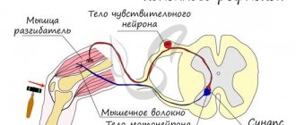
| Cross section of the left hemisphere through the sensory centers Representation of the body in the sensitive zone of the cerebral cortex. The sensitive area of each hemisphere receives information from the muscles, skin and internal organs of the opposite side of the body. | Cross section of the right hemisphere through the motor centers Representation of the body in the motor zone of the cerebral cortex. Each region of the motor zone controls the movement of a specific muscle. |
_______________
A source of information:
Biology in tables and diagrams./ Edition 2, - St. Petersburg: 2004.
Rezanova E.A. Human biology. In tables and diagrams./ M.: 2008.
First and second signaling systems
The role of the cerebral cortex in improving the first signaling system and developing the second is invaluable. These concepts were developed by I.P. Pavlov. The signaling system as a whole is understood as the entire set of processes of the nervous system that carry out perception, processing of information and the response of the body. It connects the body with the outside world.
First signaling system
The first signaling system determines the perception of sensory-specific images through the senses. It is the basis for the formation of conditioned reflexes. This system exists in both animals and humans.
In the higher nervous activity of man, a superstructure has developed in the form of a second signaling system. It is peculiar only to humans and is manifested by verbal communication, speech, and concepts. With the advent of this signaling system, abstract thinking and generalization of countless signals from the first signaling system became possible. According to I.P. Pavlov, words turned into “signals of signals.”
Functions
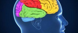
In addition to the physiological division of the brain into lobes, the need arose to divide it into areas that are responsible for certain functions.
Frontal lobes
This is the so-called command center. What is the frontal lobe responsible for? It is a point of independence, self-awareness, and initiative. The defeat of these areas or the presence of pathologies in their functioning will affect a person’s attitude towards the world - almost everything will become indifferent to him, motivation will disappear, interest in current events will disappear, and laziness will appear.
The main functions of the frontal lobes are to control human behavior. It generates responses to social phenomena. When zones are violated, the limiter is deactivated, establishing a ban on certain actions, called uncultural.
The frontal lobes also allow you to analyze, plan, and learn new skills. Repeated repetition of the same sequences of movements over time becomes automatic and does not require effort or thought to perform them. Damage will force you to repeat monotonous movements every time as if for the first time, putting a lot of effort into it.
Perseveration is another consequence of deviations in the functioning of the frontal lobes. This is looping or repetition, such as repeating one phrase or word during a conversation
On the left side (for right-handed people) there are centers that are responsible for speech and attention.
These areas of the brain are also involved in coordinating and maintaining the body in an upright position when sitting and walking.
Temporal
They are located on the sides in the upper part of the brain, in the temple area. Thanks to them, the sound perceived by sound receptors turns into images, a person understands what he heard, certain sound vibrations are associated with images and are assigned to them. With the help of this part of the brain, people understand each other, their sound vibrations are filled with meaning, they choose the necessary words to describe certain phenomena.
Usually the left, non-dominant lobe is involved in determining the intonation of speech and reads emotions from facial expressions alone. Thanks to a small formation, the hippocampus, access to long-term memory is provided. Where it is located, on what medium and in what way our memories are recorded, this should be found out. The non-dominant part is used in visual memory, but the dominant part is used in verbal memory.
With problems with the temporal lobes, deviations in the functioning of the speech apparatus appear, in particular aphasia.
Parietal
MRI has shown that for left-handers and right-handers these lobes perform different functions, in fact directly opposite.
The left one gives us the ability to create a whole from fragments, that is, it helps to form a holistic picture of the world from small, at first glance, unrelated pieces.
They allow you not only to put together a mosaic from fragments, but also from letters into words, from a sequence of actions into a dance or technique, etc.
The non-dominant part allows you to perceive the world in three dimensions by processing information coming from the occipital lobes. Due to impairments, a person loses the ability to recognize faces, outline objects, and determine the distance to them and between them. These areas are also involved in the perception of pain and cold.
Occipital
Visual information processing center. They interpret the photons reflected from obstacles arriving at the biological light sensor - the retina - and form the resulting image, rotating it 180 degrees. Data about the size, color, shape, and material of an object are processed in separate centers and then recombined to form a single three-dimensional picture.
Editorial: Are alpha rhythms indicated for Borderline Disorder or Asperger's Syndrome?