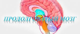
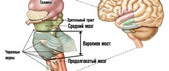
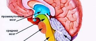
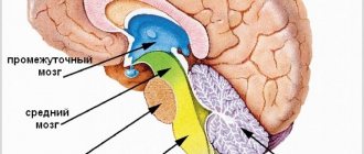
Structure of the hindbrain
The hindbrain consists of two main parts: the pons and the cerebellum located behind it. The first looks like a thick white cushion and is located above the medulla oblongata. The posterior dorsal surface of the pons is covered by the cerebellum, and the anterior ventral surface is represented by numerous transverse fibers that pass into the cerebellar middle peduncle. Nerve roots emerge into the bulbar-pontine groove. The main cerebral artery runs along the central sulcus of the pons. In general, the hindbrain has a rather complex structure.
Looking at the frontal section of the bridge, one can see both the large anterior and small posterior parts, delimited by a trapezoidal fibrous body. All interacting parts of the hindbrain provide conductive function. The cerebellum is otherwise called the small brain; it fills almost the entire space of the posterior cranial fossa. The usual weight of this organ is about 150 grams. The cerebral hemispheres are located above the cerebellum and are separated from it by a transverse fissure.
Bridge
The pons is part of the hindbrain, representing a convexity located on the ventral surface of the brain stem, between the midbrain and medulla oblongata. The length of the bridge is from 25 to 27 millimeters. In the posterior part, the pons is a continuation of the medulla oblongata. The descending and ascending pathways, as well as the reticular formation, pass through the bridge without interruption. In the place where the bridge connects to the medulla oblongata, the exit point of the vestibulocochlear VIII nerve is located. In addition, the abducens, facial and ternary nerves emerge from the bridge.
Functions of the hindbrain area
The responsible area of the nervous system, in which, thanks to the action of the nuclei, the chains of a large number of vegetative and somatic reflexes are closed, is the hindbrain. These include, for example, chewing and swallowing reflexes; regulation of the intensity of salivation as a secretion of the salivary glands.
The hindbrain, namely the suprasegmental organ, the cerebellum, is responsible for such specific actions as regulating the tone of different muscle groups; sensorimotor coordination of body position and meaningful movements; implementation of instant, purposeful movements based on impulses from the cerebral cortex of the cerebral hemispheres. When any disturbances in the functioning of the hindbrain appear, certain pathological symptoms arise: unnecessary movements, alternating paralysis, legs unnaturally wide apart when walking, swaying from side to side, etc.
Functions of Wernicke's and Broca's centers
We need to start with the fact that it is the brain that performs the highest function that determines the intelligence of a person as a species - thinking. It also processes signals received from all receptors - vision, hearing, smell, touch and taste. In addition, the brain controls sensations in the form of emotions, feelings, etc.
It is impossible not to mention that all movements of the human body are also controlled by the brain - even if these are reflex reactions that we are not always aware of.
The functional significance of the speech center for reading and perceiving spoken speech is quite great. Information that comes from outside in the form of words and sentences is ultimately processed in the auditory analyzer.
It is also where the content and idea of what a person is trying to say is formed. This is the process of initiating speech.
The functional significance of Broca's speech center lies in ensuring speech motor skills. The idea is formed at the stage of Wernicke's zone. Information along the arcuate fasciculus reaches Broca's speech-motor center, from where impulses are sent to the articulatory speech apparatus.
Functions of the pons
The hindbrain, the functions of the pons in which support the activity of the entire organism, primarily controls muscle contractions and stability when walking. The bridge contains nerve fibers that transmit impulses to the cortex and from the cerebral cortex in the opposite direction - to the spinal cord, medulla oblongata, and cerebellum. There are the main centers that control facial expressions, chewing functions of a person, hearing and vision. The pons is even responsible for some reflexes: blinking, coughing, sneezing, vomiting. Therefore, we can say that the only purpose of the bridge is to transmit the necessary information from the spinal cord to the large brain, and then to some internal organs.
Brain diseases
The brain is an organ like all others in the human body, which means it is also susceptible to various diseases. The list of such diseases is quite extensive.
It will be easier to consider it if you divide them into several groups:
- Viral diseases. The most common of these are viral encephalitis (muscle weakness, severe drowsiness, coma, confusion and difficulty thinking in general), encephalomyelitis (fever, vomiting, loss of coordination and motor skills of the limbs, dizziness, loss of consciousness), meningitis (high temperature, general weakness, vomiting), etc.
- Tumor diseases. Their number is also quite large, although not all of them are malignant. Any tumor appears as the final stage of a failure in cell production. Instead of the usual death and subsequent replacement, the cell begins to multiply, filling all the space free from healthy tissue. Symptoms of tumors include headaches and seizures. Their presence can also be easily determined by hallucinations from various receptors, confusion and problems with speech.
- Neurodegenerative diseases. By general definition, these are also disturbances in the life cycle of cells in different parts of the brain. Thus, Alzheimer's disease is described as impaired conduction of nerve cells, which leads to memory loss. Huntington's disease, in turn, is the result of atrophy of the cerebral cortex. There are other options. The general symptoms are as follows: problems with memory, thinking, gait and motor skills, the presence of convulsions, tremors, spasms or pain. Also read our article about the difference between seizures and tremors.
- Vascular diseases are also quite different, although, in essence, they come down to disturbances in the structure of blood vessels. So, an aneurysm is nothing more than a protrusion of the wall of a certain vessel - which does not make it any less dangerous. Atherosclerosis is a narrowing of blood vessels in the brain, but vascular dementia is characterized by their complete destruction.
23.09.2016
Source
Structure and functions of the cerebellum
The lower part of the cerebellum is adjacent to the medulla oblongata, which is a continuation of the spinal cord and consists of two brain substances - gray and white. Both hemispheres of the cerebellum are separated from the cerebrum by a deep horizontal fissure, and the surface is dotted with fissures and convolutions of the medulla, forming the anterior, posterior and flocnonodular lobes. The hemispheres are connected to each other by a worm, and inside them lie the nerve nuclei.
The cerebellar gray matter seems to branch out into the white, resembling a twig of thuja. Located at the edges, the gray forms a layered cortex, under which there is always white medulla. Coordination of movement and balance during movement depend on the proper functioning of the cerebellum, and the main function of the department is to connect the forebrain with the hindbrain.
Functions and significance of the medulla oblongata
The hindbrain, whose functions are so important for the functioning of the body, is in close contact with the medulla oblongata, which is responsible for many processes. Nerve impulses that pass from the cerebrum to the spinal cord and then in the opposite direction are transmitted through the pons and medulla oblongata.
The peculiarity of the structure of the latter is that it reads information from sensory organs and nerve impulses and is responsible for metabolism in the body. The centers for regulating the digestive organs and some others are located here.
In addition, the medulla oblongata plays a critical role in controlling breathing and sweating, as well as the state of the cardiovascular system. It is in this department that the responsible centers are located, excited not only reflexively by receiving nerve impulses from the periphery, but also by means of internal chemical stimuli acting on them.
The forebrain and hindbrain are in close interaction, fully responsible for the functioning of the body as a whole. It is thanks to brain cells (neurons) that impulses are transferred, and knowledge of the paths in the hindbrain helps to understand the mechanisms of the appearance of functional pathologies and disorders and, if possible, eliminate them.
content .. 100 101 102 103 104 105 ..Hindbrain (human anatomy)
The hindbrain includes the pons and cerebellum. It develops from the fourth cerebral vesicle (metencephalon).
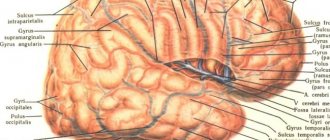
The pons borders the medulla oblongata from below, passes into the cerebral peduncles from above, and its lateral sections form the middle cerebellar peduncles. In the anterior (basilar) part of the pons there are accumulations of gray matter - the nuclei of the pons; in the posterior part of the tegmentum of the pons there are nuclei of the superior olive, reticular formation and V - VIII pairs of cranial nerves. These nerves emerge from the base of the brain lateral to and behind the pons, at the border with the cerebellum and medulla oblongata. The white matter of the pons in its anterior part is represented by transversely running fibers going to the middle cerebellar peduncles. They are penetrated by powerful longitudinal bundles of fibers of the pyramidal tracts, which then form the pyramids of the medulla oblongata and go to the spinal cord. In the rear part of the bridge (pontineum) there are ascending and descending fiber systems (Fig. 113).
Rice. 113. Brain stem (front view). 1 - anterior median fissure; 2 - pyramids of the medulla oblongata; 3 - olive; 4 - cerebellum; 5 - decussation of the pyramids (the place of transition of the medulla oblongata into the spinal cord); 6 - middle cerebellar peduncle; 7 - bridge; 8 - interpeduncular fossa; 9 - cerebral peduncle; III - XII roots of cranial nerves; C - first spinal nerve
Physiology of the medulla oblongata and pons
The medulla oblongata and the pons perform two functions - reflex and conductive. Through the sensitive fibers of the roots of the cranial nerves, it receives impulses - information from the receptors of the scalp, mucous membranes of the eyes, nose, mouth (including taste buds), from the organ of hearing, the vestibular apparatus (organ of balance), from the receptors of the larynx, trachea, lungs, as well as from interoreceptors of the cardiovascular system and digestive apparatus.
Through the medulla oblongata, many simple and complex reflexes are carried out, covering not individual metameres of the body, but organ systems, for example, the digestive, respiratory, and circulatory systems. The reflex activity of the medulla oblongata can be observed in a bulbar cat, that is, a cat in which the brain stem above the medulla oblongata has been cut. The reflex activity of such a cat is complex and diverse.
The following reflexes occur through the medulla oblongata: 1) protective: coughing, sneezing, blinking, lacrimation, vomiting; 2) food: sucking, swallowing, secretion of juice from the digestive glands; 3) cardiovascular, regulating the activity of the heart and blood vessels; 4) in the medulla oblongata there is an automatically working respiratory center that provides ventilation to the lungs; 5) the vestibular nuclei are located in the medulla oblongata and the pons.
From the vestibular nuclei of the medulla oblongata begins the descending vestibulospinal tract, which is involved in the implementation of posture reflexes, namely in the redistribution of muscle tone. A bulbar cat can neither stand nor walk, but the medulla oblongata and cervical segments of the spinal cord provide those complex reflexes that are elements of standing and walking. All reflexes associated with the standing function are called positioning reflexes. Thanks to them, the animal holds its body, usually with the crown up.
The special importance of this part of the central nervous system is determined by the fact that the medulla oblongata contains vital centers: respiratory, cardiovascular. Therefore, not only removal, but even damage to the medulla oblongata ends in death.
In addition to the reflex function, the medulla oblongata performs a conductive function. Conducting pathways pass through it, connecting the cortex, diencephalon, midbrain, cerebellum and spinal cord with a two-way connection.
The cerebellum is located dorsal to the pons and medulla oblongata. It has two hemispheres and a middle part - the worm. The surface of the cerebellum is covered with a layer of gray matter (cerebellar cortex) and forms narrow convolutions separated by grooves. With their help, the surface of the cerebellum is divided into lobules. The central part of the cerebellum consists of white matter, which contains accumulations of gray matter - the cerebellar nuclei. The largest of them is the dentate nucleus. The cerebellum is connected to the brain stem by three pairs of peduncles: the upper ones connect it to the midbrain, the middle ones to the pons, and the lower ones to the medulla oblongata. The peduncles contain bundles of fibers connecting the cerebellum to various parts of the brain and spinal cord.
During development, the isthmus of the rhombencephalon forms the boundary between the hindbrain and midbrain. From it develop the superior cerebellar peduncles, the superior medullary velum located between them and the triangles of the loop, lying outward from the superior cerebellar peduncles.
During development, the fourth (IV) ventricle (ventriculus quartus) is a common cavity of the medulla oblongata and hindbrain. At the bottom, the IV ventricle communicates with the central canal of the spinal cord, at the top it passes into the cerebral aqueduct of the midbrain, and in the roof area it is connected by three openings to the subarachnoid space of the brain. Its anterior (ventral) wall—the bottom of the fourth ventricle—is called the rhomboid fossa. The lower part of the rhomboid fossa is formed by the medulla oblongata, and the upper part by the pons and isthmus. The posterior (dorsal) wall - the roof of the IV ventricle - is formed by the superior and inferior medullary sails and is supplemented at the back by a plate of the pia mater lined with ependyma. This area contains a large number of blood vessels that form the choroid plexus of the fourth ventricle. The convergence of the superior and inferior sails protrudes into the cerebellum and forms a tent. The rhomboid fossa is of vital importance, since most of the nuclei of the cranial nerves (V - XII pairs) are located in this area.
Physiology of the cerebellum (human anatomy)
The cerebellum is a suprasegmental part of the central nervous system that does not have a direct connection with the receptors and effectors of the body. It is connected in numerous ways to all parts of the central nervous system. Afferent pathways are sent to it, carrying impulses from proprioceptors of muscles, tendons, vestibular nuclei of the medulla oblongata, subcortical nuclei and cerebral cortex. In turn, the cerebellum sends impulses to all parts of the central nervous system.
The functions of the cerebellum are studied by irritating it, partially or completely removing it, and studying bioelectrical phenomena. The Italian physiologist Luciani characterized the consequences of removal of the cerebellum and loss of its functions with the famous triad A: astasia, atony and asthenia. Subsequent researchers added another symptom: ataxia.
A dog without a cerebellum stands on widely spaced legs and makes continuous rocking movements (astasia). She has impaired proper distribution of flexor and extensor muscle tone (atony). Movements are poorly coordinated, sweeping, disproportionate, abrupt. When walking, the paws are thrown beyond the midline (ataxia), which is not observed in normal animals. Ataxia is explained by the fact that movement control is impaired. Analysis of signals from proprioceptors of muscles and tendons is missing. The dog cannot get its muzzle into the food bowl. Tilt of the head downwards or to the side causes a strong opposite movement.
The movements are very tiring: the animal, after walking a few steps, lies down and rests. This symptom is called asthenia.
Over time, movement disorders in a cerebellar dog smooth out. She eats on her own and her gait is almost normal. Only biased observation reveals some violations (compensation phase).
As E. A. Asratyan showed, compensation of functions occurs due to the cerebral cortex. If the bark of such a dog is removed, then all the violations are revealed again and are never compensated.
The cerebellum is involved in the regulation of movements, making them smooth, precise, proportionate. According to the figurative expression of L. A. Orbeli, the cerebellum is an assistant to the cerebral cortex in controlling skeletal muscles and the activity of autonomic organs. As studies by L.A. Orbeli have shown, cerebellar dogs have impaired autonomic functions. Blood constants, vascular tone, the functioning of the digestive tract and other autonomic functions become very unstable and easily shift under the influence of certain reasons (food intake, muscle work, temperature changes, etc.).
When half of the cerebellum is removed, motor functions on the side of the operation are impaired. This is explained by the fact that the cerebellar pathways either do not cross at all or cross twice.
content .. 100 101 102 103 104 105 ..