Cerebral cortex
The cerebral cortex forms a thin layer of gray matter responsible for higher mental function.
On the superficial part of the cortex you can visually see grooves, which is why all parts of the brain have a folded surface. The central organ of each person has a different shape of grooves, depth and length, thus creating an individual pattern. Studies of brain structures have made it possible to determine the most ancient cortical layer and the evolutionary development of the organ through histological analysis. The bark is divided into several types:
- Archipallium - the oldest part of the cortex, regulates emotions and instincts;
- Paleopallium is the younger part of the cortex, responsible for vegetative regulation and maintains the physiological balance of the whole organism;
- Neocortex is a new area of the cortex that forms the upper layer of the cerebral hemispheres;
- Mesocortex - consists of an intermediate old and new cortex.
All areas of the cortex are in close interaction with each other, as well as with subcortical structures. The subcortex includes the following structures:
- Varoliev bridge: structure, functions, symptoms in pathological conditions
- Structure
- Treatment
- Other GM departments and their functions
- Brain structure. Pons
- Possible pathologies and their diagnosis
- Functions
- Anatomy
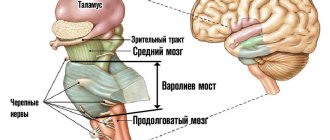
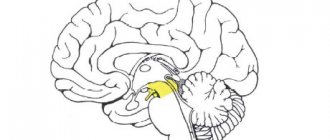
- The thalamus (visual thalamus) is a collection of large masses of gray matter. The thalamus contains sensory and motor nuclei, and nerve fibers allow it to be connected to many parts of the cortex. The visual thalamus is connected to the limbic system (hippocampus) and is involved in the formation of emotions and spatial memory;
- The basal ganglia (nuclei) are a collection of white matter in the thickness of gray matter. The layer is located on the side of the thalamus, near the base of the hemispheres. The basal ganglia carry out higher processes of nervous activity, the active phase of work occurs during the daytime, and stops during sleep. Neurons in the nuclei are activated during mental work of the organ (concentration), and produce electrochemical impulses;
- Brainstem nuclei - regulate the mechanisms of redistribution of muscle tone, and are responsible for maintaining balance;
- The spinal cord is located in the spinal canal and has a cavity filled with cerebrospinal fluid. It is presented in the form of a long cord and provides communication between the cerebrum and the periphery. The spinal cord is divided into segments and performs reflex activities. Information flows through the spinal canal to the brain.
Varoliev bridge: structure, functions, symptoms in pathological conditions
- Bridge structure
- Bridge functions
- Signs of defeat
- Conclusion
The pons is a formation of the central nervous system, located midway between the midbrain and medulla oblongata. Conductive bundles pass through it from the overlying parts of the brain and to them, arteries and veins.
The pons Varolius contains nuclei - centers of cranial nerves that are responsible for chewing movements. It also provides sensitivity to the skin of the face, mucous membranes of the eyes and nose due to the location of the trigeminal nerve. Performs connecting and conducting functions. This department is named after the Bolognese anatomist Constanzo Varolii.
The article contains information about the pons, the structure and functions of this formation, as well as symptoms of damage.
Bridge structure
The pons is part of the hindbrain. This section is a roll-like structure and makes up the trunk. Located in front of the cerebellum, it is a continuation of the midbrain and passes into the medulla oblongata.
It is separated from the midbrain by the place from which the nerve of the IV pair, which innervates the trochlear muscle of the eye, arises. The borders with the medulla oblongata are formed by the medullary striae and the transverse sulcus.
The pons is a roller that has a groove in which the nerves that provide sensitivity to the face (fifth pair) and the basilar arteries that supply blood to the hindbrain pass.
On the posterior surface of the bridge is the upper part of a depression called the rhomboid fossa. Above the brain stripes that limit it are colliculi facials - facial mounds. Above the facial hillocks is the median eminence, on the side of which there is the locus coeruleus, which is responsible for anxiety and contains many norepinephrine nerve endings.
Pathways - thick nerve fibers stretch from the pons to the cerebellum, forming the arms of the pons and the cerebellar peduncles.
The pons consists of a tegmentum, which contains accumulations of gray matter - the centers of cranial nerves, and a base containing pathways.
Thus, in the upper part there are centers from which the vestibulocochlear, facial, trigeminal and abducens nerves emerge. Of the pathways, the medial and lateral loops lie there.
The base of the pons includes pathways running from the cortex to the pons itself, the medulla oblongata and spinal cord (as part of the pyramidal tract), and the cerebellum. Blood supply is provided by the arteries of the vertebrobasilar region.
Read about diseases associated with dysfunction of the pituitary gland: adenoma, endocrine pathologies.
OBLONG BRIDGE
The medulla oblongata is a direct continuation of the spinal cord upward. The lower border of the medulla oblongata is considered to be the lower level of the decussation of the pyramids or the exit site of the first pair of spinal roots, which almost coincides.
The upper border of the medulla oblongata is formed on the ventral surface of the brain by the posterior edge of the pons, and on the dorsal surface by medullary stripes of the rhomboid fossa. Skeletotopically, the medulla oblongata extends from the upper edge of the atlas to the middle of the Blumenbach clivus. Its length is about 3 cm. The medulla oblongata basically has the same grooves and columns as the spinal cord. Between the fissura mediana anterior of the medulla oblongata and its sulcus lateralis anterior lies a pyramid - pyramis. Outside of it, in the territory of the lateral column of the medulla oblongata, between the sulcus lateralis anterior and sulcus retroolivaris, an olive is located. The posterior columns of the medulla oblongata, between the suIchs medianus posterior and sulcus lateralis posterior, contain well-demarcated bundles of Gall and Burdach. The Gaulle bundle expands at its upper end, forming an elevation—tuberculum nuclei gracilis. Burdach's bundle, located between the sulcus intermedius posterior and sulcus lateralis posterior, also forms an elevation at the top - tuberculum nuclei cuneati.
The roots of the XII cranial nerve (n. hypoglossus) emerge from the sulcus lateralis anterior. Three cranial nerves emerge from the sulcus lateralis posterior of the medulla oblongata: IX, X, XI, i.e. glossopharyngeus, a. vagus and superior bundles n. accessorii. The described elevations on the surface of the medulla oblongata determined its now forgotten name - bulbus cerebri. However, this designation formed the basis of the term characterizing damage to the medulla oblongata - bulbar syndrome. Varoliev bridge (pons). The pons is a massive fibrous cord located at the base of the brain, the anterior-posterior diameter of which is 20-30 mm, width - 30-36 mm, thickness - about 25 mm. The pons is bounded posteriorly by the medulla oblongata, in front by the cerebral peduncles, and laterally it passes into the middle cerebellar peduncles without a clear boundary. Skeletotopically, the pons extends from the middle of the Blumenbach clivus to the upper edge of the dorsum of the sella turcica. The ventral surface of the bridge is distinguished by pronounced transverse fibers. In the middle of the bridge there is its main groove - sulcus basilaris, in which a. basilaris. On the sides of the furrow, along the bridge, there are elevations formed by pyramidal bundles that run through its thickness. The trigeminal nerves emerge laterally from the pons. The dorsal surface of the bridge faces the cavity of the fourth ventricle, participating in the formation of the bottom of its rhomboid fossa. Fourth ventricle (ventncuius quartus). The cavity of the rhombencephalon forms the fourth ventricle containing cerebrospinal fluid. Through the aqueduct of Sylvius, the fourth ventricle communicates with the third ventricle, below which it passes into the central canal of the spinal cord. Three openings lead from the IV ventricle to the subarachnoid space of the brain: apertura medians (Magendie), located in the posterior part of the roof of the IV ventricle, and aperturae lateraies (Lushka), located on the side in the area of recessus lateralis. The roof of the IV ventricle is formed by the tela chorioidea ventriculi quarti, the superior and inferior medullary velum, velum medullare superius et velum medullare inferius. Above the sails lies the cerebellum. The floor of the ventricle is formed by the dorsal surface of the upper part of the medulla oblongata, the dorsal surface of the entire pons and the cerebellar peduncles to the quadrigeminal. It has the shape of a diamond, the lower and upper corners of which are pointed, and is described as the rhomboid fossa.
Structure
Varolievye formation is located on the basal surface of the brain. This is the location of the bridge in the brain.
Speaking about the internal structure, the bridge consists of accumulations of white matter, where its own nuclei (accumulations of gray matter) are located. On the posterior part of the bridge lie the nuclei of the 5th, 6th, 7th and 8th pairs of cranial nerves. An important structure lying on the territory of the bridge is the reticular formation. This complex is responsible for the energetic activation of higher-lying elements of the brain. The retinal formation is also responsible for activating the wakeful state.
Externally, the bridge resembles a cushion and is part of the brain stem. The cerebellum is adjacent to it posteriorly. Below the bridge passes into the medulla oblongata, and above into the middle brain. The structural features of the cerebral bridge are the presence of cranial nerves and many pathways in it.
On the back surface of this structure there is a diamond-shaped fossa - this is a small depression. The upper part of the bridge is limited by the medullary stripes, on which the facial hillocks lie, and even higher by the medial eminence. A little to the side of it there is a blue spot. This colored formation is involved in many emotional processes: anxiety, fear and rage.
Treatment
Constant monitoring of cyst size is necessary. If the patient has no complaints and no symptoms accompanying the anomaly, the therapy is focused on the cause that provoked the formation of the formation.
Important VSD according to the vagotonic type
Medications used:
- Drugs to normalize blood circulation;
- Medicines against adhesions;
- Immunomodulatory agents;
- Antioxidants;
- Nootropics.
Constant monitoring of the patient's condition involves regular blood clotting and cholesterol tests, and monitoring blood pressure levels.
Surgery methods
When growth of the formation is observed, surgical intervention is prescribed:
- The endoscopic method is the least invasive, but can only be used for a certain location of the cyst.
- Trepanation is a very complicated operation, but it allows you to remove formations of any location and any size. Accompanied by a high risk of injury to brain tissue.
- During bypass surgery, a puncture is made and a tube is inserted to prevent the accumulation of cerebrospinal fluid. The method is accompanied by the risk of infection.
Other GM departments and their functions
The structure of the human brain is divided into several sections.
The formation of brain activity occurs during intrauterine development thanks to the rhomboid, midbrain, and forebrain.
The parts of our brain are responsible for various tasks, characterized by the telencephalon and medulla oblongata, intermediate and middle brain, as well as the hindbrain, pons and cerebellum.
Their functions are shown in the table:
| Medulla | Another name for this zone is the bulbus, located in the back of the skull, between the cerebellar region, the pons and the dorsal segment. The bulbus is a continuation of the spinal cord. The white matter of the brain in this area is represented by neurons, and the gray matter by nuclei:
If the functioning of this department is disrupted, heart problems will arise and the transmission of impulses to the brain centers will be disrupted. |
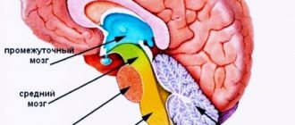
| Diencephalon | This brain region “filters” the impulses of neurons. It will accept all incoming data and decide where and how it will go next. It consists of a lower zone and a posterior zone, consisting of the epithalamus and thalamus. This department is responsible for the functioning of the endocrine system. The hypothalamus is part of the inferior region. This dense neuronal bundle regulates body temperature and the cycle of wakefulness and sleep. It synthesizes hormonal compounds that “tell” a person when to drink or eat. This is the pleasure zone, responsible for interest in the opposite sex. The medullary zone is connected to the pituitary gland, which regulates all glands. Impulses come from the hypothalamic zone to the pituitary gland, and the order to synthesize or stop the release of hormones is “executed.” The thalamus processes impulses from receptors responsible for vision, taste, hearing, and tactile sensitivity. The signals are distributed to the corresponding brain areas. The epithalamus synthesizes the melatonin hormone, which is responsible for the cyclic processes of wakefulness, the emotional sphere, and puberty. |
| Midbrain | The brain section is small in size and consists of two halves: on the roof in the subcortex there are centers of hearing and vision, and conductive pathways are located on the legs. This brain segment includes substantia nigra with red nuclei. There is a temporoparietal node and nuclei of neurons that control the ocular myofibers and temporal zones, which process sound effects that are transformed into recognizable sounds. Reflex activity and reaction to the stimulus are controlled. This organ is responsible for spatial orientation. |
| Finite brain | This is the youngest part of the brain, the main part of the brain, responsible for higher nervous activity, and has numerous grooves with convolutions. The corpus callosum separates the right and left hemisphere zones. Each hemisphere is equipped with a nucleus, mantle, and olfactory brain. |
| Pons | This anatomical formation is part of the hindbrain, which contains the cerebellar region. The functions of the bridge are similar to its name; it consists of nerve fibers. Through it there are impulses passing from the body to the GM and vice versa. It makes up the brain stem, located between the midbrain and medulla oblongata. It contains the nuclei of nerves that control chewing, facial expressions, and some ocular myofibers. It receives signals from receptors responsible for the sensory organs, skin, and inner ear. Thanks to this department, a person feels taste, maintains balance, and hears sounds. |
| Cerebellum | Consists of 2 hemispheric areas and an unpaired formation connecting them. The cerebellar surface is covered by the cortex, which forms 2 nuclei in the thickness of the hemispheric zones. In the deep layers, the lobules consist of a white substance that connects the cerebellar segment with three pairs of legs with the spinal trunk and GM. Responsible for coordinating and regulating the movements of myofibers and muscle memory. Thanks to him, a person maintains a certain body position. |
The anatomical and physiological parameters of the GM have been studied by scientists for decades; they are different for each person, because there are not even two people who think the same way. Experts will eventually reveal these and other secrets of the brain.
Brain structure. Pons
Among all body systems, the central nervous system occupies a special place. The brain regulates all the functions that a person is endowed with. Thanks to it, the relationship between the work of organs and systems is realized. Without brain regulation, a person would not be a viable being.
Thanks to the coordinated activity of the central nervous system, we move, speak, think and feel external stimuli. The brain has a complex structure, each of its components is responsible for a specific function.
Nevertheless, all its structures ensure the functioning of our body only in their entirety. Particularly important formations that make up the central nervous system are the medulla oblongata and the pons.
They contain the main vital centers (vascular, respiratory, cough, lacrimal), and also give rise to most cranial nerves.
Brain structure
The structural unit of the central nervous system is the neuron. It is this cell that is responsible for receiving, processing and storing information. The entire human brain is a collection of neurons and their processes - axons and dendrites.
They ensure the transmission of signals entering the central nervous system and back to the organs. The brain consists of gray and white matter. The first is formed by the neurons themselves, the second by their axons.
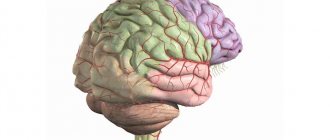
The main structures of the brain are the hemispheres (left and right), the cerebellum and the brainstem. The first are responsible for a person’s mental abilities, his memory, thinking, imagination.
Important Capgras syndrome (double syndrome). What is Capgras syndrome?
The pons is one of the parts of the hindbrain. Its length ranges from 2.4 to 2.6 cm. The pons has a mass of about 7 g. The structures that border it are the medulla oblongata and midbrain, the transverse sulcus. The main components of the pons are the superior and middle cerebellar peduncles, which are large pathways.
In front is the basilar groove, which contains the arteries that supply the brain, and nearby is the exit site of the trigeminal nerve. On the posterior side, the pons forms the upper part of the rhomboid fossa, which contains the 6th and part of the 7th cranial nerves. The upper part of the bridge contains the most cores (5, 6, 7, 8).
At the base of the bridge there are descending pathways: corticospinal, bulbar and pontine tracts.
The main functions of this body:
- Conductive - along its paths nerve impulses pass to the cerebral cortex and to the spinal cord.
- Sensory function is provided by the vestibulocochlear and trigeminal nerves. In the nuclei of the 8th pair of cranial nerves, information about vestibular stimulation is processed.
- Motor – ensures contraction of all facial muscles. This occurs thanks to the nuclei of the trigeminal nerve. In addition, its sensitive part receives information from receptors in the mucous membrane of the mouth, eyeball, part of the head and teeth. These signals are sent along the pons fibers to the cerebral cortex.
- The integrative function ensures the relationship between the forebrain and hindbrain.
- Reflexes of the brain.
Reticular formation of the bridge
The reticular formation is a branched network located in the brain and consisting of nerve cells and nuclei. It is present in almost all formations of the central nervous system and smoothly passes from one department to another. The reticular formation of the pons is located between the medulla oblongata and midbrain.
Its long processes, called axons, form white matter and pass into the cerebellum. In addition, signals can be transferred from the head to the back along the fibers of the nerve cells of the bridge. In addition, the reticular formation transmits signals to the cerebral cortex, due to which a person awakens or sleeps.
The nuclei located in this part of the pons belong to the respiratory center located in the medulla oblongata.
Reflex function of the bridge
The ability of the central nervous system to respond to external stimuli is called a reflex. An example is the appearance of salivation at the sight of food, the desire to sleep at the sound of soothing music, etc. Reflexes of the brain can be conditioned and unconditioned.
A person acquires the first ones in the course of life; they can be developed or adjusted depending on our desire. The latter are not amenable to consciousness, they are laid down at birth, and it is impossible to change them. These include chewing, swallowing, grasping and other reflexes.
How does the bridge affect reflexes?
Due to the fact that the pons is an integral part of the quadrigeminal region, it is related to the development of the auditory and statistical reflex. Thanks to the latter, we are able to hold the body in a certain position. In addition, interacting with the midbrain, it closes a significant part of muscle reflexes.
Magnetic resonance imaging (MRI) in St. Petersburg
Localization of the pathological focus with MRI of the brain begins with determining the location of the lesion in relation to the tentorium of the cerebellum. Therefore, formations above the tentorium are classified as supratentorial, and everything below is classified as infratentorial.
MRI of the brain. Midsagittal section. tentorium cerebellum (arrow).
Above the tentorium are the cerebral hemispheres. Each hemisphere of the brain consists of four lobes - frontal, parietal, occipital and temporal. If the pathology is located in the hemisphere, then it is necessary to decide which lobe it belongs to. To do this, you first need to find the grooves that serve as the boundaries of the lobes. The central sulcus (sulc.centralis) is better visible in the sagittal plane. It is located in the middle between the parallel precentral and postcentral sulci. There are many options for the structure and course of the furrow. Usually it has a significant extent and goes in the anterior-inferior direction from the interhemispheric fissure to the Sylvian fissure, which it does not always reach. The lower end of the furrow either continues in its main direction or bends back. The central sulcus may be interrupted along the way. In the transverse plane on the upper sections the groove has the greatest extent, reaching almost to the interhemispheric fissure. The lower the cut, the shorter the central groove on it. At the level of the lateral ventricles it is barely visible. The central sulcus separates the frontal and parietal lobes.
MRI of the brain. Lateral sagittal section. Central sulcus (arrow).
MRI of the brain. Axial slice. Central sulcus (arrows).
MRI of the brain. Axial section at the level of the roof of the lateral ventricles. Central sulcus (arrows).
MRI of the brain. Borders of the frontal and parietal lobes in the axial plane.
Another important groove is the Sylvian fissure (fissura cerebri lateralis). On sagittal sections it goes from bottom to top in the anteroposterior direction (Fig. 32). In the axial plane, the Sylvian fissure itself also deviates backward, while its branches are directed perpendicularly towards the interhemispheric fissure. The Sylvian fissure separates the frontal and parietal lobes from the temporal lobe.
MRI of the brain. Lateral sagittal section. Sylvian fissure (arrows).
MRI of the brain. Axial section at the level of the third ventricle. Sylvian fissure (arrows).
MRI of the brain. Borders of the frontal, parietal, temporal and occipital lobes on a sagittal section.
To delimit the parietal lobe, you also need to find the parieto-occipital sulcus (sulc. parietooccipitalis). This groove in the sagittal plane can be traced on the median and medial sections. It extends from the surface of the brain downwards, has a considerable extent and is often segmented. In the transverse plane, the parieto-occipital sulcus extends almost perpendicular to the interhemispheric fissure (Fig. 36) and gives off many small branches. Thus, the boundaries of the parietal lobe are with the frontal lobe - the central sulcus, with the occipital - the parieto-occipital sulcus, with the temporal - the Sylvian fissure and the superior temporal sulcus (angular gyrus).
MRI of the brain. Medial sagittal section. Parieto-occipital sulcus (arrow).
MRI of the brain. Axial slice. Parieto-occipital sulcus (arrow).
MRI of the brain. Borders of the parietal lobe on the medial sagittal section.
The next important dividing groove is the collateral groove (sulc.collateralis). On sagittal sections, it is visible as the inferolateral border of the parahippocampal gyrus, in the region of the pole of the temporal lobe (Fig. 38). It is easier to see in the axial plane in sections at the level of the midbrain (Fig. 39). When the axial plane of the slices is tilted backward, it is visible simultaneously with the temporo-occipital groove. The temporo-occipital groove (sulc. temporooccipitalis) on lateral sagittal sections runs sinuously backward along the border of the brain with the temporal bone and then bends upward (Fig. 40). On axial sections at the level of the Varoliev bridge, it is located in the anteroposterior direction. Thus, the border of the temporal lobe (Fig. 41) with the frontal and parietal lobes is the Sylvian fissure, with the occipital lobe - the temporo-occipital sulcus and the collateral sulcus.
MRI of the brain. Sagittal section. Collateral groove (arrow).
MRI of the brain. Axial slice. Collateral groove (arrows).
MRI of the brain. Axial section at the level of the Varoliev bridge. Temporo-occipital sulcus (arrows).
MRI of the brain. Axial section at the level of the cerebral peduncles. Borders of the temporal lobe.
To determine the boundaries of the occipital lobe, we already have all the landmarks. The border with the parietal lobe is the medially located parieto-occipital sulcus, and the border with the temporal lobe is the laterally located temporo-occipital sulcus.
MRI of the brain. Coronal section. Border sulci (SPO - parieto-occipital sulcus, STO - temporo-occipital sulcus, SCol - collateral sulcus).
MRI of the brain. Borders of the occipital lobe on the medial sagittal section.
Usually localization by lobes is sufficient to describe hemispheric pathologies. In some cases, when reference to gyri or functional areas is required, we recommend using the appropriate atlases (A.V. Kholin, 2005). With centrally (axially) located space-occupying formations, the ventricles of the brain and the subcortical (basal) nuclei located around them may be involved. The optic thalamus, hypothalamus, subthalamus, and epithalamus belong to the diencephalon, a component of the brain stem.
MRI of the brain. Axial slice. Lateral ventricles and subcortical nuclei (NC - caudate nucleus, NL - lenticular nucleus, Th - thalamus optic). Parts of the brainstem (lower midbrain, pons, and medulla oblongata) and the cerebellum are located infratentorially.
The midbrain only partially occupies the supratentorial space; a significant part of it passes through the hole in the tentorium into the posterior cranium. hole. The paired legs of the brain and roof (tectum) are always clearly visible from behind. The roof of the midbrain lies posterior to the aqueduct and consists of the quadrigeminal plate.
MRI of the brain. Midsagittal section. Brain stem (V3 - third ventricle, V4 - fourth ventricle, Q - plate quadrigeminal, Mes - midbrain, P - pons, C - cerebellum, M - medulla oblongata).
The boundary between the midbrain and the pons is the superior sulcus, and the border with the medulla oblongata is the inferior sulcus of the pons. The bridge has a characteristic protruding front part. The posterior surface of the pons is a continuation of the medulla oblongata. At the upper border of the bridge between its abdomen and the middle cerebellar peduncle, the trigeminal nerves (n. trigeminus, V pair) begin. They are clearly visible on transverse MR sections, as they run horizontally forward and have a thickness of about 5 mm. The trigeminal nerve is divided into 3 branches - optic (1), maxillary (2) and mandibular (3). They all go forward into Meckel's cavity to the trigeminal ganglion. From here the 3rd branch goes down through the foramen ovale, and the 1st and 2nd branches go through the cavernous sinus, along its lateral wall. Then, branch 1 enters the orbit through the superior foramen, and branch 2 exits the cranial cavity through the foramen rotundum. The III, IV and VI pairs of cranial nerves, which provide movement of the eyeball, are usually not visualized on MRI scans.
MRI of the brain. Axial slice. Trigeminal nerves (arrow).
The facial nerve (n. facialis, VII pair) and the vestibular-cochlear nerve (n. vestibulocochlearis, VIII pair) exit their trunk together, the facial nerve is slightly more medial, and go in one bundle, crossing the pontocerebellar cistern, and go into the internal auditory opening temporal bone. In the internal auditory canal, the vestibular branch runs in the posterior superior and inferior quadrants, the cochlear branch in the inferior, and the facial nerve in the anterior superior. The VII nerve enters the labyrinth (labyrinthine segment), runs inside the temporal bone to the geniculate body, turns back and passes under the lateral semicircular canal (tympanic segment) and exits the temporal bone through the stylomastoid foramen (foramen stylomastoideum). Next, the nerve goes to the salivary gland, where it divides into terminal branches. On MRI scans in sections 3-5 mm thick, the VII and VIII nerves are not separated and are designated as the auditory nerve. With thinner sections, the course of each nerve can be visualized separately.
MRI of the brain. Axial slice. Auditory nerve.
The medulla oblongata begins from the lower border of the pons. At the level of the foramen magnum it passes into the spinal cord. From it depart from the IX to XII pairs of cranial nerves, of which the initial part of the hypoglossal nerve (n. hypoglossus, XII pair) and, in the form of a single complex, IX, X, XI pairs are sometimes visible on transverse MRI. The IV ventricle runs from the aqueduct above to the foramen of Majendie below. It is located between the brainstem anteriorly and the velum and cerebellar peduncles posteriorly. Posterior to the pons and medulla oblongata is the cerebellum. It is connected to the brain stem by the superior, middle and inferior cerebellar peduncles. The cerebellum consists of a midline vermis and paired hemispheres.
MRI of the brain. Axial slice. Cerebellum (CV - cerebellar vermis, CH - cerebellar hemisphere).
MRI in St. Petersburg, carried out by us, always clearly indicates the localization of the lesion in the report, which is necessary for comparison with the clinic and deciding on the possibility and scope of the operation.
Leave feedback.
MRI in St. Petersburg USA
Possible pathologies and their diagnosis
The significance of the bridge can be assessed based on the influence of pathologies (syndromes) that damage individual functions of the body.
The most common of them include:
- Bonnier syndrome is accompanied by damage to the nuclei of the auditory and vestibular nerves. In this case, the patient becomes dizzy, hearing loss occurs, and trigeminal neuralgia may occur. Common symptoms include weakness, depression, and sleep disturbances.
- Locked-in syndrome (ventral pontine syndrome) is a condition in which consciousness and full sensitivity are retained, but the ability to speak is completely lost. The function of the extraocular muscles is preserved. Communication with others is possible using non-verbal gestures. The condition is preceded by signs confirming insufficiency of the blood supply artery: double vision, dizziness, unsteadiness of gait.
- Raymond-Sestan syndrome (another name is oral tegmentum syndrome) is a combination of paralysis of the muscles responsible for the movement of the eyeball on the side opposite to the lesion. Etiological factors: atherosclerotic changes in cerebral vessels, tumors, ischemic strokes.
- Millard-Hubler syndrome is manifested by paralysis of the facial muscles on the affected side, along with partial paralysis on the opposite side. This disease manifests itself in pathologies at the base of the bridge. Constriction of blood vessels or micro-stroke predisposes to pathology, for example, if there is a cavernous angioma in a given structure with subsequent damage to the structures of the vascular system. Less commonly, the cause may be neurosyphilis or diffuse glioma.
- Foville syndrome is a combined lesion of individual elements of the facial and abducens nerves. The pathology is expressed in complete paralysis of the facial muscles in combination with strabismus. Often the cause of its development is ischemic stroke, less often tumor-like formations, inflammation.
- Gasperini syndrome is caused by the occurrence of pathology in the area of the bridge tire. It affects the nuclei of several nerves at once (facial, trigeminal, vestibulocochlear abducens). From the location of the pathological focus on the opposite side, a person feels a sensitivity disorder. The clinical picture includes strabismus, dizziness, and ataxia. This condition occurs due to ischemia, tumors, and inflammation.
- Grenet's syndrome is a sensory disorder with simultaneous damage to the muscles responsible for chewing on the affected side. On the opposite side, hemihypesthesia is noted. Often, pathology can occur due to ischemic changes in the branches of the posterior cerebral artery.
- Brissot-Sicart syndrome is a set of signs of damage to the nucleus of the facial nerve with partial paralysis of the limbs. Clinically manifested by a spasm of the facial muscles, which is accompanied by peripheral paralysis of the facial nerve and hemiparesis. Its occurrence is associated with ischemia and previous infectious diseases.
Important Hebephrenic schizophrenia: symptoms, course of the disease and prognosis
Modern methods of magnetic resonance imaging help to clarify the location, duration of the lesion, volume and other parameters of the pathological process.
Like any organ in the human body, the VM can also stop functioning and this can be caused by the following diseases:
- stroke of the cerebral arteries;
- multiple sclerosis;
- head injuries. Can be obtained at any age, including during childbirth;
- tumors (malignant or benign) of parts of the brain.
In addition to the main reasons that can provoke brain pathologies, you need to know the symptoms of such a lesion:
- the process of swallowing and chewing is impaired;
- loss of skin sensitivity;
- nausea and vomiting;
- nystagmus is eye movements in one specific direction, as a result of such movements one can often feel dizzy, even to the point of loss of consciousness;
- may see double when turning the head sharply;
- disturbances in the functioning of the motor system, paralysis of certain parts of the body, muscles, or tremors in the hands;
- in case of disturbances in the functioning of the facial nerves, the patient may experience complete or partial anemia, lack of strength in the facial nerve;
- speech disorders;
- asthenia – decreased strength of muscle contraction, rapid muscle fatigue;
- dysmetria - incompatibility between the task of the movement being performed and muscle contraction, for example, when walking, a person may raise his legs much higher than necessary or, on the contrary, may stumble over small bumps;
- snoring, in cases where it has never been observed before.
Motor and sensory functions
Speaking in more detail about motor and sensory function, let's talk about cranial nerves. When mentioning cranial nerves, it should be noted the ternary or mixed nerve (V pair). This pair of nerves is responsible for the movement of the masticatory muscles, as well as the muscles that are responsible for the tension of the eardrum and the palatine curtain.
To the sensory part of the trigeminal nerve there are afferent connections of nerve cells from receptors that are located in the skin of the human face, nasal mucosa, 60% of the tongue, eyeball and teeth. The sixth pair, or the so-called abducens nerve, is responsible for the movement of the eyeballs, namely for its rotation outward.
The 7th pair has one of the most important functions for human interaction; it is responsible for the innervation of muscles that allow the production of facial expressions. In addition, the facial nerve controls three glands: salivary, sublingual and submandibular. These glands provide reflexes such as salivation and swallowing.
The bridge also has a connection with the vestibulocochlear nerve. It is clear from the name that the cochlear part reaches the cochlear nuclei, but the vestibular part ends in the triangular nucleus. The eighth pair of nerves is responsible for analyzing vestibular stimuli; it determines the degree of their severity and where they are directed.
Useful to know: Midbrain: structure, functions, development
to contents ^
Functions
The important functional significance of the cerebral pons is due, on the one hand, to the location in it of the nuclei of the cranial nerves (V, VI, VII, VIII pairs), the reticular formation, and the pons nuclei; on the other hand, the passage of efferent pathways through the pons (corticospinal and corticonuclear, tegmental-spinal cord, red nucleus-spinal cord, reticular-spinal cord, etc.) and afferent pathways (spinothalamic, pathways of proprioceptive - deep - sensitivity, etc.), which are vital for the body and carry out two-way communication between the brain (see) and spinal cord (see).
Integrating function
These pons functions connect parts of the brain called the cerebral hemispheres. Also, all other paths, both ascending and descending, pass along the bridge, connecting it with many departments of the central nervous system. Among them are the spinal cord, cerebellum and cerebral cortex.
Impulses passing through the pontocerebellar pathways of the cerebral cortex have an impact on the functioning of the cerebellum. The cortex cannot exert influence directly, so it uses the bridge as an intermediary for these purposes. The pons regulates the medulla oblongata, influencing the centers that are responsible for the respiratory process and its intensity.
to contents ^