Physiology of the medulla oblongata, cerebellum and midbrain
The medulla oblongata, like the spinal cord, performs two functions - reflex and conductive. Eight pairs of cranial nerves (V to XII) emerge from the medulla oblongata and the pons and it, like the spinal cord, has a direct sensory and motor connection with the periphery. It receives impulses through sensory fibers - information from the receptors of the scalp, mucous membranes of the eyes, nose, mouth (including taste buds), from the organ of hearing, the vestibular apparatus (organ of balance), from the receptors of the larynx, trachea, lungs, as well as from the interoceptors of the heart. -vascular system and digestive system.
Through the medulla oblongata, many simple and complex reflexes are carried out, covering not individual metameres of the body, but organ systems, for example, the digestive, respiratory, and circulatory systems. The reflex activity of the medulla oblongata can be observed in a bulbar cat, that is, a cat in which the brain stem above the medulla oblongata has been cut. The reflex activity of such a cat is complex and diverse. The following reflexes occur through the medulla oblongata: Protective reflexes: coughing, sneezing, blinking, lacrimation, vomiting. Food reflexes: sucking, swallowing, juice production (secretion) of the digestive glands. Cardiovascular reflexes that regulate the activity of the heart and blood vessels. The medulla oblongata contains an automatically functioning respiratory center that provides ventilation to the lungs. The vestibular nuclei are located in the medulla oblongata.
From the vestibular nuclei of the medulla oblongata begins the descending vestibulospinal tract, which is involved in the implementation of posture reflexes, namely in the redistribution of muscle tone. A bulbar cat can neither stand nor walk, but the medulla oblongata and cervical segments of the spinal cord provide those complex reflexes that are elements of standing and walking. All reflexes associated with the standing function are called positioning reflexes. Thanks to them, the animal, contrary to the forces of gravity, maintains the posture of its body, as a rule, with its crown upward.
The special importance of this part of the central nervous system is determined by the fact that the medulla oblongata contains vital centers - respiratory, cardiovascular, therefore not only removal, but even damage to the medulla oblongata ends in death. In addition to the reflex function, the medulla oblongata performs a conductive function. Conducting pathways pass through the medulla oblongata, connecting the cortex, diencephalon, midbrain, cerebellum and spinal cord with a bilateral connection.
The cerebellum does not have a direct connection with the body's receptors. It is connected in numerous ways to all parts of the central nervous system. Afferent (sensitive) pathways are sent to it, carrying impulses from proprioceptors of muscles, tendons, ligaments, vestibular nuclei of the medulla oblongata, subcortical nuclei and cerebral cortex. In turn, the cerebellum sends impulses to all parts of the central nervous system.
The functions of the cerebellum are studied by irritating it, partially or completely removing it, and studying bioelectrical phenomena.
The Italian physiologist Luciani characterized the consequences of removal of the cerebellum and loss of its function with the famous triad A - astasia, atony and asthenia. Subsequent researchers added another symptom: ataxia. A dog without a cerebellum stands on widely spaced legs, making continuous rocking movements (astasia). She has impaired proper distribution of flexor and extensor muscle tone (atonia). Movements are poorly coordinated, sweeping, disproportionate, abrupt. When walking, the paws are thrown beyond the midline (ataxia), which does not happen in normal animals. Ataxia is explained by the fact that movement control is impaired. Analysis of signals from proprioceptors of muscles and tendons also falls out. The dog cannot get its muzzle into the food bowl. Tilt of the head downwards or to the side causes a strong opposite movement.
The movements are very tiring; the animal, after walking a few steps, lies down and rests. This symptom is called asthenia.
Over time, movement disorders in a cerebellar dog smooth out. She eats independently and her gait is almost normal. Only biased observation reveals some violations (compensation phase).
As shown by E.A. Asratyan, compensation of functions occurs due to the cerebral cortex. If the bark of such a dog is removed, then all the violations are revealed again and are never compensated. The cerebellum is involved in. regulation of movements, making them smooth, precise, proportionate.
As studies by L.A. Orbeli have shown, cerebellar dogs have impaired autonomic functions. Blood constants, vascular tone, the functioning of the digestive tract and other autonomic functions become very unstable and easily shift under the influence of certain reasons (food intake, muscle work, temperature changes, etc.).
When half of the cerebellum is removed, motor dysfunction occurs on the side of the operation. This is explained by the fact that the cerebellar pathways either do not cross at all or cross twice.
The midbrain plays an important role in regulating muscle tone and in the implementation of righting and righting reflexes, which make standing and walking possible.
Fig. Transverse (vertical) section of the midbrain at the level of the superior colliculus.
The role of the midbrain in the regulation of muscle tone is best observed in a cat in which a transverse incision is made between the medulla oblongata and the midbrain. Such a cat has a sharp increase in muscle tone, especially extensors. The head is thrown back, the paws are sharply straightened. The muscles are so strongly contracted that an attempt to bend the limb ends in failure - it immediately straightens. An animal placed on outstretched paws like sticks can stand. This condition is called decerebrate rigidity.
If the incision is made above the midbrain, then decerebrate rigidity does not occur. After about 2 hours, such a cat makes an effort to get up. First she raises her head, then her body, then stands on her paws and can begin to walk. Consequently, the nervous apparatus for regulating muscle tone and the functions of standing and walking are located in the midbrain.
The phenomena of decerebrate rigidity are explained by the fact that the red nuclei and reticular formation are separated from the medulla oblongata and spinal cord by transection. The red nuclei do not have a direct connection with receptors and effectors, but they are connected with all parts of the central nervous system. They are approached by nerve fibers from the cerebellum, basal ganglia, and cerebral cortex. The descending rubrospinal tract begins from the red nuclei, through which impulses are transmitted to the motor neurons of the spinal cord. It is called the extrapyramidal tract. The sensitive nuclei of the midbrain perform a number of important reflex functions. The nuclei located in the superior colliculi are the primary visual centers. They receive impulses from the retina and participate in the orientation reflex, i.e. turning the head towards the light. In this case, a change in the width of the pupil and the curvature of the lens (accommodation) occurs, facilitating clear vision of the object.
The nuclei of the inferior colliculi are the primary auditory centers.
They participate in the orienting reflex to sound - turning the head in the direction of the sound. Sudden sound and light stimulation cause a complex alarm reaction, mobilizing the animal to quickly respond. (Visited 188 times, 1 visits today)
Medulla
: responsible for breathing, blood circulation, digestion;
contains reflexes of coughing, sneezing, swallowing, sucking, vomiting, etc. The cerebellum
is responsible for the coordination of movements.
The midbrain
contains subcortical visual and auditory centers and is responsible for orienting reactions to light and sound.
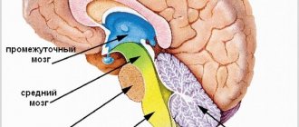
The diencephalon
regulates metabolism in the body and maintains homeostasis (constancy of the internal environment) in two ways:
- through the pituitary gland controls all other endocrine glands of the body;
- participates in the formation of feelings of hunger, cold, thirst, etc., thus influencing behavior.
Tests
1. In which part of the brain is the breathing center located?
A) medulla oblongata B) diencephalon C) cerebellum D) cerebral cortex
2. Regulation and coordination of physiological processes occurring in internal organs is ensured by
A) diencephalon B) midbrain C) spinal cord D) cerebellum
3. Which part of the brain regulates coordination of movements?
A) medulla oblongata B) diencephalon C) cerebellum D) cerebral cortex
4. Injury to the cerebellum can lead to impairment
A) vision B) coordination of movements C) activity of the respiratory organs D) activity of the circulatory system
5. The center of respiratory reflexes is located in
A) cerebellum, B) midbrain C) medulla oblongata D) diencephalon
6. The medulla oblongata, unlike the cerebellum,
A) coordinates movements B) ensures balance of the body in space C) promotes accuracy of actions D) controls cardiac activity and breathing
7. In which part of the brain are the human speech centers located?
A) medulla oblongata B) diencephalon C) cerebellum D) cerebral cortex
8. The centers of swallowing, respiratory, cardiovascular and other vital reflexes are located in
A) cerebellum B) midbrain C) medulla oblongata D) diencephalon
9. In the human brain, unlike other mammals, centers appear in the process of evolution
A) speech B) smell and taste C) hearing and vision D) movement coordination
10. Voluntary human movements provide
A) cerebellum and diencephalon B) midbrain and spinal cord C) medulla oblongata and pons D) cerebral hemispheres of the forebrain
11) The medulla oblongata of the human brain does not regulate
A) respiratory movements B) intestinal peristalsis C) heart contractions D) body balance
12) The brain stem includes
A) oblongata, pons, middle B) oblongata, middle, intermediate C) average, pons, cerebellum D) average, pons, intermediate
Basic information about the brain
Traditionally, the brain is considered an organ whose task is to receive and process information.
Brain functions
Brain functions include processing sensory information from the senses, planning, decision making, coordination, motor control, positive and negative emotions, attention, memory. The human brain performs the highest function - thinking. One of the most important functions of the human brain is the perception and generation of speech.
The flow of signals to and from the brain occurs through the spinal cord, which controls the body, and through the cranial nerves. Sensory (or afferent) signals arrive from the sense organs to the subcortical (that is, preceding the cerebral cortex) nuclei, then to the thalamus, and from there to the higher section - the cerebral cortex.
Main parts of the human brain
The brain is located in the cranial cavity. The shape of the skull is difficult to describe, which is why it is called “complex.”
The mass of the adult human brain is approximately one and a half kilograms, and to be more precise, it ranges from 1100 to 2000 grams.
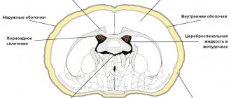
The human brain, due to its fragility and other features, is securely “hidden” in the cranium. Inside the skull, the brain is covered with membranes of connective tissue - hard and soft, between which is the choroid, or arachnoid membrane. Between the membranes and the surface of the brain and spinal cord is cerebrospinal fluid (often called cerebrospinal fluid). Cerebrospinal fluid is also contained in the ventricles of the brain.
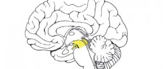
The brain is usually divided into 3 sections: posterior, middle and anterior.
Hindbrain (diamond-shaped)
The hindbrain (diamond-shaped) consists of:
- medulla oblongata,
– hindbrain (actually hindbrain)
– pons (contains mainly projection nerve fibers and groups of neurons, is an intermediate link in the control of the cerebellum)
– cerebellum (consists of the vermis and hemispheres; on the surface of the cerebellum, nerve cells form the cortex)
Medulla
The medulla oblongata is a direct continuation of the spinal cord, so their structure and functions have much in common.
Of the vital functions that the medulla oblongata performs, the main ones, perhaps, are those related to breathing, digestion, and the activity of the cardiovascular system.
The medulla oblongata also controls a number of protective reflexes: such as coughing, sneezing, and lacrimation.
The nerves that control the activity of the tongue, pharynx, larynx, thyroid gland, and internal organs depart from the medulla oblongata. Here are the centers that regulate the position of the body in space.
Bridge
The pons , in turn, is a continuation of the medulla oblongata. Nerve pathways pass through it, connecting the forebrain and midbrain with the medulla oblongata and spinal cord.
The facial and auditory nerves depart from the pons.
The auditory nerves transmit signals to the brain not only from auditory receptors, but also from the balance organs.
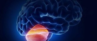
Cerebellum
The cerebellum is located in the occipital part of the brain and consists of paired hemispheres and an unpaired part connecting them.
The surface of the cerebellum has various grooves and convolutions.
The cerebellum takes part in the coordination of movements, making them precise and purposeful.
If this part of the brain is damaged, human movements are also disrupted. It becomes difficult for a person to maintain balance, the gait becomes “drunk,” and internal sensations of loss of orientation arise.
| Interesting to know: To touch your nose with the tip of your index finger, you need to move 33 muscles. The order and sequence of their contraction is determined by the cerebellum. |
Midbrain
The midbrain is involved in the reflex regulation of various types of movements that arise under the influence of auditory and visual impulses.
For example, the midbrain ensures a change in the size of the pupil, the curvature of the lens, depending on the brightness of the light, as well as turning the head towards the light source.
The midbrain performs several functions:
The first function is motor. The midbrain is responsible for coordination of movements, as well as muscle tone. He keeps under control all automatic movements from walking to breathing, chewing, swallowing.
It is important to understand: if you have problems with muscles, the muscles suddenly become flabby and weak, if you begin to have problems with breathing, swallowing, the culprit is the midbrain.
The next function is orientation and coordination. Together with the medulla oblongata, the midbrain is responsible for orientation in space and reaction to various external stimuli: light, sound.
The third function is sensory. The midbrain contains subcortical centers that are responsible for hearing and vision.
Please note that, as a rule, with age all unpleasant processes occur: weakening of muscle tone, vision, and hearing.
And finally, the fourth function is conductive - at the base of the midbrain there are fibers that connect the cerebral cortex with the cerebellum. The conductors of conscious motor impulses also pass here.
Thus, the midbrain is responsible for beauty (read: muscle tone), information coming through hearing and vision.
Forebrain
The forebrain consists of two sections: the diencephalon and the cerebral hemispheres.
In the diencephalon there are a huge number of centers that are involved in partial processing of nerve impulses that are sent to the cerebral hemispheres.
The centers of the diencephalon control the functions of internal organs, regulate body temperature, and are responsible for feelings of thirst, hunger, and satiety.
The cerebral hemispheres of the forebrain are the largest structures of the brain.
They tower above all its departments.
The hemispheres are connected by bundles of nerve fibers, which are also called the corpus callosum.
Each hemisphere is formed by gray and white matter.
The gray matter is thin, 3-4 mm, the surface layer is the cerebral cortex.
Cerebral cortex
The total surface of the hemispheres of the cortex in an adult is 1700 – 2000 cm2.