A retrocerebellar cyst is often a congenital formation in the brain structures of the cerebellar-stem localization. The pathology is manifested by stem (nausea, vomiting, dizziness and weakness, tinnitus, increased sweating and hyperemia of the skin) and cerebellar symptoms (ataxia - impaired motor coordination).
Characteristics of the pathology
A retrocerebellar cyst is a cavity in the brain that has dense walls and often liquid contents, which makes it look like a capsule. A retrocerebral cyst is a cavity formed in the brain tissue, mostly congenital. The walls are formed from cells of the arachnoid membrane or fragments of gray matter.
The contents are often a liquid fraction, sometimes with fragments of soft tissue. The prevalence of the pathology is about 1-4% of all space-occupying formations localized in the brain. Retrocerebellar location means that the cyst is located behind the cerebellum.
Often the formation is localized in the area of the cerebral-cerebellar cistern - a large natural cavity filled with cerebrospinal fluid. The cistern is limited by the medulla oblongata and the cerebellum. A retrocerebellar arachnoid cyst formed in the brain becomes dangerous when its size increases significantly, causing compression of the surrounding brain structures.
How is a retrocerebellar cyst treated?
For the recovery of a patient with such a diagnosis, an endoscopic session is considered an effective operation, since it provides a minimum number of injuries. During the operation, using surgical equipment, doctors puncture the skull, remove the cyst and suck out the substance. This method of therapy may not be carried out in every situation, since the location of the tumor significantly influences the choice of treatment. Shunt surgery may be indicated if hydrocephalus or persistent fluid influx develops. In this case, surgeons perform intracranial shunting, when during the intervention a clinical shunt is used, which helps drain the natural cavities of the department. When the cyst is localized in an accessible area for trepanation, surgery is prescribed to completely remove the anomaly. This procedure is quite traumatic for the patient.
Classification
In the ICD-10 list, a retrocerebellar cyst is listed under the code “Q04.6”. In the ICD-10 list, an arachnoid cyst formed in the brain is also listed under the code “Q04.6”; this section is devoted to congenital cystic formations localized in the brain. Depending on the dynamics of development, progressive (gradually increasing in diameter) and stable (maintaining original dimensions) are distinguished. Taking into account the peculiarities of the formation of a cystic cavity, the following types are distinguished:
- True (isolated, represented by a dense shell with contents).
- Diverticular (communicating, formed in the late stages of embryogenesis as a result of disruption of the process of liquor dynamics).
- Valvular (partially communicating with natural liquor ducts and cavities, formed due to changes in the morphological structure of the arachnoid membrane).
Arachnoid cysts located in the brain are divided into small, medium and large, taking into account their size - the presence of cystic formations that do not entail undesirable consequences, in particular, the appearance of neurological symptoms, can be considered a normal variant.
Cystic formations of large diameter are often associated with restriction and deformation of reserve intracranial spaces. In severe cases, mass effect (compression of brain tissue) and dislocation of brain structures develop. How dangerous a retrocerebellar cyst formed in the brain is depends on its size and location. Small cystic formations are often asymptomatic and do not cause discomfort to the patient.
The progressive growth of the cystic formation leads to compression of the surrounding brain structures, which causes disruption of its functions. It is incorrect to say that a cyst-shaped cavity of a certain size can be considered dangerous. The prognosis for each patient is made individually. In some cases, large cysts located away from vital centers of the brain do not cause neurological symptoms.
Conversely, a retrocerebellar cerebrospinal fluid cyst with a diameter of several millimeters can provoke hypertensive-hydrocephalic syndrome, the development of convulsions and epilepsy attacks, if it is located in such head regions as the trunk, medulla oblongata or cerebellum. Arachnoid forms localized in the area of the brain cisterns with a significant increase in diameter interfere with the circulation of cerebrospinal fluid, which leads to the development of hydrocephalic syndrome and increased intracranial pressure values.
How does the disease manifest itself?
The symptoms of the anomaly have a direct relationship with the size of the tumor. Localization and educational factors are important. If the cyst continues to increase in volume or the pressure of the substance in it increases, the patient may experience pronounced manifestations. At the same time, the formation of small cysts that stop their growth does not provoke symptoms. The main reasons for the progression of the disease are:
- neuroinfectious microorganisms;
- long infectious process;
- malfunctions in the functioning of the circulatory system of a chronic nature;
- autoimmune disorders.
The patient exhibits the following signs of an unstable condition:
- pain in the head area;
- discomfort from a feeling of pulsation inside the skull;
- temporary hearing problems;
- presence of noise in the ear canals;
- a feeling of fullness in the head and a surge in pressure;
- disturbances in visual abilities (formation of spots, blurriness, bifurcation);
- paralysis of the arms or legs, complete or partial;
- seizures;
- sudden loss of consciousness;
- numbness of body parts of a temporary or permanent nature.
Diagnostic measures
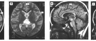
To prevent the development of an anomaly and establish a timely correct diagnosis, it will be necessary to rely on all the patient’s existing complaints along with the clinical picture. Medical staff use clinical tests such as magnetic resonance imaging.
Causes
Primary cystic formations are congenital anomalies of brain formation, which are usually associated with adverse external influences on the fetus (intoxication, infectious diseases suffered by the mother during gestation, hypoxia, birth injuries) or hereditary predisposition. Causes of secondary forms:
- Past inflammatory processes and infections of the central nervous system (meningitis, encephalitis).
- History of surgical interventions in the brain area.
- Injuries, hemorrhages in the head area.
- Neurodegenerative processes.
- Cerebral blood flow disorders occurring in acute or chronic form (TIA, ischemia, cerebral infarction, stroke).
Statistics show that in 40-60% of cases after TBI, complications arise in the form of a cerebral cyst. Post-traumatic cystic formation can appear as a result of a concussion, compression or contusion of the brain of varying severity.
How to study a retrocerebellar cyst?
MRI testing with an additional enhancer in the form of a contrast agent helps to distinguish a neoplasm from a tumor. An anomaly of malignant origin can easily accumulate a contrast agent, while a benign cyst is not capable of such a reaction. Doctors identify the causes of the formations to further prevent the formation of new objects and the increase in size of old ones. To achieve this, various diagnostic techniques are used to help identify the sources of infections, problems with the functioning of the circulatory system and autoimmune disorders. These include:
- Doppler diagnostics of the vessels of the head and cervical region - the procedure is prescribed by doctors to identify narrowing of the vessels that serve the function of supplying the brain with arterial blood cells. Due to blood deficiency, gray matter may begin to die.
- Heart check - necessary to determine heart rhythm disturbances and look for insufficiency.
- Donating blood to analyze coagulation and cholesterol levels - an increase in cholesterol and coagulability can become a factor in blocking blood vessels, which will subsequently lead to the formation of a cyst.
- Controlling blood pressure - pressure surges often become a source of strokes and the appearance of post-stroke neoplasms.
- Testing blood cells for infections and autoimmune diseases - prescribed if there is a likelihood of developing a neuroinfection, multiple sclerosis, and other things.
You can make an appointment at any local clinic for diagnostics on the search portal piter-mrt.ru. Here you can apply for an appointment online, find out the current price lists of the centers, the number of vacancies for the coming days and read reviews from visitors.
Symptoms
The clinical course of a progressive cyst occurs in 3 stages - compensation, subcompensation, decompensation. At the 1st stage, the pathology is asymptomatic, at the 2nd stage, pronounced signs of damage to brain structures are observed, at the 3rd stage, the increasing development of hydrocephalus occurs. Symptoms of a retrocerebellar cyst of post-traumatic origin that forms in the head of an adult include cephalalgia and increased intracranial pressure. Other signs:
- Symptoms of damage to the cranial nerves (paresis, paralysis of the facial muscles, speech dysfunction due to impaired innervation of the organs of the speech apparatus, rarely oculomotor disorders).
- Transient short-term clouding of consciousness.
- Visual, auditory dysfunction.
- Motor dysfunction (impaired motor coordination, pyramidal insufficiency - increased skeletal muscle tone with the development of paresis and paralysis). In infancy, it manifests itself with a weak grasping reflex, tremor of the hands and chin, standing on tiptoes when walking (without support on the heel), throwing the head back.
- Deterioration of cognitive abilities.
- Behavior change.
- Convulsive, epileptic seizures are often of a generalized type.
- Astheno-neurotic syndrome (emotional lability, muscle and general weakness, deterioration in performance, irritability).
Disturbances in the functioning of the autonomic system are often observed - vegetative-vascular dystonia (dizziness, nausea, heart rhythm disturbances, increased heart rate, increased fatigue, sleep disturbance), sometimes accompanied by panic attacks. A retrocerebellar arachnoid cerebrospinal fluid cyst formed in the brain is manifested by symptoms:
- Feeling of pulsation and bursting in the area of the skull.
- Pain in the head area, resistant to drug therapy.
- Poor balance, difficulty keeping the body upright.
- Noise, hum, ringing in the ears, deterioration of hearing acuity.
- Disturbances in sleep and wakefulness (daytime sleepiness, nighttime insomnia).
- Fainting conditions.
- Deterioration of visual acuity.
- Paresthesia (skin sensitivity disorder - burning, itching, tingling sensation in the skin area).
In infants, signs are often observed: frequent regurgitation, bouts of vomiting, loss of appetite, crying for no reason, throwing the head back. In children of the 1st year of life, deformation of the bone structures of the skull, divergence of cranial sutures, and delayed mental and physical development are possible.
Preventive measures
You can prevent the appearance of retrobulbar and retrocerebellar cysts of the brain if you follow the recommendations of doctors:
- Protect your head and brain from damage and injury, take safety measures seriously during sports activities, driving vehicles, and during work situations.
- Constantly monitor your condition if you have suffered a concussion.
- Treat infectious diseases in a timely manner and avoid the development of complications.
- Control cholesterol levels.
- Regularly examine the brain and monitor the condition.
During pregnancy, a woman should lead a healthy lifestyle, not abuse medications, take prescribed tests and other tests in a timely manner, and appear on time for gynecological examinations at the clinic where she is registered.
Diagnostics
Diagnostic measures include collecting anamnesis, determining the somatic and neurological status of the patient. Consultations with a neurologist, otoneurologist, and ophthalmologist are indicated. Instrumental examination is carried out in the format of MRI and CT (neuroimaging); in parallel, EEG (electroencephalography) is prescribed to determine the bioelectrical activity of the brain and identify foci of epileptic activity.
Examination of newborn children is carried out using neurosonography. In doubtful cases, an MRI with contrast is performed. In the presence of a retrocerebellar cyst, an expansion of the cerebral-cerebellar cistern is often detected. Small cystic cavities that are asymptomatic often become an incidental finding during a diagnostic examination prescribed for another reason.
Treatment methods
Treatment of a retrocerebellar cyst formed in the brain of a child or adult is carried out taking into account its size and nature of its course. If the pathology is asymptomatic, therapeutic measures are limited to regular visits to a neurologist for observation. If neurological symptoms are present, medical or surgical treatment is performed.
Drug therapy
Drug treatment is carried out taking into account the symptoms and causes. If the formation of a cystic cavity is associated with an inflammatory process, antibiotics, antivirals, and immunomodulators are prescribed. Nootropic drugs and cerebral blood flow correctors improve blood supply to brain tissue, increase the energy status of neurons, regulate the processes of glucose utilization in nervous tissue, and activate synaptic transmission of nerve impulses. Anticonvulsants and antiemetics are prescribed if appropriate symptoms are present.
Surgery
Indications for neurosurgical surgery are divided into absolute and relative. In the first case, we are talking about such evaluation criteria as an increase in neurological deficit, hypertension syndrome (sustained increase in intracranial pressure values), provoked by a retrocerebellar arachnoid cyst or hydrocephalus, which developed against its background.
Relative indications include large cyst sizes, which cause deformation of adjacent parts of the brain, progressive growth of the cystic cavity, and disruption of the cerebrospinal fluid tract associated with the cyst. When considering the need for surgery, the individual characteristics of the patient are taken into account, for example, the elastic properties of brain tissue and the parameters of resorption (absorption) of cerebrospinal fluid. Contraindications to surgery:
- Gross disturbance of vital functions (cardiac, respiratory activity).
- Coma.
- Extreme physical exhaustion (in children).
- Acute inflammatory process.
The main surgical methods include microsurgical and endoscopic surgery, as well as bypass surgery. Surgical procedures are carried out under the control of intraoperative (continuous examination during surgery) ultrasound or neuronavigation.
Endoscopic intervention involves making a burr hole in the cranial bones, after which a cruciform opening of the dura mater is performed. The walls of the cyst are dissected, and a communication is created between the cystic cavity and the brain cisterns. The formed anastomosis with liquor ducts ensures the outflow of liquid contents, which leads to a reduction in the size of the cystic formation and elimination of the mass effect.
The microsurgical method involves performing a craniotomy - opening the skull to form a trepanation window through which access to the space-occupying lesion is provided. The dura is dissected linearly for the purpose of subsequent hermetically suturing of the hole. The wall of the cystic cavity is dissected and partially excised for histological examination.
Shunt operations are considered low-traumatic surgical methods. During the operation, a drainage system is installed, with the help of which the contents of the cyst are drained into a cavity located outside the skull. Implantation of a drainage system involves placing a catheter equipped with valves into the cystic cavity. The distal part of the drainage system is often placed in the abdominal cavity.
Dimensions of education
The sizes of education vary, but three groups are conventionally distinguished. It is worth noting that the size of the cyst may be within normal limits, that is, not regarded as dangerous. There are the following education parameters:
- Initial stage cyst - up to 2 mm;
- Moderate cyst - from 2 to 10 mm;
- Large cyst - from 10 mm in thickness and at least 13 cm in length.
REFERENCE! The cyst may have minimal dimensions for a long time, and then suddenly begin to grow, or immediately increase at a high speed, or remain small and not grow at all.
Useful video on the topic:
Possible complications
A retrocerebellar cyst is a formation that can be large in size and clinically dangerous in location, which is associated with difficulties in treatment. Common complications:
- Ineffectiveness of therapy (medical, surgical).
- Formation of liquor-type hydromas (secondary cystic formation).
- Formation of intracranial hematomas with subsequent development of cerebral edema.
- Decompensation of impaired circulation of cerebrospinal fluid.
Early postoperative complications include liquorrhea (leakage of cerebrospinal fluid), necrosis (tissue death) of the edge of the skin flap, and dehiscence of the surgical wound. Late complications include infectious lesions associated with the penetration of viral or bacterial agents into the wound.
Cystic retrocerebellar expansion is a formation most often located in the brain space behind the cerebellum. Symptoms of pathology depend on the size and exact location. It may be asymptomatic and then does not require treatment. In the presence of severe, progressive neurological symptoms, surgical intervention is usually performed.
210
Symptoms
When a cyst just begins to form, it does not manifest itself in any way.
Symptoms are detected when the formation has already taken a certain shape, size, and begins to put pressure on the brain.
Symptoms include:
- Intense constant headaches , which are localized in a certain place and do not disappear after taking painkillers;
- Increased intracranial pressure , which leads to nausea, dull diffuse headaches, visual disturbances;
- Motor disorders (paresis, paralysis);
- Tactile perception disorders;
- Hyperreflexia and anisoreflexia;
- Problems with coordination in space and time, with coordination of movements;
- Possible disturbances in the mental sphere ;
- Possible psycho-emotional disorders : poor sleep, increased nervousness, anxiety.
The symptoms of a cyst directly depend on the location, because it is this fact that determines which lobe of the brain is affected. Only a doctor can detect a cyst and identify its location using special methods. Self-diagnosis and self-medication can lead to serious consequences.