The term syringomyelia is a collective term that includes various conditions that are characterized by damage to the spinal cord with the formation of pathological cavities filled with fluid.
In 1827, the French physician Charles Prosper Olivier Angers (1796-1845) proposed the term syringomyelia, as syring from Greek means cavity (tube) and myelo means brain. Later, the term hydromyelia was used to denote enlargement of the spinal canal, and syringomyelia to denote cystic cavities without communication with the spinal canal.
Cavities in the spinal cord can be the result of a spinal cord injury, a spinal cord tumor, or a congenital abnormality. The idiopathic form of syringomyelia (a form of the disease without a specific cause) has also been described in the clinic. The fluid-filled cavities slowly expand and lengthen over time, causing progressive damage to the nerve centers of the spinal cord due to the pressure exerted by the fluid. This damage results in pain, weakness, and stiffness in the back, shoulders, arms, or legs. Patients with syringomyelia may have a varying combination of symptoms. In many cases, syringomyelia is combined with an abnormality of the foramen magnum, where the lower part of the medulla oblongata is located, which connects the brain and spinal cord. In addition, syringomyelia is often combined with Chiari malformation, in which part of the brain is displaced down to the medulla oblongata, thus reducing the spinal canal. Familial cases of syringomyelia are also sometimes observed, although this is rare.
Types of syringomyelia include:
- syringomyelia with communication with the fourth ventricle
- syringomyelia due to CSF block (without connection with the fourth ventricle)
- syringomyelia due to spinal cord injury
- syringomyelia and spinal dysraphism (incomplete closure of the neural tube)
- syringomyelia due to intramedullary tumors
- idiopathic syringomyelia
Syringomyelia occurs in about eight out of every 100,000 people. The onset of the disease is most often noted between the ages of 25 and 40. Rarely, syringomyelia can develop in childhood or in old age. Men get this disease more often than women. No geographic or racial dependence was noted. Cases of familial syringomyelia have been described.
Causes and symptoms
Most patients with syringomyelia experience headaches, often in combination with periodic pain in the arms or legs, usually more severe in half of the body. The pain may begin as dull and mild and gradually increase in intensity, or it may appear suddenly, often as a result of coughing or straining. Pain in the limbs often becomes chronic. You may also experience numbness and tingling in your arm, chest, or back. An inability to feel the ground under your feet, or tingling in the legs and feet, may also occur. Weakness in the limbs with syringomyelia leads to improper motor skills of hand movements or impaired walking. Eventually, functional use of the limbs may be lost.
The causes of syringomyelia remain unknown. No theory at present can correctly explain the basic mechanisms of the formation of cysts and cystic enlargements. One theory suggests that syringomyelia occurs as a result of pulsating cerebrospinal fluid pressure between the fourth ventricle of the brain and the spinal canal. Another theory suggests that cysts develop due to differences in intracranial pressure and spinal pressure, especially in the presence of a Chiari malformation. A third theory suggests that the formation of cysts is caused by the cerebellar tonsils acting like a piston and causing a large pressure drop of the cerebrospinal fluid in the subarachnoid space, and this action of fluid forces affects the spinal cord. Syringomyelia usually progresses slowly, and the course of the disease lasts for years. But sometimes there is an acute course of the disease, especially when the brain stem is involved.
Syringomyelia
To begin with, I would like to immediately decide - what is syringomyelia? In simple words, this medical category consists of several components that are in one way or another associated with damage to the spinal cord, as well as the formation of excess fluid that collects there.
In general, this term became known to the world back in 1827. It was then that the French doctor Charles Anger managed to patent just such a name, because it was not invented just like that, but carries certain knowledge. So, “syringa” is the Greek name for the cavity, the second part of the word “myelo” means the brain. This is exactly how, after a certain evolutionary period, the medical term “syringomyelia” began to refer to the bones that communicated with the spinal canal.
Unfortunately, now many patients are treated with the problem of cavities in the spinal cord. But numerous studies in this area have made it clear that this disease occurs as a consequence of previous injuries to the spinal cord, such as tumors or other anomalies. Syringomyelia can manifest itself in several forms. By the way, some may occur without any reason. So, a striking example of this is the disease of filling cavities with fluid. This problem may not bother you at all at first, but over time the disease will only progress and expand, which may be a consequence of damage to the nerve centers of the spinal cord. Physiologically, this can be described as a process of pressure, because the liquid, in the literal sense, exerts force on other vital organs.
In the future, the disease will begin to remind itself, and severe pain, constant weakness of the body, discomfort in the back joints and shoulders will arise very sharply. These injuries will place heavy strain on the legs.
It is noteworthy that patients diagnosed with syringomyelia cannot be compared; they may all have different symptoms and causes of the disease. The progression of this disease may even entail the displacement of one part of the brain bones to another. These processes result in a decrease in the total length of the spinal canal. In very rare cases, syringomyelia affects all family members.
So, what types of syringomyelia can you encounter? Varieties of this disease are considered as communication with other organs. So, it could be related to the fourth ventricle, spinal cord injury, incomplete neural tube closure, tumors, and idiopathic syringomyelia.
Statistics show that out of one hundred thousand people, eight have a similar disease. This disease progresses in the “prime of life” of a person. This range falls from twenty-five to forty years. The male part of the population is more prone to syringomyelia than the female part.
However, how can one recognize this difficult disease in the early stages? The most striking signs include a very acute headache, hand and leg pain, and it most often manifests itself in only one half of the body. The pain threshold may differ in its intensity. So, at first you may feel a slight tingling sensation, and over time, severe pain or, on the contrary, a very intense manifestation. And if, for example, it is still possible to cope with headaches in the future and completely recover from them, then the limbs will suffer for the rest of their lives, because the disease wounds them chronically. Sometimes syringomyelia can manifest itself as tingling in the legs, arms and temporary numbness. As a result of all these symptoms, general motor skills of movement and walking are impaired. In the worst cases, you can completely lose all functions of your limbs.
But if the symptoms of syringomyelia can still be somehow determined, then the causes are somewhat more difficult. To this day they are completely unknown. Of course, there are many theories, each of which explains the occurrence of the disease in its own way, but none of them has been practically proven. So, for example, one of them says that syringomyelia appears as a result of strong pressure of the cerebrospinal fluid between the 4th ventricle and the brain, which in turn is connected to the spinal canal.
How to detect this disease in time? First of all, when the first symptoms appear, you should definitely undergo a diagnostic examination. The clinic will immediately check the sensitivity of your limbs, motor skills of movement of the whole body, as well as the efficiency of the spinal cord. It will be possible to obtain a final diagnosis only after a special tomography that examines the spine. It is this medical procedure that can identify all the extra formations and the degree of their pressure on the spinal cord, which, in fact, is a symptom of syringomyelia. It is also possible to determine the extent to which the entire disease has spread to the body.
To be fair, it should be noted that curing this disease is not so easy. In order to achieve a positive result, a whole team of professionals will have to work hard, including neurologists, radiologists, neurosurgeons, and orthopedists. Unfortunately, syringomyelia is unlikely to be cured without surgery. Therefore, you should immediately prepare yourself for the operation. Doctors are doing everything possible to stop the progression of spinal cord damage, and most importantly, to preserve all the natural functions of the body. In particularly severe cases, several operations will have to be performed. By the way, even surgical intervention in the final stages of syringomyelia will not help you completely. Yes, naturally, doctors will do everything possible to smooth out all the symptoms that appear, but in this way they will only “cure” the disease, but will not cure it in any case. Therefore, under no circumstances should you neglect your disease; as soon as you feel the first manifestation and symptoms, immediately run for diagnosis. It is better to make sure that everything is in order several times than to find out once about severe syringomyelia.
In the initial stages, it is likely that it will be possible to get out of it with drug treatment without surgery. Its essence is that doctors prescribe special drugs that can reduce the swelling that appears around the spinal cord. It is very important, at the first suspicion of syringomyelia, to immediately eliminate as much as possible unnecessary physical activity from your daily diet; there should be as little of it as possible.
Despite the fact that doctors always talk about the complete restoration of the body’s functionality after surgery, in reality this happens very rarely or after a considerable period of time. If syringomyelia occurs in children, their pain is much stronger than in an adult, so this should be taken into account. In some cases, the child may even lose all of his sensory abilities. Most often, a whole year is allocated for the patient’s adaptation period. Moreover, during this time, it’s not that his body should recover much, no, most likely, the patient will have to come to terms with some functional losses and learn to live without them, which is not so simple. It is very important to follow all medical recommendations regarding rehabilitation after surgery. That is, if you are supposed to do exercises twice a day, then it should be done exactly this way and no other way. The rehabilitation process in children should be monitored, since this process is especially difficult for them. Syringomyelia in a child can lead to the development of scoliosis in the future.
Many patients are always interested in one single question - what is the prognosis for possible survival?! So, according to statistics, more than a third of victims of syringomyelia live happily ever after. But in many ways this information determines the cause of the disease and the degree of its severity at the time of treatment. With surgical intervention, you are more likely to do the opposite: you remove all the pain that previously bothered you and continue to live on, without the previously existing irritants
Therefore, it is very important not to start the treatment process itself.
As soon as you begin to suspect that you may have syringomyelia, immediately run to the hospital. You should not put off this procedure until tomorrow, sparing your money or time. Remember that there is nothing more valuable than your health and happy life. After all, your comfort and well-being will provide joyful conditions for your entire family. Author: K.M.N., Academician of the Russian Academy of Medical Sciences M.A. Bobyr
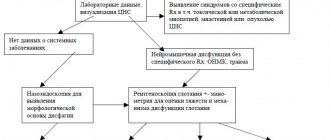
Diagnostics
An examination by a neurologist may reveal loss of sensation or movement caused by compression of the spinal cord. Diagnosis is usually made using magnetic resonance imaging (MRI) of the spine, which can confirm the presence of syringomyelia and determine the exact location of the cystic lesions and the extent of damage to the spinal cord. The most common location of cysts is the cervical or thoracic spine. The least likely location for cysts is the lumbar spine. An MRI of the head can be of diagnostic value to determine the presence of any changes such as hydrocephalus (excess cerebrospinal fluid in the ventricles of the brain). As cystic formations increase, spinal deformity (scoliosis) may occur, which can be easily diagnosed using radiography. The degree of conduction disturbance in syringomyelia is determined using EMG.
Are there any other effects of spinal cord traction?
Tension of the filum terminale not only provokes syringomyelia , but also generates a flexion reflex in the spinal column, leading to scoliosis . To avoid strain on the spinal cord, it forces the lower part of the brain, the so-called tonsils of the cerebellum, to descend down the foramen magnum, through which the cranium communicates with the spinal column. This causes Arnold-Chiari malformation , a disease described 100 years ago, the cause of which remains unknown.
Treatment
Diagnosis and treatment of syringomyelia requires a whole team of medical specialists, including neurologists, radiologists, neurosurgeons, and orthopedists.
Treatment, usually surgical, is aimed at halting the progression of spinal cord damage and preserving functionality. Surgical procedures are often performed when there is spinal cord compression. In addition, surgical procedures are performed to correct deformities or apply various shunts. Surgeries were also performed to implant fetal tissue to close the cystic formations. Surgery results in stable or moderate improvement of symptoms in most patients. Many doctors believe that surgical treatment is necessary only for patients with progressive neurological symptoms. Delaying surgical treatment in cases where the disease progresses can lead to irreversible damage to the spinal cord and severe permanent neurological impairment.
Drug treatments (vasoconstrictors) are often prescribed to reduce swelling around the spinal cord. It is often recommended to avoid strenuous physical activity, which can increase venous pressure. Certain exercises, such as bending your torso forward, can reduce the risk of cystic growths expanding. Life expectancy in patients with progressive symptoms without surgical treatment ranges from 6 to 12 months.
Complications of the disease
In most cases, syringomyelia has a sluggish course, which does not lead to disability of the sick person. However, if the disease progresses and many new cavities form, the risk of death increases and immediate surgical intervention is required. In addition, this disease can cause the following complications:
- Attachment of a secondary infection (lungs, genitourinary system, etc. are affected).
- Suppuration of long-term non-healing wounds, which can lead to sepsis.
- Bulbar paralysis, and as a result, respiratory arrest, which in the absence of immediate help will lead to death.
- The resulting injuries and burns can also cause serious complications in the form of burn disease or other skin lesions.
Recovery and rehabilitation
Despite reports of complete neurological recovery after surgery, most patients experience stable or only moderate improvement in symptoms. Syringomyelia in children is characterized by much less pronounced sensory and pain disturbances than in adults, but a much greater risk of developing scoliosis, which is more favorable for surgical treatment. In addition, syringomyelia does not progress equally in all patients. Some patients, usually those with milder symptoms, experience stabilization of symptoms over the course of a year. A frequent complication of the progression of symptoms is the patient’s need to adapt to life due to impairment or loss of some functions. These adjustments can result in a loss of quality of life. The goal of rehabilitation is to preserve functionality as much as possible using exercises and adaptive equipment, or, especially in the case of syringomyelia in children, activities should be aimed at correcting scoliosis.
Bibliography
- Dr. Miguel B. Royo Salvador (1996), Siringomielia , escoliosis y malformación de Arnold-Chiari idiopáticas, etiología común (PDF). REV NEUROL (Barc); 24 (132): 937-959.
- Dr. Miguel B. Royo Salvador (1996), Platibasia , impresión basilar, retroceso odontoideo y kinking del tronco cerebral, etiología común con la siringomielia , escoliosis y malformación de Arnold-Chiari idiopáticas (PDF). REV NEUROL (Barc); 24 (134): 1241-1250
- Dr. Miguel B. Royo Salvador (1997), Nuevo tratamiento quirúrgico para la siringomielia , la escoliosis , la malformación de Arnold-Chiari , el kinking del tronco cerebral, el retroceso odontoideo, la impresión basilar y la platibasia idiopáticas (PDF). REV NEUROL; 25 (140): 523-530
- M. B. Royo-Salvador, J. Solé-Llenas, J. M. Doménech, and R. González-Adrio, (2005) “Results of the section of the filum terminale in 20 patients with syringomyelia , scoliosis and Chiari malformation .” (PDF). Acta Neurochir (Wien) 147:515–523.
- M. B. Royo-Salvador (1992), “Aportación a la etiología de la siringomielia ,” Tesis doctoral (PDF). Universidad Autónoma de Barcelona.
- M. B. Royo-Salvador (2014), “Filum System® Bibliography” (PDF).
- M. B. Royo-Salvador (2014), “Filum System® Guía Breve.”
Forecast
The prognosis for patients with syringomyelia depends on the underlying cause of the cyst and the type of treatment. Untreated cases of syringomyelia in 35-50% of cases have a prognosis for long-term survival. Patients who underwent bypass surgery for spinal cord injury experienced long-term pain relief and increased survival. Recent studies have shown poor long-term prognosis due to high rates of cyst recurrence in other forms of syringomyelia. Surgical treatment (posterior decompression) for syringomyelia combined with Chiari malformation is considered a fairly effective treatment with a high chance of clinical improvement. For syringomyelia in children, surgical treatment is effective in stabilizing scoliosis.
Material and methods
The study included 40 patients with SM who did not undergo surgery for various reasons (lack of indications, patient refusal to undergo surgery, etc.). The diagnosis of SM was made at the prehospital stage based on MRI data of the spinal cord.
Patients were divided into two groups: without clinical manifestations of SM and with clinical manifestations of the disease.
Based on the analysis of the data obtained from studying these patients, it was planned to determine in which patients the disease progresses over time, how the size of the cyst changes, and does this affect the condition of the patients, what symptoms appear first as the disease progresses?
Until now, there are no clear indications for surgery for SM, so it is also necessary to determine which patients require surgery, and in which case follow-up should be continued?
Criteria for inclusion in the study group:
- the presence of a syringomyelic cyst with a diameter of more than 3 mm, of any location, with a length of more than 2 segments;
— observation period from the moment of diagnosis is more than 3 years;
— presence of at least two MRI studies with a range of 3 years or more;
— absence of mental illness;
— absence of tumors of the central nervous system;
- absence of severe concomitant diseases of the nervous system that could affect the clinical picture.
All patients were interviewed using questionnaires, MRI data were studied, and available medical documentation was analyzed. Particular attention was paid to the clinical manifestations of the disease at the time of its onset, their dynamics and assessment of how the dynamics correlated with the size of the syringomyelic cyst.
Based on the presence of clinical manifestations of SM at the time of diagnosis, patients were divided into two groups: 28 (70%) had neurological symptoms (progressive or stable over time), and 12 (30%) had no neurological symptoms. On control MRIs, the localization of the cyst, its extent, size, and Vacuero index were determined [11], and changes in the size of the syringomyelic cyst over time were studied. Comparisons were made between baseline MRI data and the MRI data that showed the greatest changes. There were 17 (42.5%) men, 23 (57.5%) women. The average age of the patients was 40±21 years. The reasons for the development of SM in these patients are presented in Table. 1 .
Table 1. Distribution of patients depending on the cause of syringomyelia.
The functional status of patients at the time of diagnosis was assessed using the Karnofsky, McCormick, and mJOA scales. The average duration of follow-up for patients was 84±47 months. At the time of detection of SM, surgical treatment was offered to 12 (30%) patients, but they refused it for personal reasons. The remaining 28 (70%) patients were not offered surgery at the time the disease was diagnosed.
see also
- Syringobulbia
| Spinal cord diseases | |
| Inflammatory |
|
| Non-inflammatory |
|