Hydrocephalus (ICD code 10 G91) is a disease of the central nervous system, which is accompanied by the accumulation of excess cerebrospinal fluid in the ventricles or spaces between the membranes of the brain. The disease does not always manifest itself with symptoms of increased intracranial pressure. At the Yusupov Hospital, doctors use innovative methods for diagnosing hydrocephalus using modern devices from leading companies in Europe, the USA and Japan. Neurologists prescribe individual treatment depending on the cause, type and severity of hydrocephalus.
All complex cases of the disease are discussed at a meeting of the expert council with the participation of candidates and doctors of medical sciences, neurologists of the highest category, who are leading experts in the field of diseases of the central nervous system. Patients requiring surgical treatment are consulted by neurosurgeons. Surgical interventions are performed in partner clinics. The staff of the neurology clinic is highly professional and attentive to the wishes of patients.
Causes
Hydrocephalus can be congenital or acquired. Congenital hydrocephalus debuts in childhood. Acquired hydrocephalus occurs under the influence of various provoking factors.
Depending on the mechanism of development of the disease, there are 3 main forms of hydrocephalus:
- occlusive hydrocephalus (ICD 10 code - G91.8);
- communicating (open, disresorptive) hydrocephalus (code G91.0);
- hypersecretory hydrocephalus (code G91.8 - other types of hydrocephalus).
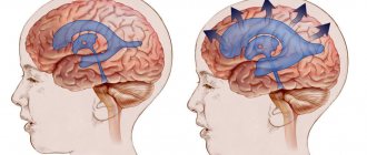
Disruption of the flow of cerebrospinal fluid in occlusive (closed, non-communicating) hydrocephalus occurs due to the closure (occlusion) of the cerebrospinal fluid pathways by a blood clot, a voluminous neoplasm, or an adhesive process that develops after inflammation. If the blockage occurs at the level of the ventricular system (Aqueduct of Sylvius, foramen of Monroe, foramina of Magendie and Luschka), proximal occlusive hydrocephalus occurs. If the block in the path of cerebrospinal fluid flow is at the level of the basal cisterns, a distal form of occlusive hydrocephalus develops. Communicating (open, disresorptive) hydrocephalus occurs when the processes of reabsorption of cerebrospinal fluid are disrupted, due to damage to the structures involved in the resorption of cerebrospinal fluid into the venous bed (Pachionian granulations, arachnoid villi, cells, venous sinuses). Hypersecretory hydrocephalus develops due to excess production of cerebrospinal fluid.
Based on the rate of progression of the disease, there are 3 forms of the disease:
- acute hydrocephalus, when no more than 3 days pass from the first symptoms of the disease to severe decompensation.
- subacute progressive hydrocephalus, developing within a month from the onset of the disease;
- chronic hydrocephalus, which develops within a period of 3 weeks to 6 months.
Depending on the level of cerebrospinal fluid pressure, hydrocephalus is divided into the following groups: hypertensive, normotensive, hypotensive. In hypertensive hydrocephalus, intracranial pressure is increased, in the case of hypotensive hydrocephalus, it is decreased. Normal pressure hydrocephalus (ICD code 10 – G91.2) is accompanied by normal values of cerebrospinal fluid pressure.
Hydrocephalus can develop after traumatic brain injury and various diseases. Hydrocephalus is formed due to the following diseases of the central nervous system:
- brain tumors localized in the brain stem or ventricles;
- acute cerebrovascular accidents;
- subarachnoid and intraventricular hemorrhages;
- encephalopathy of various origins (chronic hypoxic conditions, alcohol intoxication).
Elderly people often develop replacement hydrocephalus. Its cause is atrophy of brain tissue. When the volume of the brain decreases, the vacated space is filled with cerebrospinal fluid. Background diseases that provoke the development of hydrocephalus are arterial hypertension and diabetes mellitus. In case of thrombosis of cerebral vessels, the outflow of cerebrospinal fluid is blocked and hydrocephalus occurs. Intracranial pressure increases and hydrocephalus develops with instability of the cervical spine.
At the Neurology Clinic of the Yusupov Hospital, priority is given to the problems of diagnosis and treatment of acute and chronic hydrocephalus in non-traumatic subarachnoid hemorrhages due to disruption of arteriovenous connections and rupture of arterial vascular aneurysms, post-traumatic hydrocephalus.
Meningoencephalitis of newborns
The most common pathogen is viruses. Intrauterine infection of a baby occurs from a mother suffering from glandular fever, measles, rubella, herpetic infection, and mumps.
The most common symptoms are focal disorders, hyperkinesis, hydrocephalus. Nonspecific manifestations of meningoencephalitis in newborns:
- Eye twitching;
- Difficulty feeding from the breast;
- High fever;
- Intoxication syndrome;
- Vomiting reflex;
- Diarrhea;
- Strabismus;
- Increased heart rate (tachycardia);
- Muscle twitching.
Neurologists define neurological disorders in the form of Kernig's sign (the inability to bring the head to the chest due to stiff neck muscles). Cerebrospinal fluid contains an increased amount of lymphocytes and protein.
Symptoms and diagnosis
Acutely developed occlusive hydrocephalus is manifested by symptoms of increased intracranial pressure:
- headache;
- nausea and vomiting;
- drowsiness;
- congestion of the optic discs;
- symptoms of axial displacement of the brain.
Headache is most pronounced upon awakening in the morning due to an additional increase in intracranial pressure during sleep. This is facilitated by the expansion of cerebral vessels due to the accumulation of carbon dioxide, which is accompanied by blood flow, stretching of the dura mater of the brain in the area of the base of the skull and the walls of blood vessels. Nausea and vomiting worsen and sometimes lead to a decrease in headaches. The most dangerous sign of increased intracranial pressure is drowsiness. It appears on the eve of a sharp and rapid deterioration of neurological symptoms.
With increased pressure in the subarachnoid space, congestion of the optic discs develops. Manifestations of dislocation syndrome are rapid depression of the patient’s consciousness to a deep coma, oculomotor disorders, and forced head position. When the medulla oblongata is compressed, breathing and cardiac activity are inhibited.
The main signs of chronic dysresorptive hydrocephalus are a triad of symptoms: dementia, paresis of both lower extremities and impaired walking, urinary incontinence. The first symptoms appear 3 weeks after a traumatic brain injury, hemorrhage, or meningitis. Initially, the sleep cycle is disrupted - patients become drowsy during the day with disturbances in night sleep. Over time, their overall activity level drops sharply. Patients become spontaneous, lacking initiative, and inert. Short-term memory is impaired, patients lose the ability to remember numbers. In the later stages of the disease, intelligence is impaired, patients cannot take care of themselves, they answer questions asked inadequately, in monosyllables with long pauses.
Walking impairment is manifested by apraxia. The patient can freely pretend to walk or ride a bicycle in a lying position, but in an upright position this ability is immediately lost. A person walks uncertainly, with his legs spread wide apart, and his gait becomes shuffling. In the later stages of hydrocephalus, paresis of the lower extremities develops. The most late and variable symptom is urinary incontinence.
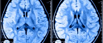
Neurologists at the Yusupov Hospital diagnose occlusive hydrocephalus using computed tomography and magnetic resonance imaging. In chronic dysresorptive hydrocephalus, tomograms reveal a symmetrical expansion of the ventricular system with a balloon-like enlargement of the anterior horns, the subarachnoid fissures are not visualized, and there is a diffuse bilateral change in the white matter of the cerebral hemispheres in the form of a decrease in its density, most often around the lateral ventricles. Computed tomography also makes it possible to clarify the presence and extent of concomitant ischemic brain damage in patients with subarachnoid hemorrhages.
Patients undergo a lumbar puncture and at least 40 ml of cerebrospinal fluid are removed. She is sent to the laboratory for research. Improvement in patients' condition after the procedure is a good predictor of the patient's recovery after surgery.
Meningoencephalitis viral, bacterial, parasitic
The term “meningoencephalitis” includes two nosological forms: “encephalitis” and “meningitis”.
The definition describes the morphological changes that occur against the background of pathology - damage to the white matter and meninges. The pathology is characterized by high mortality, disability, and a large number of disorders. Diagnosis of the symptoms of the disease at the beginning of its development, prevent dangerous consequences, eliminate damage to functional centers. The effectiveness of treatment depends on the cause, pathogen, and extent of the inflammatory focus.
The initial signs of pathology are neurological disorders. Neurologists conduct differential diagnostics that allow one to suspect meningoencephalitis and timely prescribe neuroimaging methods (brain MRI and CT).
Treatment
With an advanced clinical picture of the disease, conservative treatment is ineffective. Patients at the Yusupov Hospital are consulted by a neurosurgeon to decide on immediate neurosurgical intervention. In case of hemorrhage and thrombosis, the operation consists of applying external ventricular drains followed by the introduction of streptokinase into the ventricular cavity - a drug that dissolves blood clots and thereby ensures normal outflow of cerebrospinal fluid.
If the symptoms of chronic hydrocephalus do not progress in patients, they are prescribed diuretics - diacarb, mannitol, furosemide or lasix. To prevent hypokalemia, patients take asparkam. When the symptoms of occlusive hydrocephalus increase, neurosurgeons perform shunt operations. Timely surgical intervention for hydrocephalus allows for the recovery of all patients. Currently, neurosurgeons prefer to perform endoscopic operations for hydrocephalus.
If you have signs of occlusive hydrocephalus, call the Yusupov Hospital. Neurologists take an individual approach to choosing a treatment method.
ICD 10 code for meningoencephalitis
The international classification of the tenth revision identifies the following types of brain inflammation with code “G04”:
- Meningomyelitis;
- Meningoencephalitis;
- Acute ascending myelitis.
The category excludes multiple sclerosis (“G95”), toxic, alcoholic encephalopathy “G31.2”, “G92”.
Classification of meningoecephalitis by course:
- Chronic – long-term development with a slow increase in symptoms;
- Subacute - erased signs of nosology increase over two to three years;
- Acute – rapid progression of symptoms helps early diagnosis;
- Fulminant – rapid cerebral damage causes death.
The difficulties of verifying pathology are complicated by the variety of etiological factors.
Consequences of meningoencephalitis
In addition to high mortality, the disease is characterized by dangerous conditions leading to disability. Severe consequences of inflammation of the white matter and meninges:
- Paralysis of limbs;
- Epileptic seizures;
- Mental retardation in children;
- Hydrocephalus;
- Psychoses;
- Hallucinosis.
The conditions are irreversible. Early verification and competent therapy prevent negative consequences in bacterial types of encephalitis and meningitis. For other forms of nosology, the prognosis is unfavorable.
Manifestations of meningoencephalitis in adults
Various symptoms of the disease in an adult are caused by different pathogens and characteristics of the course. The incubation period of the disease lasts several weeks.
Symptoms of the clinical stage:
- Muscle stagnation;
- Decreased appetite;
- Constant fatigue;
- Headaches without the effectiveness of painkillers.
Inflammatory changes in the meninges lead to meningeal syndrome with special manifestations:
- Nausea;
- Speech disorders;
- Interruptions of heart activity;
- Respiratory disorders.
The occurrence of the described manifestations leads to death. The progression of individual symptoms becomes a cause of disability.