The cerebellum is the part of the brain that controls the musculoskeletal system. The human cerebellum is not directly connected to the receptors that convert the influence of external and internal factors into nerve impulses. This part of the brain interacts with all other parts of the central nervous system. Impulses coming from receptors of tendons, muscles, joint capsules, ligaments, cerebral cortex, subcortical nuclei and medulla oblongata are directed to it.
In turn, impulses depart from the cerebellar medulla in the direction of other parts of the central nervous system. In this way, two-way interaction is carried out. The functions of the cerebellum, located in the human brain, are associated with maintaining balance and coordinating movements of the limbs and other parts of the body.
General information about the organ
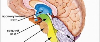
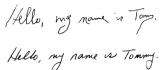
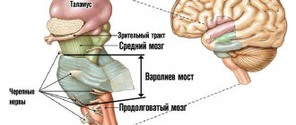
The cerebellum, also called the small brain, is located above the pons and the medulla oblongata. Outwardly it looks like other brain structures with characteristic convolutions, depressions, and folds on the surface. The condition of the organ and the presence of disturbances in its functioning can be clearly seen when performing fast movements - playing a musical keyboard instrument, typing using a typewriter or laptop keyboard, running.
The organ controls the consistency, clarity, and measuredness of actions performed by different parts of the body. It does not cause muscle contractions, but determines the sequence of movements and the time frame for their execution. Information about the desired sequence of actions enters the cerebellum from other parts of the brain and parts of the body.
Feedback information allows you to evaluate the consistency of planned and executed movements. The cerebellum adjusts the motor activity program taking into account deviations. Correction occurs by increasing or decreasing the activation of individual muscles. The organ, in cooperation with the cortical structures of the cerebrum, plans a new movement in advance. Irritation of this part of the brain provokes dilation of the pupils and an increase in blood pressure.
Serious damage to the cerebellar fibers leads to malfunctions in the body:
- Astasia. Impaired ability to hold the body in a standing position.
- Atony. Abnormally decreased tone of skeletal muscles and muscle fibers of internal organs.
- Asthenia. A state of powerlessness, general weakness.
- Ataxia. Impaired fine motor functions, lack of coordination of movements.
Autonomic functions are impaired when the structures that make up the cerebellum are damaged. The mechanisms of natural regulation of breathing, blood circulation, and maintaining normal body temperature are slowed down. Damage to all or part of the cerebellum does not provoke the development of paralysis and has virtually no effect on cognitive abilities.
A person experiences difficulties when it comes to learning and remembering new movements. To understand the questions of what the cerebellum is needed for and what it is responsible for, it is worth remembering that the organ is the control center for gross and fine motor skills. It coordinates complex and simple movements on a subconscious level without involving thought processes and involving the work of consciousness.
Complications after a stroke
If the threatening condition is already over, doctors try not only to maintain life with drugs, but also to minimize the consequences of stroke. The cerebellum is an extremely important functional element. If a hemorrhage occurs and the brain tissue begins to swell, squeezing the cerebellum, then the following functions will be affected first:
- coordination of movements;
- regulation of body balance;
- muscle tone;
- coordination of actions of antagonist muscles.
As a result of ischemia, ataxia may develop, the type of which depends on the location of the lesion. If the cerebellar vermis is affected, static-locomotor ataxia develops. With this disorder, walking is affected, and patients cannot stand independently. If the cerebellar hemispheres are affected, then in this case they speak of dynamic ataxia. In this case, voluntary movements of the limbs are impaired.
Traditionally, when the cerebellar area is damaged, muscle tone decreases and problems with speech appear. Patients begin to speak much more slowly, draw out their words, and sometimes their speech becomes abrupt, as if slogans are being chanted. This leads to a misunderstanding of patients and their needs, but after a while caregivers can find contact with the patient.
- Consequences
- Treatment and prognosis
- Structure of the cerebellum
- Symptoms of tissue damage in the frontal lobe of the brain
- Structure
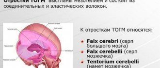
- The tentorium cerebellum, or the cerebellar tent, or tentorium cerebelli.
The symptoms of disorders after a stroke are quite diverse and are not limited to the manifestations described above. Some patients develop hemiparesis, hemihypersthesia, and problems with cognitive functions arise. Headaches often occur and severe migraines develop. Such complications are a consequence of changes after stroke in the fronto-pontocerebellar tract and the occipito-temporo-pontocerebellar tract.
Due to the fact that the cerebellum affects cognitive functions, it is not difficult to predict problems with neuronal networks, as a result of which dementia or dementia develops. Not only the elderly, but also young people who have suffered a stroke can suffer from it. Patients experience the so-called cerebellar cognitive-affective syndrome. This term provides for the following violations:
- problems with spatial thinking;
- speech deficit;
- personality change;
- executive function disorder.
Life expectancy after a stroke and its quality depend on many factors and, first of all, on the timeliness of medical care. How the patient will continue to live and recover largely depends on properly organized home care, good nutrition, and obtaining the necessary medications.
Structure of the cerebellum
The structure of the cerebellum is complex. The organ consists of many parts - lobes of the hemispheres, vermis, cortex, nuclei and legs, which perform certain functions and connect the cerebellum with parts of the brain. The anatomy of the cerebellum is represented by two hemispheres, lying on either side of the vermis. The cerebellar cortex is formed from three areas:
- Archicerebellum. Otherwise called the flocculonodular lobe.
- Paleocerebellum. Bilaterally interacts with the receptors of the spinal cord and the sensorimotor area of the cortex covering the cerebral hemispheres.
- Neocerebellum. Bilaterally interacts with cortical neurons that make up the cerebral hemispheres, receptors of the visual and auditory apparatus.
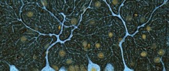
The cortex consists of an inner (granular), outer (molecular) and middle layer formed from Purkinje cells. The layered structure of the organ indicates integration processes that reflect the active interaction of different parts of the brain. The layers of the cerebellum provide its connection with different parts of the central nervous system. The thickness of the cerebellum contains 3 pairs of nuclei - lateral dentate, median tent nuclei, rounded and cork-like in the space between the first two pairs.
The cerebellum is connected to the parts of the brain through the peduncles. The fiber composition of the peduncles located in the cerebellum includes the nuclei of the vestibular nerve, as well as nerve bundles - the gentle and sphenoid. The lower legs connect the organ with a portion of the medulla oblongata. The middle legs in the form of a spacious, protruding part of the cerebellum are located laterally and pass into the pons. The cortical structures of the hemispheres located in the cerebrum control the functioning of the cerebellum using the middle peduncles. The flattened tufts of the upper legs are directed in both directions.
Worm
The structure and functions of the cerebellum cannot be imagined without the vermis, which connects both hemispheres of the organ together. The vermiform structure is composed of white matter nerve fibers. Pathological changes in the tissues of the worm cause ataxia.
Slices
The lobes of the hemisphere located in the cerebellum are delimited by grooves. The worm and hemispheres contain 8 lobules. The anterior, as well as the flocculonodular lobes, located in the cerebellum, are connected by working functions with the brain of the spinal column and the vestibular apparatus. The cerebellar hemispheres receive impulses from muscle and joint tissue receptors. On the front side, a small formation is adjacent to the hemisphere - a shred.
Cores
The nuclei located in the cerebellum control the axial and proximal muscles of the limbs. The idea of movement appears in the associative cortical structures of the brain, then is transmitted through afferent pathways towards the elements of the musculoskeletal system. The program of planned movement is formed in the dentate nucleus and simultaneously in the cerebellar hemispheres, then redirected to the motor centers of the cortex using the thalamic nuclei. The reproduced movement cannot be controlled using the somatosensory feedback system. Damage to the nuclei causes convulsive contraction of the muscles that control the limbs.
Functions of the small brain
The physiology of the cerebellum is mainly related to motor activity and regulation of the autonomic system. Increased heart rate and breathing during sports activities are triggered by the cerebellum’s reaction to increased physical activity. As a result, blood supply to tissues, their saturation with oxygen and nutrients improves. The organ plays a crucial role in maintaining balance. Functions of the cerebellum include:
- consistency, synchronization of movements;
- maintaining balance and a given posture;
- ensuring the accuracy of targeted movements with timing and taking into account clearly marked coordinates;
- interaction of agonist and antagonist muscles;
- regulation of the activity of the autonomic system.
The organ takes part in memorizing motor information. Its activities are associated with the correct execution of physical techniques, poses and exercises in sports.
Symptoms of cerebellar damage
Pathological processes are often accompanied by dysmetria. Limbs cannot move smoothly to a given position due to incorrect force and incorrect direction of movement. Another sign of clinical damage is dysdiadochokinesis, which is expressed in awkward, angular execution of movements that alternate rapidly. To identify the disorder, the patient is asked to tap the fingertips of one hand on the back of the other hand or quickly turn the hand with the palm up and down.
An attempt to reproduce a complex movement leads to fragmentation of the action into separate stages. As a result, the movement is performed intermittently. If the patient tries to touch an object, an intention tremor of the limb occurs. The amplitude of the jitter increases as the distance to the target decreases. Patients with cerebellar dysfunction when walking move their legs too far apart, lose balance and often fall. Words and sentences are spoken at a slow pace, indistinctly. The speech takes on a chanting character.
Hypotonia is a decrease in muscle tone, manifested in combination with a decrease in muscle strength. Muscle resistance to passive movements decreases. Palpation reveals increased muscle softness. A pendulum-shaped knee reflex appears. When the doctor hits the knee tendon below the cap with a special hammer, the leg swings for a while due to weakened muscle tone. Normally, long-term vibrations of the limb do not occur. Other symptoms:
- Dizziness, nausea, headache. The conditions develop due to a breakdown in the functional connection between the vestibular apparatus and the cerebellum.
- Increased fatigue due to physical and mental stress.
- Malfunctions in the functioning of the cardiovascular system.
- Dysfunction of the gland that produces gastric juice.
- Neurological abnormalities.
The location of the cerebellum in the posterior cranial fossa determines the characteristics of symptoms in some diseases. For example, when a tumor forms, the disease in the first stages manifests itself as symptoms of damage to the brain stem or signs of blocking the drainage of cerebrospinal fluid.
Consequences
A brain stem stroke is dangerous not only due to death, but also to a large percentage of patients’ disability. Often people who have had an attack cannot walk or even sit on their own. Their speech is impaired and they become completely dependent on the people caring for them. In addition, patients may experience a second attack at any time or develop dangerous complications. With this course of the disease, patients most often die.
The most common cause of death after a stroke is swelling of the brain stem. The resulting hematoma infringes on the trunk, and as a result, cardiac or respiratory arrest occurs. This complication develops in the first days after the attack.
In a later period, the patient may develop complications such as:
- Pneumonia.
- Vein blockage.
- Infectious diseases of the kidneys and urinary tract.
- Internal bleeding.
- Heart attack.
These are only those complications that can lead to the death of the patient, but there are others that significantly complicate patient care and bring the patient a lot of suffering, among them:
- Bedsores.
- Difficulty swallowing.
- Psycho-emotional disorders.
Those patients who can ambulate a little are often injured as a result of falls. Caring for post-stroke patients requires special patience from relatives. Treatment can last for years and often does not bring the desired results.
Diseases
Diseases that affect the cerebellum - cyst, malignant tumor, malformations and anomalies, abscess, olivopontocerebellar degenerative processes, cerebellar ataxia caused by hereditary factors, known as Pierre Marie's ataxia. Pathologies of the cerebellum cause disorders in the functioning of the musculoskeletal system. Violations affect:
- coordination of movements (dynamic ataxia);
- maintaining balance (static ataxia);
- muscle tone.
The defeat of a specific lobe is reflected in symptoms. For example, damage to the tissue of the flocculo-nodular area leads to atony of the muscles running along the spinal column.
Forecast and prevention of cerebellar dysarthria
The prognosis for regression of the disease is good. But subject to timely and comprehensive treatment.
The first condition is to identify the cause of the disease and direct therapy towards its elimination.
The second condition is speech therapy work aimed at restoring and improving speech skills.
How pronounced the residual symptoms of cerebellar dysarthria will be depends on the degree of damage to the cerebellum. The most difficult disease to treat is one that develops against the background of oncology or hereditary diseases.
Diagnosis of pathologies
Instrumental diagnostics are carried out using ultrasound, MRI and CT methods. Other research methods are cerebrospinal fluid puncture and vascular Dopplerography. To identify disturbances in the functioning of the organ, vestibulometry is performed - a series of neurological tests:
- Romberg's test. The patient is in a standing position, the feet are moved together, the eyes are closed, the upper limbs are extended forward, then spread to the sides.
- Romberg test with complication. The body position is the same as in the usual Romberg test. The only difference is that the feet are on the same line. In this case, the right foot is located in front of the left foot.
- Walking along a straight line. The test is performed with eyes open, then with eyes closed.
- Unterberger's test. The patient walks in one place with his eyes closed, raising his knees high. The angle of deviation from the initial position after taking 50 steps normally does not exceed 30°.
- Finger test. An attempt to touch your own nose with your index finger with your eyes closed ends in a miss or trembling of the finger.
A blood test will show the presence of specific markers of inflammation or stroke. A lumbar puncture is performed if the neurologist suspects an infectious disease occurring in the cerebrospinal fluid.
The cerebellum is an organ that regulates the functions of the musculoskeletal system. Failures in the functioning of this part of the brain are accompanied by multiple disorders of motor activity. Organ dysfunction impairs quality of life. With significant damage to the cerebellar tissue, a person is unable to hold the body in the desired position, walk smoothly, or make precise movements with the arms and legs.
150