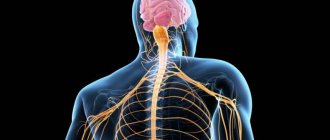
What is the sympathetic nervous system
This is part of the autonomic nervous system, which covers the upper lumbar and thoracic spinal cord, mesenteric nodes, cells of the sympathetic border trunk, and solar plexus. In fact, this section of the nervous system is responsible for the vital activity of cells and maintaining the functionality of the whole organism. In this way, a person is provided with an adequate perception of the world and the body’s reaction to the environment. The sympathetic and parasympathetic departments work together and are structural elements of the central nervous system.
Meaning of MHC
In medicine, the study of ganglion nodes of internal organs is important for the study of diseases associated with impaired organ development. One such disorder is Hirschsprung's disease.
MHC is responsible for feeding organ cells and blood circulation in the internal muscle layers of organs.
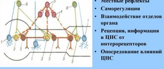
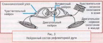
One more important detail. Due to the fact that reflex arcs are present in the intraorgan system, it has the ability to work without the constant “guidance” of the central nervous system. What is a reflex arc? This is a chain of neurons that allows you to quickly transmit a pain signal and receive an immediate response to receptor irritation.
Structure
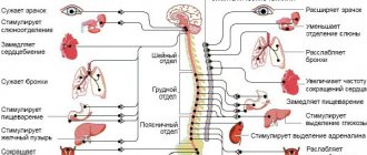
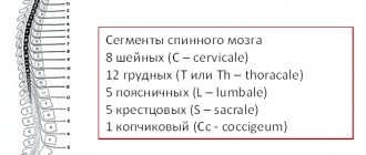
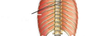
On both sides of the spine there is a sympathetic trunk, which is formed from two symmetrical rows of nerve ganglia. They are connected to each other using special bridges, forming a so-called “chain” with an unpaired coccygeal node at the end. This is an important element of the autonomic nervous system, which is characterized by autonomous operation. To ensure the required physical activity, the design distinguishes the following departments:
- cervical of 3 nodes;
- thoracic, which includes 9-12 nodes;
- area of the lumbar segment of 2-7 nodes;
- sacral, consisting of 4 nodes and one coccygeal.
From these sections, impulses move to the internal organs and support their physiological functionality. The following structural links are distinguished. In the cervical region, the nervous system controls the carotid arteries, in the thoracic region - the pulmonary and cardiac plexuses, and in the peritoneal region - the mesenteric, solar, hypogastric, and aortic plexuses. Thanks to postganglionic fibers (ganglia), there is a direct connection with the spinal nerves.
Prevention
The best treatment for any disease is, of course, prevention. To prevent functional failures in the innervation of a particular organ, experts recommend that people follow the basic principles of a healthy lifestyle:
- give up bad habits – consumption of tobacco and alcohol products;
- get a good night's sleep - at least 8-9 hours of sleep in a ventilated, darkened, quiet room;
- adjust the diet - the predominance of vegetables, various fruits, herbs, cereals;
- compliance with the water regime - taking at least 1.5–2 liters of purified water, juices, fruit drinks, compotes, so that toxins and waste are removed from the tissues;
- daily activity - long walks, visiting the pool, gym, mastering yoga, Pilates.
A person who carefully monitors his health and visits a doctor for an annual medical examination will have calm nerves at any level. Therefore, they know about such problems as sweating, tachycardia, shortness of breath, high blood pressure only by hearsay, from their relatives.
Functions
The sympathetic system is an integral part of human anatomy, located closer to the spine, and is responsible for the proper functioning of internal organs. Controls the flow of blood through vessels and arteries, filling their branches with vital oxygen. Among the additional functions of this peripheral structure, doctors highlight:
- increasing the physiological abilities of muscles;
- decrease in the absorption and secretory capacity of the gastrointestinal tract;
- increased blood sugar and cholesterol;
- regulation of metabolic processes, metabolism;
- providing increased strength, frequency and rhythm of the heart;
- the flow of nerve impulses to the fibers of the spinal cord;
- dilated pupils;
- innervation of the lower extremities;
- increased blood pressure;
- release of fatty acids;
- decreased tone of smooth muscle fibers;
- adrenaline rush in the blood;
- increased sweating;
- stimulation of sensitive centers;
- dilation of the bronchi of the respiratory system;
- decreased saliva production.
How is it formed
The initiation begins in the ectoderm. The main inclusions are formed in the spine, hypothalamus, and brain stem. Peripheral inclusions originate in the lateral vertebrae of the spinal cord. From this moment on, connecting branches are formed that approach the nodes of the sympathetic system. Already from the third week of embryonic growth, neural trunks and nodes are formed from neuroblasts, which serve as a prerequisite for the subsequent formation of internal organs. Initially, the trunks are formed in the intestinal walls, then in the heart tube.
The trunks of the sympathetic system consist of the following nodes - 3 cervical, 12 thoracic, 5 abdominal and 4 pelvic. The plexuses of the heart and carotid artery are formed from the cells of the cervical ganglion. The thoracic nodes trigger the work of the lungs, blood vessels, bronchi, pancreas, and the lumbar nodes are involved in the transmission of nervous reactions to the bladder and male and female genital organs.
The entire process of formation of the sympathetic system takes about four to five months of embryonic growth and fetal development.
Sympathetic and parasympathetic nervous system
The interaction of both structures supports the vital functions of the whole organism; dysfunction of one of the departments leads to serious diseases of the respiratory, cardiovascular, and musculoskeletal systems. The effect is exerted through nerve tissues consisting of fibers that provide excitability of impulses and their redirection to internal organs. If one of the diseases predominates, the choice of high-quality medications is made by the doctor.
- The battery on Android is draining quickly - what to do. Reasons and how to increase battery power for Android
- Triglycerides are elevated - what does this mean in a blood test, causes and treatment
- How to lose weight in your thighs
Any person should understand the purpose of each department, what functions it provides to maintain health. The table below describes both systems, how they can manifest themselves, and what effect they can have on the body as a whole:
| Nervous sympathetic structure | Parasympathetic nervous structure | |
| Department name | Functions for the body | Functions for the body |
| Cervical region | Dilated pupils, decreased salivation | Constriction of pupils, control of saliva secretion |
| Thoracic region | Bronchial dilatation, decreased appetite, increased heart rate | Narrowing of the bronchi, decreased heart rate, increased digestion |
| Lumbar | Inhibition of intestinal motility, production of adrenaline | Possibility of gallbladder stimulation |
| Sacral section | Bladder relaxation | Bladder contraction |
Embryology
The ganglion plate is the embryonic source for the sympathetic system. It is divided into somites, which differentiate into the sympathetic and parasympathetic systems. The sympathetic level includes the cervical and thoracic somites.
During embryogenesis, the peripheral part of the sympathetic nervous system is formed as a result of the migration of sympathetic neuroblasts. Migration occurs along the fibers of the spinal cord segmentally. There are several “waves of migration”. As a result of the first wave, a “primary” sympathetic trunk is formed, represented in the neck by the superior (cranial), inferior (posterior), cervical and stellate ganglia. The second wave arises from the primary one, mainly from the dorsal sections. As a result, a “secondary” sympathetic trunk is formed.
The embryonic source for the parasympathetic system is the ganglion plate. The parasympathetic ganglia of the head are formed by the migration of cells from the midbrain and medulla oblongata. The peripheral parasympathetic ganglia of the alimentary canal originate from two sections of the ganglion plate - the “vagal” and the lumbosacral.
The ganglion plate is the embryonic source for the sympathetic system. It is divided into somites, which differentiate into the sympathetic and parasympathetic systems. The sympathetic level includes the cervical and thoracic somites.
During embryogenesis, the peripheral part of the sympathetic nervous system is formed as a result of the migration of sympathetic neuroblasts. Migration occurs along the fibers of the spinal cord segmentally. There are several “waves of migration”. As a result of the first wave, a “primary” sympathetic trunk is formed, represented in the neck by the superior (cranial), inferior (posterior), cervical and stellate ganglia. The second wave arises from the primary one, mainly from the dorsal sections. As a result, a “secondary” sympathetic trunk is formed.
Differences between the sympathetic and parasympathetic nervous systems
Sympathetic nerves and parasympathetic fibers can be located in a complex, but at the same time they provide different effects on the body. Before contacting your doctor for advice, it is recommended to find out the differences between the sympathetic and parasympathetic systems in structure, location and functionality in order to approximately understand the potential focus of pathology:
- Sympathetic nerves are located locally, while parasympathetic fibers are more discrete.
- Preganglionic sympathetic fibers are short and small, and parasympathetic fibers are often elongated.
- The nerve endings of the sympathetic are adrenergic, while the parasympathetic are cholinergic.
- The sympathetic system is characterized by white and gray connecting branches, but these are absent in the parasympathetic nervous system.
Notes[ | ]
- Sympathetic nervous system // Great Medical Encyclopedia: 30 volumes / chapter. ed. B.V. Petrovsky. — 3rd ed. - M.: Soviet Encyclopedia, 1984. - T. 23: Sucrose - Vascular tone. — 544 p. : ill.
- Sympathetic nervous system // Great Russian Encyclopedia: [in 35 volumes] / ch. ed. Yu. S. Osipov. — M.: Great Russian Encyclopedia, 2004—2017.
- Kandror V.I.
Sympathoadrenal system // Great Medical Encyclopedia: in 30 volumes / chapter. ed. B.V. Petrovsky. — 3rd ed. - M.: Soviet Encyclopedia, 1984. - T. 23: Sucrose - Vascular tone. — 544 p. : ill.
What diseases are associated with the sympathetic system?
With increased excitability of the sympathetic nerves, nervous conditions develop that cannot always be eliminated by self-hypnosis. Unpleasant symptoms are reminiscent of themselves already in the primary form of the pathology and require immediate medical attention. The doctor recommends to beware of the following diagnoses and to consult your doctor in time for effective treatment:
- reflex sympathetic dystrophy syndrome;
- peripheral autonomic failure;
- Raynaud's phenomenon;
- nocturnal enuresis.
Physiology
The neurons of the parasympathetic nervous system are predominantly cholinergic. Although it is known that, along with the main transmitter, postganglionic axons simultaneously release peptides (for example, vasoactive intestinal peptide (VIP)). In addition, in birds, in the ciliary ganglion, along with chemical transmission, electrical transmission is also present.
It is known that parasympathetic stimulation in some organs causes an inhibitory effect, in others - an excitatory response. In any case, the action of the parasympathetic system is opposite to the sympathetic one (the exception is the effect on the salivary glands, where both the sympathetic and parasympathetic nervous systems cause activation of the glands).
The parasympathetic nervous system innervates the iris, lacrimal gland, submandibular and sublingual gland, parotid gland, lungs and bronchi, heart (decreased heart rate and force), esophagus, stomach, large and small intestine (increased secretion of glandular cells). Constricts the pupil, enhances the secretion of sebaceous and other glands, constricts coronary vessels, improves peristalsis. The parasympathetic nervous system does not innervate the sweat glands and blood vessels of the extremities.
Treatment
If the sympathetic nerves are excited, it is necessary to contact the attending physician and promptly begin intensive therapy that can stabilize the general condition of the clinical patient. Pathology can occur under the influence of provoking factors, which must first be identified and eliminated. In order not to bring the situation to a critical limit, and to obtain a positive treatment result, it is recommended to pay attention to the following pharmacological groups:
- benzodiazepine tranquilizers (Phenazepam, Alprazolam);
- neuroleptics (Thioridazine, Periciazine, Azaleptin);
- antidepressants (Amitriptyline, Trazodone, Escitalopram, Maprotiline, Fluvoxamine);
- anticonvulsants (Carbamazepine, Pregabalin).
Anatomy
The sympathetic nervous system is divided into central, located in the spinal cord, and peripheral, which includes numerous nerve branches and nodes connected to each other. The centers of the sympathetic system (Jacobson's spinal center) are located in the lateral horns of the thoracic and lumbar segments. Sympathetic fibers exit the spinal cord from the I-II thoracic to the II-IV lumbar segment.
The peripheral part of the sympathetic nervous system is formed by efferent and sensory neurons with their processes located in the paravertebral and prevertebral nodes remote from the spinal cord.
The sympathetic nervous system is activated during stress reactions. It is characterized by a generalized influence, with sympathetic fibers innervating all organs without exception.
The main transmitter released by preganglionic fibers, as in the parasympathetic nervous system, is acetylcholine, and by postganglionic fibers - norepinephrine.
In mammals, the parasympathetic nervous system is divided into central and peripheral divisions. The central includes the nuclei of the brain, the nuclei of the sacral spinal cord, according to the results of recent studies, no longer belong to the central region.[1]
Literature
- Biological encyclopedic dictionary. - M.: Sov. encyclopedia, 1989. - P. 450
- Nozdrachev A.D. Physiology of the autonomic nervous system. - L-d: Medicine, 1983
- Physiology of the autonomic nervous system. - L-d: Nauka, 1981. - P. 181-211
| Central | |
| Peripheral | |
This page was last edited on November 17, 2019 at 3:53 pm.
- Nozdrachev A.D. Physiology of the autonomic nervous system. - L-d: Medicine, 1983
- Physiology of the autonomic nervous system. - L-d: Nauka, 1981. - P. 181-211
| Nervous system | |
| Normal human anatomy | |
| Central | Brain | Brain structures | Spinal cord |
| Peripheral | Somatic Nervous System Autonomic Nervous System: Sympathetic Nervous System | Parasympathetic Nervous System | Enteric nervous system |
- Nozdrachev A.D. Physiology of the autonomic nervous system. - L-d: Medicine, 1983
- Physiology of the autonomic nervous system. - L-d: Nauka, 1981. - P. 181-211
This page was last edited on September 6, 2019 at 03:37.