Cardiometasympathetic system
The metasympathetic autonomic nervous system, as mentioned, consists of several divisions.
The ganglion system of the heart is already quite well studied, so we can look at how it works. Protection of the heart occurs due to reflex cycles that have a “base” in the intramural ganglia.
- MNS
- Localization of enteral NS
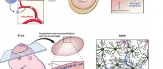
- Microstructure and functional organization
- Parasites, viruses and the central nervous system
- Peripheral department
- What side effect do we get?
- Autonomic ganglia
- Enterometasympathetic system
- What is a synapse?
- Intrauterine development of the central nervous system
- Functions of the metasympathetic division of the ANS
- Features of the structure of the PNS
- Law of Opposites
- Life is change
Thanks to the work of G.I. Kositsky, we know about one very interesting reflex. Stretching of the right atrium always affects the functioning of the right ventricle. He works harder. The same thing happens in the left side of the heart.
When the aorta is stretched, the contractility of both ventricles reflexively decreases. These effects occur through the metasympathetic nervous system. The Goltz reflex occurs when, when struck in the abdomen, the heart may stop contracting for a while. The reaction is associated with the activation of the abdominal nerve, with its afferent part.
The heart rate also decreases under other influences. The Aschner-Dagini reflex is the reaction of the heart when pressure is applied to the eyes. Cardiac arrest also occurs when the vagus nerve is irritated. But with subsequent irritation of the nerve, this effect goes away.
Cardiac reflexes are designed to maintain blood filling of the arteries at a single constant level. The autonomy of the nervous intracardiac system proves the ability of the heart to survive after transplantation. Although all of the cardiac major nerves have been cut, the organ continues to contract.
Features of the metasympathetic system
What makes MNS special? What properties distinguish it from the sympathetic and parasympathetic systems? Scientific data have confirmed assumptions that the system:
- It has its own sensory link and afferent pathway.
- Innervates exclusively the muscles of the internal organs.
- Receives signals from the sympathetic and parasympathetic systems through incoming synapses.
- It has no direct connection with the efferent link of the somatic reflex.
- Those internal organs in which the metasympathetic nervous system (MNS) is disrupted lose their coordinated motor function.
- The network has its own neurotransmitters.
As you can see, the entire nervous system is subordinated to hierarchy. “Senior” departments regulate the work of subordinate connections. The intraorgan network is the “lowest” one, but not the simplest.
Localization of enteral NS
The metasympathetic nervous system, intramural nerve plexuses are found in the heart and all hollow organs, but are more deeply studied using the example of the innervation of the stomach and intestines. In these parts of the gastrointestinal tract, the intragastric and enteric nervous system is represented so abundantly that the number of neurons (108 units) is comparable to the spinal cord. This gives rise to the figurative name of its “abdominal brain.”
acetylcholine, norepinephrine, serotonin, dopamine, nucleotides such as adenosine triphosphate and many neuropeptides: vasoactive intestinal peptide, substance P, somatostatin, enkephalin, gastrin-cholecystokinin-like substance, bombesin, neurotensin and others.
The intramural nervous regulation of intestinal motility is based on the peristaltic reflex. Physiology of the autonomic nervous system It ensures the movement of chyme in the aboral direction and represents a coordinated contraction of the circular muscles at the site of action of a mechanical stimulus (stretching a loop of intestine with chyme or, in experimental conditions, a balloon), and relaxation of the muscle layers caudal to the site of the stimulus on the mucosa. Similar effects are achieved when using chemical irritants.
Based on their responses to a long-term impulse of depolarizing current, all enteric neurons of the intermuscular plexus can be divided into two types: the first is type S and the second is type AN. Neurons of type S respond to this stimulation with a long series of spikes, and neurons of type AN - with only one or two spikes, which are accompanied by a strong and long-lasting (4-20 s) trace hyperpolarization, which is absent in type S. The spike in type S neurons is caused by sodium, and in neurons of the AN type - sodium and calcium conductivity of the membrane.
Microstructure and functional organization
The activity of the MNS is based on a functional module: a cluster of neurons connected in a special way, where oscillatory cells are distinguished, such as sensory neurons, tonic neurons, motor neurons, interneurons. The oscillator cell is the key cell of the module. It is excited spontaneously in a certain rhythm, transmitting action potentials through interneurons to the motor neuron, the axon of which is in contact with the muscle cell. The more active the oscillator cell, the more pronounced the inhibition of the motor neuron becomes. The oscillator-motoneuron system is modulated by:
- afferent neurons acting on the motor neuron through activation through the cholinergic synapse or at its termination (axo-axonal inhibition), removing the inhibitory effect on the muscle cell;
- parasympathetic and sympathetic postganglionic fibers, by influencing interneurons.
Oscillatory cells are extremely stable, and their function does not change under the action of neurotransmitters or ganglion blockers. Through an interneuron, an impulse from an oscillator cell triggers slave cells, which, according to the structure of their connections, represent sequentially organized chains. The sensory elements included in the neural ensemble activate special tonic neurons, causing the appearance of a prolonged discharge in them. In turn, tonic neurons form excitatory or inhibitory synaptic input to slave cells. Activation of a tonic neuron depends on the nature of the connection and can create either supportive excitation or, conversely, inhibition, which determines the direction of the responses of smooth muscles, epithelial cells, endocrine and other elements.
Structure and organization
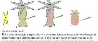
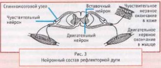
A reflex arc is neurons connected to each other that transmit impulses and receive a response to the excitation of receptors.
The arc of the reflex of the metasympathetic nervous system closes at the level of the microganglion, which is located in the walls of the hollow organs.
Otherwise, the axon enters the paravertebral or prevertebral ganglia, passing in this area to other neurons, or the axons enter the spinal ganglia and pass to other neurons.
Intramural ganglia, located in the thickness of the hollow organs, are the area of localization of the entire MNS. The physiology of the intramural ganglia divides them into sensory, intercalary and effector neurons. Neurons are connected into reflex arcs through synapses. Impulses pass through synapses with the help of neurotransmitters.
The autonomous functional module of the MNS includes the following types of intramural nerve plexuses:
- Efferent neurons (motor) are located in direct contact with the muscle cell of the organ. The efferent neuron has a huge number of small dendrites, the axons of which end in muscle fibers. This nerve plexus is the end point to the effector and is capable of exciting or inhibiting its actions.
- Axons of afferent (sensitive) neurons are capable of switching to neurons of the previous type or reaching the spinal cord, that is, the sensory response coming from microganglia is capable of closing at various levels. Afferent cells have a nucleus and issue from them several large dendrites and axons of varying sizes.
- Interneurons of the metasympathetic nervous system are located in the ganglia - they perform the function of closing the reflex from inside the organ, and their axons come to an end on the processes of nerve cells of the two previous types. These cells are also called intercalary cells and have an oval or curved appearance with a long axon and many dendrites. They have not yet been fully studied and are rare in the NS.
In addition to these three links, the MNS has its own mediators - these are formations that contribute to the transmission of impulses from one neuron to the next. In this section of the ANS, all the mediators that exist in the central nervous system are found.
The central ones are the pituitary peptide and ATP, which are aimed at transmitting the relaxation and tension reactions. The MHC includes the following neurotransmitters: histamine, serotonin, ACH, norepinephrine, dopamine, oligopeptides and gamma-aminobutyric acid.
Each type has its own receptors with neurons. For example, ATP interacts at synapses with purinoceptors, which helps relax the muscles of various organs and systems: the gastrointestinal tract, urinary system, etc. Translation of excitation in neurons is realized by norepinephrine and ACh.
Parasites, viruses and the central nervous system
A large number of “interventionists” live in the brain. Various viruses: cytomegalovirus, herpes virus, papillomavirus. Toxoplasma enters the human body, for example, through cat scratches, resulting in the formation of toxoplasmosis gumma. If someone is diagnosed with epilepsy, can you somehow relate this to the fact that they have worms? Hardly. If a child has epilepsy, will you go to a helminthologist? You 100% won't go. And in vain
It is very important to establish the direction in which to go. It is necessary to carry out certain actions: antiparasitic programs, or at least be examined for the presence of toxoplasma, cytomegalovirus
Important How to diagnose and treat schizoaffective psychosis
Peripheral department
This section is represented by nerve cells and fibers located outside the spinal cord and brain. This part of the visceral nervous system accompanies the vessels, weaving around their wall, and is part of the peripheral nerves and plexuses (related to the normal nervous system). The peripheral department also has a clear division into the sympathetic and parasympathetic parts. The peripheral department ensures the transfer of information from the central structures of the visceral nervous system to the innervated organs, that is, it carries out the implementation of what is “planned” in the central autonomic nervous system.
Sympathetic department
Represented by the sympathetic trunk, located on both sides of the spine. The sympathetic trunk is two rows (right and left) of nerve ganglia. The nodes are connected to each other in the form of bridges, moving between parts of one side and the other. That is, the trunk looks like a chain of nerve lumps. At the end of the spine, two sympathetic trunks unite into one unpaired coccygeal ganglion. In total, there are 4 sections of the sympathetic trunk: cervical (3 nodes), thoracic (9-12 nodes), lumbar (2-7 nodes), sacral (4 nodes and plus one coccygeal).
The cell bodies of neurons are located in the area of the sympathetic trunk. Fibers from the nerve cells of the lateral horns of the sympathetic part of the central part of the autonomic nervous system approach these neurons. The impulse can switch on the neurons of the sympathetic trunk, or it can transit and switch on intermediate nodes of nerve cells located either along the spine or along the aorta. Subsequently, the fibers of the nerve cells, after switching, form weaves in the nodes. In the neck area it is the plexus around the carotid arteries, in the chest cavity it is the cardiac and pulmonary plexuses, in the abdominal cavity it is the solar (celiac), superior mesenteric, inferior mesenteric, abdominal aortic, superior and inferior hypogastric. These large plexuses are divided into smaller ones, from which autonomic fibers move to the innervated organs.
Parasympathetic Division
Represented by nerve ganglia and fibers. The peculiarity of the structure of this department is that the nerve nodes in which the impulse switches occur are located directly next to the organ or even in its structures. That is, the fibers coming from the “last” neurons of the parasympathetic department to the innervated structures are very short.
From the central parasympathetic centers located in the brain, impulses go as part of the cranial nerves (oculomotor, facial and trigeminal, glossopharyngeal and vagus, respectively). Since the vagus nerve is involved in the innervation of internal organs, its fibers reach the pharynx, larynx, esophagus, stomach, trachea, bronchi, heart, liver, pancreas, and intestines. It turns out that most internal organs receive parasympathetic impulses from the branching system of just one nerve: the vagus.
From the sacral sections of the parasympathetic part of the central visceral nervous system, nerve fibers go as part of the pelvic splanchnic nerves and reach the pelvic organs (bladder, urethra, rectum, seminal vesicles, prostate gland, uterus, vagina, part of the intestine). In the walls of organs, the impulse is switched in the nerve ganglia, and short nerve branches are in direct contact with the innervated area.
Metasympathetic division
It stands out as a separate separately existing department of the autonomic nervous system. It is detected mainly in the walls of internal organs that have the ability to contract (heart, intestines, ureter and others). It consists of micronodes and fibers that form a nerve plexus in the thickness of the organ. The structures of the metasympathetic autonomic nervous system can respond to both sympathetic and parasympathetic influences. But, in addition, their ability to work autonomously has been proven. It is believed that the peristaltic wave in the intestine is the result of the functioning of the metasympathetic autonomic nervous system, and the sympathetic and parasympathetic divisions only regulate the force of peristalsis.
Criticism of the conditional allocation of the MNS
Professor Pavel Aleksandrovich Motavkin argues in his scientific work (Motavkin P.A. Course of lectures on histology. - “Medicine DV”, 2007 - P. 168) critical views in the research world in relation to the theory of the MNS by the fact that in the metasympathetic department of the autonomic The nervous system includes the intramural ganglia of the digestive and respiratory organs, heart, ureter and others.
This nervous system, according to physiologists, is quite peripheral, independent and independent of the functionality of the central nervous system. Its functioning is possible due to autonomous reflexes, which are based on personal neurons of the ANS, which do not interact with the central cells.
The expressed independence of the MNS is real if in the intramural region the presence of motor neurons that are not controlled by preganglionic tissues is supported by evidence.
To date, 6 types of cells have been discovered in this area, differing in mediator direction and morphology. One cannot discount the potential fact that among these six species there are autonomous effector cells that are independent of the CNS.
However, the dominant belief in science so far places intramural neurons in the parasympathetic division.
What side effect do we get?
Our nervous system forgets how to slow down on its own. What does this mean?
When external disturbance or change occurs, we change and cannot stop for a long time. We get used to “drinking the problem down” or “smoking.”
If we don't have access to a drug (alcohol), we become overstimulated. The motors of the psyche continue to work at full speed when this is no longer necessary. Imagine, the conflict has long ended or the problem has been resolved, but you continue to be in an overexcited state for several hours or even several days (this is quite realistic).
And all because your psyche has forgotten how to use “inhibition”.
This is also the difficulty of getting out of addiction.
If you want to learn even more about this, read my previous article “The Effects of Alcohol on the Nervous System.”
- Now it seems to you that in order to “relieve tension” (slow down), you need to “drink.”
- By drinking, you even more forget how to “slow down” and calm down naturally without the use of alcohol.
- The situation is getting worse.
Autonomic ganglia
Ganglia are nerve nodes. The autonomic ganglia help distribute electrical signals efficiently. One or more preganglionic nerve fibers approach one ganglion, which transmit signals from the “higher” system. And postganglionic neurons depart from the ganglion, transmitting excitation or inhibition further along the network. This universal system allows you to fully control all processes in the body.
In the ganglia of the excitatory nerve network, the presynaptic fiber regulates up to 30 nerve cells connected to the ganglion. And in the parasympathetic there are only 3 or 4 neurons.
Vegetative nodes are found in all tissues and organs, as well as in the glands of internal and external secretion. The neurons of the MHC network are extremely diverse, but each consists of an axon, a nucleus, and a dendrite.
Important Trimethine (trimethadione): instructions for use, reviews, analogues
Dendrite - from Latin - tree-like. From the name it is clear that this part of the neuron transmits signals through a highly branched network of small fibers. In the enteric system, for example, each neuron has many dendrites.
Some fibers have a myelin sheath, which improves conductivity and speeds up the signal.
Types of MNS
There are several systems. They are divided depending on the location of the microganglia:
- cardiometasympathetic system;
- vesiculometasympathetic;
- enterometasympathetic;
- urethrometasympathetic;
- ganglion system of the uterus.
It is known that the parasympathetic and sympathetic systems interact with the organ ganglion system and correct their functioning when necessary. And also many organs have intersecting reflexes. For example, the Goltz reflex.
Enterometasympathetic system
The enteric nervous system is a unique mechanism where thousands of neurons are completely coordinated with each other. This mechanism, created by nature, is rightfully considered the second human brain. Because even if the vagus nerve, which is connected to the brain, is damaged, the system continues to perform all its functions, namely: digesting food and absorbing nutrients.
But it turns out that the digestive tract is not only responsible for the digestion of food, but, according to the latest data, also for the emotional background of a person. It has been established that the intestines produce 50% of dopamine, the hormone of joy, and about 80% of serotonin. And this is even more than is produced in the brain. Therefore, the intestines can safely be called the emotional brain.
In the enteric autonomic metasympathetic system, several types of neurons are distinguished:
- primary sensory afferents;
- ascending and descending interneurons;
- motor neurons.
Motor neurons, in turn, are divided into moving muscles, excitatory and inhibitory.
Metasympathetic nervous system - what is it?
This section of the nervous system was described for the first time by A. D. Nozdrachev.
The term is relatively new - many old textbooks do not mention it. It was noted that any organ has its own motor skills and pace of activity. The ganglia of the metasympathetic network are located inside the muscular walls of organs, as a result of which this system is intraorgan.
The MNS differs in that it innervates only those organs that have autonomous motor activity. It contains personal sensory formations that transmit impulses into their neural networks with information about the state of a particular structure in the body. Such signals come from the MNS and the central nervous system, although the former works independently of brain impulses.
The location of this structure has its own reflex arcs. But the metasympathetic division of the autonomic nervous system does not directly contact the nerve arches of the somatic nervous system - synaptic outputs are carried out only through the sympathetic and parasympathetic network.
The work of this NS is carried out using reflex arcs located in the septa of hollow organs. The intramural section of all visceral organs is equipped with independent motor activity, which is provided by the MNS.
The areas of this system are named according to their location: cardiometasympathetic - in the cardiovascular system, enteromestasympathetic - in the gastrointestinal tract, and so on.
The enterometasympathetic system can be called the human second brain. It is here that about 45% of dopamine and 80% of serotonin are released - more than in the brain. This system of the alimentary tract, even with pathologies of the vagus nerve, continues its activity of digesting the food eaten and processing useful substances. This entire process is also regulated by the metasympathetic network.
What is a synapse?
A synapse is a special structure that ensures the transmission of a nerve impulse from a nerve fiber to another nerve fiber or nerve cell, and in order for a nerve fiber to be affected by a receptor cell (the area of contact between nerve cells and another nerve fiber), two nerve cells are required .
A synapse is a small section at the end of a neuron. With its help, information is transferred from the first neuron to the second. The synapse is located in three areas of nerve cells. Also, synapses are located in the place where the nerve cell enters into connection with different glands or muscles of the body.
Intrauterine development of the central nervous system
The importance of the nervous system is to ensure the functioning of internal organs, intellectual function, motor skills, sensitivity and reflex activity. The child’s central nervous system develops not only during the prenatal period, but also during the first year of life. Ontogenesis of the nervous system begins from the first week after conception.
The basis for brain development is formed already in the third week after conception. The main functional nodes are identified by the third month of pregnancy. By this time, the hemispheres, trunk and spinal cord have already been formed. By the sixth month, the higher parts of the brain are already better developed than the spinal part.
By the time a baby is born, the brain is the most developed. The size of the brain in a newborn is approximately an eighth of the child’s weight and ranges from 400 g.
The activity of the central nervous system and PNS is greatly reduced in the first few days after birth. This may include an abundance of new irritating factors for the baby. This is how the plasticity of the nervous system manifests itself, that is, the ability of this structure to be rebuilt. As a rule, the increase in excitability occurs gradually, starting from the first seven days of life. The plasticity of the nervous system deteriorates with age.
Functions of the metasympathetic division of the ANS
The MHC is a relatively independent department of interconnected neurons that regulates the functioning of internal organs:
- preserves the absorption capacity of the intestines and intestines;
- regulates heart contraction;
- affects contractions of the ureter, cervix, etc.;
- innervates and controls the activity of all organs in standard conditions and in pathologies of the spinal cord;
- provides constant regulation of organ functions.
MS affects the activity of cardiac muscle tissue, smooth muscle structures and glandular epithelium. Areas or strips of the uterus, bladder, and gall bladder contract with the range and amplitude that is characteristic of each organ.
Microglia of this system are found in the stomach, kidneys, intestines, bronchi and other areas. The structure plays a significant role in lesions and dysfunctions of organs. In case of damage to the spinal nerve canal, many internal organs undergo failure and are restored after six months due to the presence of the metasympathetic network.
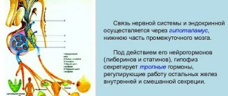
All reflexes of the system are controlled by the autonomic centers of the telencephalon - the striatum, as well as the hypothalamus, the structure of the midbrain.
The gastrointestinal tract, more clearly than other systems, displays the main characteristics of the MNS - it is there that it has been studied to a greater extent. Here it regulates complex intestinal motility - peristalsis.
The structure of the gastrointestinal tract includes various formations - muscle tissue, mucosal surfaces, endocrine and lymph nodes.
The local MHC regulates all these structures with the participation of other parts of the autonomic system. Thanks to this, the functionality of the gastrointestinal tract is not greatly affected by disruptions in the activity of parasympathetic and sympathetic nerve tissues.
Meaning of MHC
The main role of the entire ANS is the preservation and support of homeostasis, that is, the constancy of the functioning of the human body.
In medicine, studies of the metasympathetic nervous system are important for the study of various pathologies associated with organ dysfunction. In the cardiovascular and gastrointestinal systems, the independent MHC is of important physiological importance in stabilizing the dynamics of activity and coordinated innervation of all organs. The metasympathetic system is an intermediary between other parts of the ANS and organ tissues, since it is involved in the transmission of impulses emanating from the extraorganic NS.
The presence of “independent” functions of nervous regulation makes it easier for the central nervous system to process numerous signals coming from internal systems and organs. This doubles the correct regulation of functions.
Due to the reflex arcs of this autonomous structure, internal organs are able to work without the direct participation of the central nervous system. Most organs continue to function even after the sympathetic and parasympathetic nerve connections are cut or removed from the body.
Such independence is the result of the presence of a metasympathetic department, which has all the necessary independent links.
An important unit of the entire network is the oscillator cell, which sends its impulses to the motor neuron. This is how it interacts with muscle cells and regulates muscle activity.
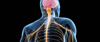
Features of the structure of the PNS
Thanks to the PNS, the activity of the entire human body is regulated. The PNS consists of cranial and spinal neurons and fibers that form ganglia.
The human peripheral nervous system has a very complex structure and functions, so any slightest damage, for example, damage to blood vessels in the legs, can cause serious disruptions to its functioning. Thanks to the PNS, all parts of the body are controlled and the vital functions of all organs are ensured. The importance of this nervous system for the body cannot be overestimated.
The PNS is divided into two divisions - the somatic and autonomic PNS systems.
The somatic nervous system performs double duty - collecting information from the sensory organs, and further transmitting this data to the central nervous system, as well as ensuring the motor activity of the body by transmitting impulses from the central nervous system to the muscles. Thus, it is the somatic nervous system that is the instrument of human interaction with the outside world, as it processes signals received from the organs of vision, hearing and taste buds.
The autonomic nervous system ensures the functions of all organs. It controls the heartbeat, blood supply, and breathing. It contains only motor nerves that regulate muscle contraction.
Important Midbrain
To ensure the heartbeat and blood supply, the efforts of the person himself are not required - this is controlled by the autonomic part of the PNS. The principles of the structure and function of the PNS are studied in neurology.
Mediators MNS
Neurotransmitters are substances that help transmit impulses from one neuron to another. The mediators of the metasympathetic nervous system are as follows:
- histamine;
- serotonin;
- adenosine triphosphoric acid;
- acetylcholine;
- somatostanin;
- catecholamines.
In total, about 20 mediators and modulators in the neural network were discovered in laboratory conditions. A mediator such as acetylcholine, which belongs to the group of catecholamines, is a mediator of the sympathetic system, that is, it helps transmit the excitation signal. An excess of catecholamines in the body leads to overexcitation of the central nervous system. Heart failure often begins due to constant stress and releases of norepinephrine. Therefore, the body needs a restorative parasympathetic system.
Mediators such as pituitary peptide and ATP are designed to transmit the impulse of relaxation and recovery. Parasympathetic centers are located in the autonomic nuclei of the cranial nerves.
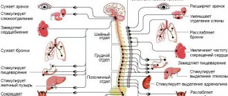
Law of Opposites
Ensuring the existence of the human body requires the ability to adapt. Different situations may require opposite actions. For example, when it’s hot you need to cool down (sweating increases), and when it’s cold you need to warm up (sweating is blocked). The sympathetic and parasympathetic sections of the autonomic nervous system have opposite effects on organs and tissues; the ability to “turn on” or “turn off” one or another influence allows a person to survive. What effects does activation of the sympathetic and parasympathetic divisions of the autonomic nervous system cause? Let's find out.
Sympathetic innervation provides:
- dilation of the pupil, widening of the palpebral fissure, “protrusion” of the eye forward;
- decreased salivation, saliva becomes thick and viscous;
- increased heart rate;
- increased blood pressure;
- dilatation of the bronchi, decreased mucus secretion in the bronchi;
- increased breathing rate;
- slowing down intestinal motility;
- decreased secretion of digestive glands (stomach, pancreatic juice);
- stimulation of ejaculation;
- vasoconstriction;
- raising of skin hairs (“goose bumps”).
Parasympathetic innervation acts as follows:
- constriction of the pupil, narrowing of the palpebral fissure, “retraction” of the eyeball;
- increased salivation, there is a lot of saliva and it is liquid;
- reduction in heart rate;
- decreased blood pressure;
- narrowing of the bronchi, increased mucus in the bronchi;
- decreased breathing rate;
- increased peristalsis up to intestinal spasms;
- increased secretion of the digestive glands;
- causes erection of the penis and clitoris.
There are exceptions to the general pattern. There are structures in the human body that have only sympathetic innervation. These are the walls of blood vessels, sweat glands and the adrenal medulla. Parasympathetic influences do not apply to them.
Typically, in the body of a healthy person, the influences of both departments are in a state of optimal balance. There may be a slight predominance of one of them, which is also a variant of the norm. The functional predominance of excitability of the sympathetic department is called sympathicotonia, and the parasympathetic department is called vagotonia. Some periods of human age are accompanied by an increase or decrease in the activity of both departments (for example, activity increases during adolescence, and decreases during old age). If there is a predominant role of the sympathetic department, then this is manifested by sparkle in the eyes, wide pupils, a tendency to high blood pressure, constipation, excessive anxiety and initiative. The vagotonic effect is manifested by narrow pupils, a tendency to low blood pressure and fainting, indecision, and excess body weight.
Thus, from the above it becomes clear that the autonomic nervous system with its oppositely directed sections ensures human life. Moreover, all structures work in harmony and coordination. The activity of the sympathetic and parasympathetic departments is not controlled by human thinking. This is exactly the case when nature turned out to be smarter than man. We have the opportunity to engage in professional activities, think, create, leave ourselves time for small weaknesses, being confident that our own body will not let us down. Internal organs will work even when we are resting. And this is all thanks to the autonomic nervous system.
Educational film “The Autonomic Nervous System”
https://youtube.com/watch?v=BT6h2aiwVg4