The striatum helped diagnose schizophrenia automatically
Ang Li et al.
/ Nature Medicine, 2020 Scientists from China have developed an algorithm that, based on the characteristics of striatal activity, diagnoses schizophrenia with an accuracy of more than 80 percent.
- Symptoms of basal ganglia dysfunction
- Diagnosis and prognosis of pathology
- What are the basal ganglia?
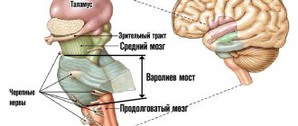
- Brain stem
- How does the midbrain develop?
- Physiology
- Pathologies in case of damage
- Functions
- Pathological conditions of the basal ganglia
This analysis also allows one to predict the patient’s susceptibility to antipsychotic drug therapy.
Abnormalities in the functioning of the striatum correlate with the functioning of the dopaminergic system and the expression of genes that are associated with the risk of developing schizophrenia. in the journal Nature Medicine
.
Diagnosing schizophrenia is complicated by the very wide range of symptoms and the fact that the causes of this disease are poorly understood. The main hypothesis about the mechanism of schizophrenia is an imbalance of dopamine balance.
Dopamine is a neurotransmitter with a wide range of functions and is closely linked to the reward system in the brain.
For schizophrenia, antipsychotics are mainly prescribed, which block dopamine receptors and reduce the activity of this neurotransmitter.
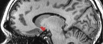
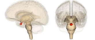
Scientists believe that the structure that plays a central role in the development of schizophrenia is the striatum, a group of basal ganglia of the cerebral hemispheres. In patients with this structure, dopaminergic activity is often increased.
An Li and his colleagues from the Chinese Academy of Sciences decided to use abnormalities in the functioning of the striatum as a marker for diagnosing schizophrenia. To do this, fMRI of the brain was performed on 560 patients with schizophrenia and 540 healthy people.
Then the scientists trained a classifier based on the support vector machine to determine the diagnosis (healthy or sick with schizophrenia) based on three characteristics of the striatum: the amplitude of low-frequency oscillations, the activity of connections within the striatum and with external structures of the brain (more than 12 thousand elements in total).
In addition, the scientists cross-validated the algorithm, testing it on data from different medical centers and different MRI machines.
The striatal abnormality coefficient was also calculated for patients with other mental illnesses.
The scientists tested whether abnormalities in the striatum were associated with increased activity of the dopaminergic system and the expression of genes that are associated with schizophrenia.
For this purpose, we used the results of positron emission tomography and single-photon emission computed tomography of healthy volunteers. Data on the activity of 43 genes that are associated with schizophrenia were also analyzed.
The spatial expression of dopaminergic system markers and selected genes was compared with the distribution of amplitudes of low-frequency oscillations in the striatum.
Researchers have found a number of differences in striatal function between people with schizophrenia and healthy volunteers. For example, the amplitude of low-frequency oscillations was increased in patients, and the connections of the striatum with other areas also differed (compared to the control group).
The algorithm, which was trained on this data, distinguished patients with schizophrenia with an accuracy of more than 80 percent. The rate did not differ from control groups for all mental illnesses studied except for bipolar disorder—people with this diagnosis had a lower rate than controls.
Ang Li et al. / Nature Medicine, 2020
Differences in external projections of the striatum of patients with schizophrenia and healthy people. Blue – projections in patients were less pronounced, red – stronger
Ang Li et al. / Nature Medicine, 2020
The rate of striatal abnormality varied significantly among patients. It turned out that this indicator is associated with susceptibility to treatment with antipsychotic drugs. This observation may help tailor therapy individually, since often antipsychotic resistance is discovered only after failure of several drugs and their concentrations.
Both markers of the dopaminergic system and genes that are associated with the risk of schizophrenia were active in the same areas of the striatum in which the amplitude of low-frequency oscillations was high. This means that both of these mechanisms may underlie changes in striatal activity in schizophrenia.
Scientists have long been searching for the structural and molecular features of the brain that are characteristic of schizophrenia. A total of 413 genes have been identified that are associated with this disease.
In addition to the striatum, the corpus callosum is suspected of participating in the development of schizophrenia, and this disease also causes changes in a number of other parts of the brain.
You can read in detail about schizophrenia, its causes, symptoms and treatment in the material “Inheritance of Madness” (here is its continuation).
Alisa Bakhareva
Consequences of basal ganglia pathologies
Further prognosis depends on a number of factors: gender, age, degree of development of the disease, genetic characteristics, physiology of the body. Each case is individual. But the statistics are not reassuring - on average, more than half of the pathological conditions of the basal ganglia have an unfavorable course.
Symptoms of the lesion accompany a person in later life and become causes of disability. The progression of the disease can be stopped with appropriate medications, physiotherapeutic procedures, sports exercises, and a lack of stress.
The body's adaptive forces are great. Correctly selected rehabilitation techniques are necessary. With them, the patient's life can become complete. Or reach a higher quality level.
Symptoms of basal ganglia dysfunction
When the basal ganglia are damaged or dysfunctional, symptoms associated with impaired coordination and precision of movements occur. Such phenomena are called the collective concept of “dyskinesia,” which, in turn, is divided into two subtypes of pathologies: hyperkinetic and hypokinetic disorders. Symptoms of basal ganglia dysfunction include:
- akinesia;
- impoverishment of movements;
- voluntary movements;
- slow movements;
- increase and decrease in muscle tone;
- muscle tremor in a state of relative rest;
- desynchronization of movements, lack of coordination between them;
- poor facial expressions, scanned language;
- erratic and arrhythmic movements of small muscles of the hand or fingers, the entire limb or part of the whole body;
- pathological postures unusual for the patient.
Most manifestations of the pathological functioning of the basal ganglia are based on disturbances in the normal functioning of the neurotransmitter systems of the brain, in particular the dopaminergic modulating system of the brain. In addition, however, the causes of symptoms are previous infections, mechanical brain injuries or congenital pathologies.
Hyperkinesis
The disease is caused by uncontrolled spontaneous movements of a muscle group. The disease occurs against the background of damage to the nerve cells of the basal ganglia, in particular the caudate body and the internal capsule. Provoking factors:
- cerebral palsy;
- intoxication;
- stress;
- encephalitis;
- congenital pathologies;
- head injuries;
- diseases of the endocrine system.
General symptoms:
- involuntary muscle contraction;
- tachycardia;
- frequent blinking;
- closing the eyes;
- facial muscle spasms;
- tongue sticking out;
- pain in the lower abdomen.
Complications of hyperkinesis lead to limited joint mobility. The disease is incurable, but with the help of medications and physical therapy, symptoms can be reduced and the person’s condition can be alleviated.
Diagnosis and prognosis of pathology
In addition to neurologists, diagnostics is carried out by doctors from other offices (functional diagnostics). The main methods for identifying diseases of the basal ganglia are:
- analysis of the patient’s life, his anamnesis;
- objective external neurological examination and physical examination;
- magnetic resonance and computed tomography;
- study of the structure of blood vessels and the state of blood circulation in the brain;
- Ultrasound;
- visual methods for studying brain structures;
- electroencephalography;
Prognostic data depends on many factors, such as gender, age, general constitution of the patient, the moment of the disease and the moment of diagnosis, his genetic predispositions, the course and effectiveness of treatment, the pathology itself and its destructive properties. According to statistics, 50% of diseases of the basal ganglia have an unfavorable prognosis. The remaining half of cases have a chance for adaptation, rehabilitation and normal life in society.
Didn't find a suitable answer? Find a doctor and ask him a question!
Tourette's syndrome
Tourette's disease is a psychogenic disorder of the nervous system. The disease is characterized by motor and vocal tics that are uncontrollable.
Causes:
- damage to the brain structure due to oxygen deficiency or during childbirth;
- maternal alcoholism during pregnancy;
- severe toxicosis in the first trimester of pregnancy, which negatively affects the unborn child.
Symptoms
Simple tics are short twitches of one muscle group. These include:
- twisting of the mouth;
- frequent blinking;
- involuntary eye movements;
- sniffling;
- twitching of the head.
Complex tics include a variety of actions performed by several muscle groups:
- pronounced gestures;
- hyperkinesis;
- eccentric gait;
- jumping;
- copying people's movements;
- body rotation;
- sniffing surrounding objects.
Vocal tics:
- coughing;
- shouts;
- barking;
- repetition of phrases;
- grunting.
Before an attack, the patient experiences tension and itching in the body; after the attack, this condition disappears. Drug therapy does not completely cure Tourette's syndrome, but it can reduce symptoms and reduce the frequency of tics.
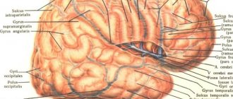
What are the basal ganglia?
The basal ganglia of the brain are functionally and anatomically related accumulations of gray matter in the deep parts of the brain. These structures are deepened into the white matter, which functions as an information transmitter. Even in the embryo, the basal ganglia develop from the ganglion tubercle, then forming into mature brain structures that perform strictly specific functions in the nervous system.
Important Gray matter of the brain: what it consists of and what it is responsible for
The basal ganglia are located at the base of the brain, lateral to the thalamus. Anatomically highly specific nuclei are part of the forebrain, which is located on the border of the frontal lobes and the brainstem. Often, by the term “subcortex”, experts mean precisely the set of basal ganglia of the brain.
Anatomists distinguish three concentrations of gray matter:
- Striped body. This structure means a set of two not entirely differentiated parts: The caudate nucleus of the brain. It has a thickened head, forming in front one of the walls of the lateral ventricle of the brain. The thin tail of the nucleus is adjacent to the bottom of the lateral ventricle. The caudate nucleus also borders the thalamus.
- Lenticular nucleus. This structure runs parallel to the previous accumulation of gray matter and, closer to the end, merges with it, forming the striatum. The lenticular nucleus consists of two white layers, each of which has its own name (globus pallidus, shell).
Corpus striatum received its name due to the alternating arrangement of white stripes on its gray matter. Recently, the lenticular nucleus has lost its functional meaning, and it is called exclusively in a topographical sense. The lenticular nucleus, as a functional compilation, is called the striopallidal system.
- The fence or claustrum is a small thin gray plate located near the shell of the striatum.
- Amygdala. This core is located under the shell. This structure also belongs to the limbic system of the brain. The amygdala usually means several separate functional formations, but they were combined due to their close location. This area of the brain has multiple connections with other brain structures, in particular with the hypothalamus, thalamus and cranial nerves.
The concentration of white matter is:
- Internal capsule - white matter between the thalamus and the lentiform nucleus
- Outer capsule - white substance between the lentil and the fence
- The outermost capsule is the white substance between the enclosure and the insula.
The internal capsule is divided into 3 parts and contains the following pathways:
Front leg:
- Frontothalamic tract - connection between the frontal cortex and the mediadersal nucleus of the thalamus
- Frontopontine tract - connection between the frontal cortex and the pons
Knee:
Corticonuclear tract - connection between the nuclei of the motor cortex and the nuclei of the motor cranial nerves
Rear leg:
- Corticospinal tract - conducts motor impulses from the cerebral cortex to the nuclei of the motor horns of the spinal cord
- Thalamo-parietal fibers - Axons of thalamic neurons are connected to the postcentral gyrus
- Temporo-parieto-occipital-pontine fasciculus - connects the pontine nuclei with the lobes of the brain
- Auditory radiance
- Visual radiance
Structure and location
The basal ganglia of the brain is a collection of gray matter in the white matter, located at the base of the brain and part of its anterior lobe. As we can see, the gray matter not only forms the hemispheres, but is also located in the form of separate clusters called ganglia. They have a close connection with the white matter and cortex of both hemispheres.
The structure of this region is based on a slice of the brain. It includes:
- amygdala;
- striatum (composed of the caudate nucleus, globus pallidus, putamen);
- fence;
- lenticular nucleus.
Between the lenticular nucleus and the thalamus there is a white substance called the internal capsule, and between the insula and the fence is the external capsule. Recently, a slightly different structure of the subcortical nuclei of the brain has been proposed:
- striatum;
- several nuclei of the midbrain and diencephalon (subthalamic, pedunculopontine and substantia nigra).
Together they are responsible for motor activity, motor coordination and motivation in human behavior. This is all that can be said for sure about the function of the subcortical nuclei. Otherwise, they, like the brain as a whole, are poorly understood. Absolutely nothing is known about the purpose of the fence.
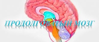
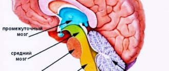
Brain stem
The brainstem consists of the medulla oblongata, pons, and midbrain and contains motor and sensory nuclei that perform motor and sensory functions for the face and head in the same way that the spinal cord performs these functions for the neck, trunk, and limbs. At the same time, the brain stem performs many special functions (including control functions: respirationcardiovascular systemGastrointestinal tractmany stereotypical body movementsbalanceeye movements) and serves as a hub for “command signals” from higher-lying centers. The vestibular and reticular nuclei of the brainstem play an important role in controlling body movements and balance.
Reticular nuclei
.
In Fig. Figure 14-3B shows the location of the reticular nuclei. They are divided into the reticular nuclei of the pons and the reticular nuclei of the medulla oblongata. These two systems of nuclei function antagonistically in relation to each other: the pons nuclei excite anti-gravity muscles
,
the nuclei the medulla oblongata inhibit them
.
Pontine reticular nuclei
transmit excitatory signals to the spinal cord through
the mostoreticulospinal tract
, localized in the anterior column of the spinal cord. The fibers of this tract activate spinal cord motor neurons, which send excitatory impulses to the muscles of the spinal column and extensor muscles of the limbs. The reticular nuclei of the bridge have high excitability. In addition, they receive excitatory impulses from both the vestibular nuclei and the deep cerebellar nuclei. Thus, the excitatory pontine reticular system causes a powerful activation of the anti-gravity muscles throughout the body.
Reticular nuclei of the medulla oblongata
transmit inhibitory signals to the same anti-gravity neurons of the spinal cord, but through a different pathway -
the reticulospinal tract of the medulla oblongata
, located in the lateral columns of the spinal cord. The reticular nuclei of the medulla oblongata receive collaterals from the corticospinal tract, rubrospinal tract and other motor pathways. The normal activity of the inhibitory reticular system of the medulla oblongata maintains a balance with the activity of the excitatory system of the reticular formation of the pons, as a result of which the muscles of the body do not have excessive tension. Commands from the upper parts of the brain can interrupt the inhibitory influence of the medulla oblongata system when the brain needs to excite the pontine system to control the vertical position of the body. Excitation of the reticular system of the medulla oblongata can inhibit the anti-gravity muscles in some parts of the body to perform any necessary movements.
The excitatory and inhibitory reticular nuclei are an essential part of the control system, which is controlled by signals from the motor cortex; In addition, these nuclei provide the primary level of tonic contraction to counteract gravitational forces and can inhibit specific muscle groups to provide other functions.
Vestibular nuclei
functionally connected with the reticular nuclei of the bridge, stimulating anti-gravity muscles.
The lateral vestibular nuclei
transmit strong excitatory signals to the lateral and medial vestibular tract. Without the participation of the vestibular nuclei, the reticular system of the bridge significantly weakens its exciting influence on the gravitational muscles of the neck, back, upper and lower extremities. The specific role of the vestibular nuclei is to selectively control the excitatory signals coming from the vestibular apparatus to various anti-gravity muscles to maintain balance.
Important Assessment of neurological status in emergency care
It is important for every person to know how he works. And one of the most interesting organs to study is the brain, which has not yet been fully understood
Few people, after a school biology course, remember the functions of the midbrain and its purpose. The need to understand complex medical terms comes in adulthood, when a person begins to visit doctors or is about to enter a medical university.
If you want to know what the midbrain is and its location, you don't have to study complex medical encyclopedias or attend medical school. Conscientious patients, before going to a medical facility, want to learn more about the disease and what functions the diseased organ performs. Then hospital procedures will not seem so scary and incomprehensible.
Anatomy of the human brain
The brain is divided into two cerebral hemispheres, the surface of which is covered with many convolutions. The cerebellum is located posteriorly. Below is the trunk, which passes into the spinal cord. The trunk and spinal cord, using the nervous system, send commands to the muscles and glands. And in the opposite direction they receive signals from external and internal receptors.
The cranium covers the top of the brain, protecting it from external influences. Blood flowing through the carotid arteries supplies the brain with oxygen. If for some reason the functioning of the main organ is disrupted, this leads to the person entering a vegetative (vegetative) state.
How does the midbrain develop?
Children in their mother's womb must go through many stages of development. During the embryonic stage, the midbrain grows from a small vesicle and remains intact throughout life. Throughout development, more and more new cells appear in this part, they compress the cerebral aqueduct. If there are disturbances at this stage, problems with the cerebral aqueduct may develop - partial or complete blockage. One of the most dangerous consequences is such a dangerous disease as hydrocephalus.
Helpful information.
Every time a person remembers information, neural connections are formed. This means that the structures of various parts, including the midbrain, are constantly changing; it does not freeze in a certain state.
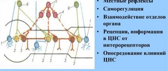
Afferent connections of the basal ganglia.
Most of the afferent signals coming to the basal ganglia enter the striatum. These signals come almost exclusively from three sources:
- from all areas of the cerebral cortex;
- from the intralamellar nuclei of the thalamus;
- from the substantia nigra (along the dopaminergic pathway).
Efferent fibers from the striatum go to the globus pallidus and substantia nigra. From the latter, not only the dopaminergic path to the striatum begins, but also the paths going to the thalamus.
The most important of all efferent tracts of the basal ganglia originates from the internal part of the globus pallidus, ending in the thalamus, as well as in the roof of the midbrain. Through the stem formations with which the basal ganglia are connected, centrifugal impulses follow to the segmental motor apparatus and muscles along descending conductors.
- from the red nuclei - along the rubrospinal tract;
- from the Darkshevich nucleus - along the posterior longitudinal fasciculus to the nuclei of nerves 3, 4,6 and through it to the nucleus of the vestibular nerve;
- from the nucleus of the vestibular nerve - along the vestibulospinal tract;
— from the quadrigeminal region — along the tectospinal tract;
- from the reticular formation - along the reticulospinal tract.
Thus, the basal ganglia play mainly the role of an intermediate link in the chain connecting the motor areas of the cortex with all its other areas.
Physiology
All subcortical nuclei are again conventionally combined into two systems. The first is called the striopallid system, which includes:
- pale globe;
- caudate nucleus of the brain;
- shell.
The last two structures consist of many layers, which is why they are grouped under the name striatum. Ballus pallidus is a brighter, lighter color and is not laminated.
The lenticular nucleus is formed by the globus pallidus (located inside) and the shell, which forms its outer layer. The amygdala and the amygdala are components of the limbic system of the brain.
Let's take a closer look at what these brain nuclei are.
Caudate nucleus
Paired component of the brain related to the striatum. The location is in front of the thalamus. They are separated by a strip of white matter called the internal capsule. Its anterior part has a more massive thickened structure, the head of the structure is adjacent to the lenticular core.
Structurally, it consists of Golgi neurons and has the following characteristics:
- their axon is very thin, and dendrites (processes) are short;
- nerve cells have reduced physical dimensions compared to normal ones.
The caudate nucleus has close connections with many other distinct brain structures and forms a very wide network of neurons. Through them, the globus pallidus and thalamus interact with sensory areas, creating pathways with closed circuits. The ganglion also interacts with other parts of the brain, and not all of them lie in its vicinity.
Experts do not have a consensus on what the function of the caudate nucleus is. This once again confirms the scientifically unfounded theory that the brain is a single structure, any of its functions can be easily performed by any part. And this has been repeatedly proven in studies of people injured due to accidents, other emergencies and diseases.
It is certainly known that it takes part in autonomic functions and plays an important role in the development of cognitive abilities, coordination and stimulation of motor activity.
The striatal nucleus consists of layers of white and gray matter alternating in a vertical plane.
Black substance
The component of the system that is most involved in the coordination of movements and motor skills, maintaining muscle tone and controlling postures. Participates in many autonomic functions, such as breathing, cardiac activity, and maintaining vascular tone.
Physically, the substance is a continuous strip, as was believed for decades, but anatomical sections have shown that it consists of two parts. One of them is a receiver that sends dopamine to the striatum, the second - a transmitter - serves as a transport artery for transmitting signals from the basal ganglia to other parts of the brain, of which there are more than a dozen.
Lenticular body
Its location is between the caudate nucleus and the thalamus, which, as stated, are separated by the external capsule. In front of the structure, it merges with the head of the caudate nucleus, which is why its frontal section has a wedge-shaped shape.
This nucleus consists of sections separated by a thin film of white matter:
- shell – darker outer part;
- pale ball.
The latter is very different in structure from the shell and consists of type I Golgi cells, which predominate in the human nervous system, and are larger in size than their type II. According to neurophysiologists, it is a more archaic brain structure than other components of the brain nucleus.
Other nodes
The fence is the thinnest layer of gray matter between the shell and the island, around which there is a white substance.
The basal ganglia are also represented by the amygdala, located under the shell in the temporal region of the head. It is believed, but not known for sure, that this part belongs to the olfactory system. It is also where the nerve fibers coming from the olfactory lobe end.
Hypokinesia
Damage to the caudate nucleus of the brain is a common cause of the development of a disease associated with a decrease in human motor function.
Symptoms and consequences:
- hypotension;
- intestinal malabsorption;
- deterioration in the functioning of the senses;
- decreased lung ventilation;
- atrophy of the heart muscle;
- stagnation of blood in the capillaries;
- bradycardia;
- degenerative changes in posture.
A drop in blood pressure leads to a decrease not only in physical activity, but also in mental activity. Against the background of hypokinesia, working capacity is lost, and the person completely drops out of society.
Pathologies in case of damage
In 1896, Ch. S. Sherrington described extreme muscle tension in an animal when the descending connections of the red nucleus are broken. If you cut the brain stem between the red and vestibular nuclei, then maximum tension occurs in the extensor muscles of the limbs, neck, and back.
Important Anankasty personality disorder
These muscles counteract gravity, which means this picture must be associated with the vestibular system. Indeed, the Deiters vestibular nucleus activates the extensors. The influence of the red nucleus on these neurons and the Deiters nucleus inhibits their activity. That is, muscle tone is created by the joint work of several nuclei.
Decerebrate rigidity in humans occurs after severe or traumatic brain injury and is a bad sign.
It looks like this: the arms are extended, as if stretched, and brought towards the body, the palms are turned outward (pronated), the fingers are bent, but the thumbs are abducted. The legs are extended and brought towards each other, the feet are turned inward. The toes are bent, as if in a suspended position. The jaws are clenched. It was described in 1912 by Dutch doctors R. Magnus and A. Klein (R. Magnus, A. de Klein).
Brain function can be disrupted due to injuries, infectious and vascular lesions of the brain, tumor processes, and aggression of the immune system.
Damage to the red nucleus and its connections in humans is manifested not only by decerebrate rigidity, but also by less severe pathology. The midbrain contains structures that give rise to nerves that control the muscles of the eyeball, pupil, and levator palpebral muscle. Therefore, damage to the red nucleus can be combined with “ocular” symptoms.
This happens with Claude syndrome and Benedict syndrome. They often develop after vascular accidents (stroke).
Claude syndrome was described by French neurologist and psychiatrist Henri Claude in 1912. In Claude's syndrome, the inferior part of the red nucleus, efferent fibers from the cerebellum to the thalamus, and the oculomotor nerve are affected.
Due to the damaged oculomotor nerve on the affected side, the upper eyelid droops, the pupil dilates and divergent strabismus appears. On the other side of the body, hand tremors occur when moving towards a goal (intention tremor), muscle weakness.
Eyelid drooping and strabismus in Claude syndrome
In this syndrome, the lesion involves the red nucleus, its connections with the cerebellum and the structures of the oculomotor nerve. On the damaged side the pupil dilates, on the opposite side intentional and erratic or writhing movements of the limbs (choreoathetosis) appear.
Latin name: nucleus ruber.
In the midbrain, the red nuclei are located in the very center. If we make a horizontal slice through the midbrain, then diagonally between and we will see two pale pink spots. These will be the red kernels. It is believed that they owe their color to iron, which they contain in two different forms - hemoglobin and ferritin.
In the following screenshot you can see a sagittal section of the brain stem. The bottom of the red nucleus lies on the ascending fibers of the superior cerebellar peduncles at the level of the top of the inferior cerebellum. From above - they reach the level of the hypothalamus.
You can learn more about where the red core is located on ours.
The red nucleus is motor, responsible for muscle tone and reflexes.
There are two parts:
- posterior large cell (magnocellular) - less developed in humans than in other vertebrates, because In humans, the cerebral cortex is much more developed, which takes away some of the functions from the magnocellular part.
- anterior small cell (parvocellular) - transmits information from the motor cortex to the cerebellum through the olives.
Some researchers isolate the posteromedial part separately.
Fahr's disease
The syndrome is characterized by the accumulation of calcium in the vessels of the brain, which are responsible for providing oxygen to the internal capsule and caudate nucleus. The rare disease manifests itself in adolescence and middle age.
Provoking factors:
- carbon monoxide poisoning;
- dysfunction of the thyroid glands;
- Down syndrome;
- radiation therapy;
- microcephaly;
- tuberous sclerosis;
- disturbance of calcium metabolism.
Symptoms:
- trembling of limbs;
- convulsions;
- facial asymmetry;
- episyndrome;
- slurred speech.
Fahr syndrome is not fully understood and has no specific treatment. Progression of the disease leads to mental retardation, deterioration of motor functions, disability and death.
Functions
In humans, the rubrospinal tract coming from the red nucleus partly controls gait and movements of the shoulder girdle. "Partly" means that it only controls large movements. The corticospinal tract is responsible for fine motor skills. If you “turn it off” and leave only the rubrospinal one, then the movements of such a person will become sharp and sweeping.
I also note that the rubrospinal tract is responsible for reflex movements.
Animal experiments show that electrical stimulation of the rubrospinal tract leads to excitation of flexor motor neurons and inhibition of extensor motor neurons. Thus, when the tract is cut at the level of the midbrain, the limbs are straightened and remain tense in this position. the head is thrown back.
Parkinson's disease
The disease causes degenerative changes in neurons, which leads to loss of control over movements. Cells stop producing dopamine, which is responsible for transmitting impulses between the caudate nucleus and the substantia nigra. The disease is considered incurable and chronic.
Initial symptoms:
- change in handwriting;
- slowness of movements;
- tremor of the limbs;
- depression;
- muscle tension;
- slurred speech;
- disturbance of gait, posture;
- frozen facial expression;
- forgetfulness.
If one of the symptoms appears, you should consult a neurologist.
Lenticular nucleus: possible pathologies
When the striatum is damaged, a specific type of movement disorder develops - dyskinesia. There are two possible variants of dyskinesia: hypo- and hyperkinesis.
Hypokinesis means paleness and inexpressiveness of movements. They arise with an increase in the inhibitory, that is, inhibitory, influence of the striatum on the globus pallidus.
Hyperkinesis - sweeping, disordered, unfocused movements. They occur in the absence of an inhibitory influence of the striatal system on the globus pallidus.