Brain tissue is represented by a wide range of formations. In terms of its structure, it is perhaps the most complex part of the human body, which determines the wide nature of the activity of the central nervous system. When assessing the structure, several areas of the central nervous system can be identified in this localization.
At the base of the cerebral structures is the so-called brain stem. It provides a group of vital functions: from breathing and cardiac activity to thermoregulation. Any damage or malfunction results in severe disability or death.
The pons is a component of the brainstem located between the midbrain and medulla oblongata, which ensures normal conduction of nerve impulses and makes it possible to perform a number of voluntary actions.
Responsible for some functions of higher activity. Its damage, for example, due to injury or stroke, leads to critical disruptions in the functioning of the entire body.
Diagnosis of lesions of this anatomical structure presents certain difficulties due to the deep and “inconvenient” localization. The only reliable method of examination is MRI or, less commonly, computed tomography.
General information
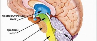
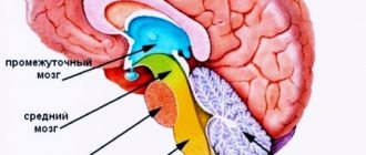
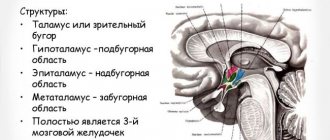
The pons is a formation in the nervous system that is located between the midbrain and medulla oblongata. The bundles of the upper parts of the brain, as well as veins and arteries, stretch through it. The pons itself contains the nuclei of the central nerves in the cranial brain, which are responsible for the chewing function of humans. In addition, it helps ensure sensitivity of the entire face, as well as the mucous membranes of the eyes and sinuses. Education performs two functions in the human body: connecting and conducting. The bridge got its name in honor of the Bolognese anatomist Constanzo Varolius.
Formation on the basal surface of the brain
The spinal cord and brain are independent structures, but in order for them to interact together, one formation is required - the pons. This element of the central nervous system acts as a collector, a connecting structure that links the brain and spinal cord together. Therefore, the formation is called a bridge , because it connects two key organs of the central and peripheral nervous system. The pons is part of the structure of the hindbrain, to which the cerebellum is also attached.
The structure of the Varoliev formation
The formation is located on the surface of the brain. If we talk about the internal structure of the bridge, then it contains an accumulation of white matter, where the nuclei of gray matter are located. In the posterior part of the formation there are nuclei consisting of 5, 6, 7, and 8 pairs of nerves. One of the most important structures located on the bridge is the reticular formation. It performs a particularly important function; it is responsible for activating all the departments located above. The pathways are represented by thickened nerve fibers that connect the pons to the cerebellum, forming the streams of the formation itself and the cerebellar peduncles.
Saturates the Varoliev bridge artery of the vertebrobasilar region with blood. Outwardly, it looks like a roller that is attached to the brain stem. The cerebellum is attached to it on the posterior side. In its lower part there is a transition to the medulla oblongata, and from the upper part to the middle brain. The main characteristic feature of the Varoliev formation is that it contains a mass of pathways and nerve endings in the brain.
Four pairs of nerves diverge directly from the pons:
- ternary;
- abductor;
- facial;
- auditory.
Brain structure. Pons
Among all body systems, the central nervous system occupies a special place. The brain regulates all the functions that a person is endowed with. Thanks to it, the relationship between the work of organs and systems is realized. Without brain regulation, a person would not be a viable being.
Thanks to the coordinated activity of the central nervous system, we move, speak, think and feel external stimuli. The brain has a complex structure, each of its components is responsible for a specific function.
Nevertheless, all its structures ensure the functioning of our body only in their entirety. Particularly important formations that make up the central nervous system are the medulla oblongata and the pons.
They contain the main vital centers (vascular, respiratory, cough, lacrimal), and also give rise to most cranial nerves.
Brain structure
The structural unit of the central nervous system is the neuron. It is this cell that is responsible for receiving, processing and storing information. The entire human brain is a collection of neurons and their processes - axons and dendrites.
They ensure the transmission of signals entering the central nervous system and back to the organs. The brain consists of gray and white matter. The first is formed by the neurons themselves, the second by their axons.
The main structures of the brain are the hemispheres (left and right), the cerebellum and the brainstem. The first are responsible for a person’s mental abilities, his memory, thinking, imagination.
Important Capgras syndrome (double syndrome). What is Capgras syndrome?
The pons is one of the parts of the hindbrain. Its length ranges from 2.4 to 2.6 cm. The pons has a mass of about 7 g. The structures that border it are the medulla oblongata and midbrain, the transverse sulcus. The main components of the pons are the superior and middle cerebellar peduncles, which are large pathways.
In front is the basilar groove, which contains the arteries that supply the brain, and nearby is the exit site of the trigeminal nerve. On the posterior side, the pons forms the upper part of the rhomboid fossa, which contains the 6th and part of the 7th cranial nerves. The upper part of the bridge contains the most cores (5, 6, 7, 8).
At the base of the bridge there are descending pathways: corticospinal, bulbar and pontine tracts.
The main functions of this body:
- Conductive - along its paths nerve impulses pass to the cerebral cortex and to the spinal cord.
- Sensory function is provided by the vestibulocochlear and trigeminal nerves. In the nuclei of the 8th pair of cranial nerves, information about vestibular stimulation is processed.
- Motor – ensures contraction of all facial muscles. This occurs thanks to the nuclei of the trigeminal nerve. In addition, its sensitive part receives information from receptors in the mucous membrane of the mouth, eyeball, part of the head and teeth. These signals are sent along the pons fibers to the cerebral cortex.
- The integrative function ensures the relationship between the forebrain and hindbrain.
- Reflexes of the brain.
Reticular formation of the bridge
The reticular formation is a branched network located in the brain and consisting of nerve cells and nuclei. It is present in almost all formations of the central nervous system and smoothly passes from one department to another. The reticular formation of the pons is located between the medulla oblongata and midbrain.
Its long processes, called axons, form white matter and pass into the cerebellum. In addition, signals can be transferred from the head to the back along the fibers of the nerve cells of the bridge. In addition, the reticular formation transmits signals to the cerebral cortex, due to which a person awakens or sleeps.
The nuclei located in this part of the pons belong to the respiratory center located in the medulla oblongata.
Reflex function of the bridge
The ability of the central nervous system to respond to external stimuli is called a reflex. An example is the appearance of salivation at the sight of food, the desire to sleep at the sound of soothing music, etc. Reflexes of the brain can be conditioned and unconditioned.
A person acquires the first ones in the course of life; they can be developed or adjusted depending on our desire. The latter are not amenable to consciousness, they are laid down at birth, and it is impossible to change them. These include chewing, swallowing, grasping and other reflexes.
How does the bridge affect reflexes?
Due to the fact that the pons is an integral part of the quadrigeminal region, it is related to the development of the auditory and statistical reflex. Thanks to the latter, we are able to hold the body in a certain position. In addition, interacting with the midbrain, it closes a significant part of muscle reflexes.
Formation in the prenatal period
Varolievye formation begins to form in the embryonic period from the rhomboid bladder. The bladder, in the process of its maturation and formation, is also divided into oblong and posterior. During the formation process, the hindbrain gives rise to the cerebellum, and the floor and its walls become components of the pons. The cavity of the diamond-shaped bladder will subsequently be common. At the stage of formation, the nuclei of the cranial nerves are located in the medulla oblongata and only over time do they move directly to the bridge.
Article on the topic: Ointment for foot fungus - a review of effective drugs for the treatment of mycosis with description, composition and prices
When a baby is born, the bridge is located just above the back of the sella turcica. Only after 2-3 years does it begin to rise and thereby become fixed in its permanent place - the upper part of the skull.
At the age of 8, the child’s spinal fibers begin to become overgrown with a myelin sheath.
Treatment
Constant monitoring of cyst size is necessary. If the patient has no complaints and no symptoms accompanying the anomaly, the therapy is focused on the cause that provoked the formation of the formation.
Important VSD according to the vagotonic type
Medications used:
- Drugs to normalize blood circulation;
- Medicines against adhesions;
- Immunomodulatory agents;
- Antioxidants;
- Nootropics.
Constant monitoring of the patient's condition involves regular blood clotting and cholesterol tests, and monitoring blood pressure levels.
Surgery methods
When growth of the formation is observed, surgical intervention is prescribed:
- The endoscopic method is the least invasive, but can only be used for a certain location of the cyst.
- Trepanation is a very complicated operation, but it allows you to remove formations of any location and any size. Accompanied by a high risk of injury to brain tissue.
- During bypass surgery, a puncture is made and a tube is inserted to prevent the accumulation of cerebrospinal fluid. The method is accompanied by the risk of infection.
VM functions
As mentioned earlier, the Varoliev bridge contains a lot of different functions necessary for the normal functioning of the human body.
Functions of the Varoliev formation:
- controlling function for purposeful movements throughout the human body;
- perception of the location of the body in space and time;
- sensitivity of taste, skin, as well as mucous membranes of the nose and eyeballs;
- facial expression;
- eating: chewing, salivating and swallowing;
- conductor, through its paths nerve endings pass to the cerebral cortex, as well as the spinal cord; interactive.
- the VM carries out the relationship between the anterior and posterior parts of the brain;
- hearing perception.
It contains the centers from which the cranial nerves emerge. They are responsible for swallowing, chewing and the perception of skin sensitivity. The nerves extending from the bridge contain motor fibers (provide rotation of the eyeballs).
The triple nerves of the fifth pair affect the tension of the muscles of the palate, as well as the eardrum in the cavity of the auricle.
The Varoliev formation contains the nucleus of the facial nerve, which is responsible for motor, autonomic and sensory function. In addition, the center of the respiratory system of the medulla oblongata depends on its normal functioning.
Research methods
Diagnosis of the functional state and activity of the trunk is carried out using clinical and instrumental laboratory methods. The first includes:
- neurological study of the activity of cranial nerves;
- study of voluntary movements;
- diagnostics of coordination of limbs and body;
- sensitivity study;
- Laboratory methods include:
- spinal puncture and examination of cerebrospinal fluid;
- X-ray of the skull;
- ventriculography;
- pneumoencephalography;
- Dopplerography;
- electroencephalography;
- magnetic resonance imaging;
- computed tomography.
CM pathologies
Like any organ in the human body, the VM can also stop functioning and this can be caused by the following diseases:
- stroke of the cerebral arteries;
- multiple sclerosis;
- head injuries. Can be obtained at any age, including during childbirth;
- tumors (malignant or benign) of parts of the brain.
In addition to the main reasons that can provoke brain pathologies, you need to know the symptoms of such a lesion:
- the process of swallowing and chewing is impaired;
- loss of skin sensitivity;
- nausea and vomiting;
- nystagmus is eye movements in one specific direction, as a result of such movements one can often feel dizzy, even to the point of loss of consciousness;
- may see double when turning the head sharply;
- disturbances in the functioning of the motor system, paralysis of certain parts of the body, muscles, or tremors in the hands;
- in case of disturbances in the functioning of the facial nerves, the patient may experience complete or partial anemia, lack of strength in the facial nerve;
- speech disorders;
- asthenia – decreased strength of muscle contraction, rapid muscle fatigue;
- dysmetria - incompatibility between the task of the movement being performed and muscle contraction, for example, when walking, a person may raise his legs much higher than necessary or, on the contrary, may stumble over small bumps;
- snoring, in cases where it has never been observed before.
Article on the topic: Akrikhin - instructions for use, composition, indications, analogues and price
Pathologies that impair the functions of the bridge and their symptoms
There is a group of diseases that are typically characterized by disruption of the normal functioning of the body as a result of the destruction of the tissues in question.
Among them:
Brissot-Sicard syndrome
Accompanied by a disorder of cranial nerve activity. It is determined by unilateral paresis or complete paralysis of half the body.
The ability to control the muscles of the facial area is also lost, and ptosis (drooping of the eyelid) with impaired visual function is possible.
This disorder occurs against the background of an infectious, autoimmune or tumor lesion. Less commonly, it results from cerebral ischemia. After a transient attack or a full-fledged stroke.
Bonnier syndrome
Characterized by damage to a group of cranial nerves. In this case, the auditory and vestibular nuclei ultimately suffer.
Symptoms are nonspecific. Problems arise with the perception of sound stimuli. Patients constantly experience dizziness, nausea, and weakness.
Insomnia is also part of the clinic. The patient becomes irritable, and emotional instability is noted. Up to sudden changes in phases, as, for example, with bipolar affective psychosis.
Grenet's syndrome
A typical feature of this pathological process is a violation of the sensitivity of facial muscles, which ultimately leads to problems with the manifestation of non-verbal signals and emotions.
There is partial paresis of the masticatory muscles on one side. On the other hand, controllability impairment is also present, but to a much lesser extent.
Ventral syndrome
An extremely difficult condition. It is characterized by at least loss of speech function. This is the easiest case.
The classic situation is determined by the complete loss of the ability to move. The man cannot move. Communication is possible only through the eyes.
This disorder persists for a long time. Quickly leads to stagnation and death of the patient. Restoration is not possible.
Raymond-Sestan syndrome
Characterized by a key manifestation of the oculomotor nerves. A person loses the ability to voluntarily focus his gaze and move it from one object to another.
Spontaneous relief of the condition and its subsequent return for unclear reasons is possible.
Hübler's syndrome
There are no specific manifestations of blood paralysis of facial muscles. The facial expression is characterized as a mask.
The patient is unable to adequately non-verbally express emotions and respond to surrounding stimuli.
Skin sensitivity also decreases, which is detected by the results of functional tests and physical examination.
Foville syndrome
There is paralysis of facial muscles and strabismus with visual impairment.
Gasperini disease
Combined pathological process. Characterized by mixed symptoms.