Magnetic resonance imaging (MRI) of the brain has become widespread due to its safety and accuracy. In the article you will learn everything about how MRI of the brain is done, features and contraindications to the procedure and the results of its implementation.
The study is carried out using a special device - a magnetic tomograph, which is a huge magnet. The procedure is absolutely painless and fairly quick. The patient is placed in the tube of the device, which takes a series of photographs of the organ being examined in three projections. Next, the images are sent to a computer screen, where they are deciphered by a doctor.
What is MRI of the brain based on?
The diagnostic effect of brain MRI is based on nuclear magnetic resonance. In response to the powerful radiation created by the generator, the hydrogen atomic nuclei contained in the tissues line up along the electromagnetic field lines and begin to vibrate. Each atom becomes like a spinning mini-spinning top, emitting energy waves.
Different structures emit different amounts of energy - some give it out more intensely, while others less so. The difference is recorded by a device that takes pictures (slices) in different projections.
To do this, the patient is placed inside a tomograph in which generators maintain a high-frequency electromagnetic field. Special radio emitters generate pulses, and coils record the energy sent by vibrating atoms.
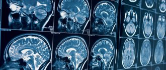
The resulting slice images are combined into a three-dimensional matrix using a special computer program, in which dark or light unhealthy areas are visualized against a gray background.
Equipment
The oncology department is equipped with a German-quality magnetic resonance imaging scanner, Siemens Magnetom Skyra (64 slices). The fundamental difference between this device and others is the double magnetic field strength (3 Tesla), which allows diagnostics to be carried out with high accuracy while maintaining safety.
It is also important that the equipment has an acoustic noise suppression system. Thanks to it, the comfort of MRI for the patient increases. Convenience is also ensured by a wide 7 cm open tunnel. The lightweight design of the coils and MoodLight create a pleasant, calming atmosphere during diagnostics. The examination can be performed even on patients weighing up to 250 kg and those suffering from claustrophobia.
Advantages of magnetic resonance imaging over other methods
An MRI examination gives results much more accurately than x-rays, echoencephalography (EchoEG), ultrasound and other diagnostic options. It allows you to obtain maximum data about existing tumors, diseases, post-traumatic and post-stroke changes. Unlike CT and X-rays, in this case the body is not irradiated.
Only soft tissues are visualized in the finished images. The bones of the skull are not visible, so they do not interfere with analysis and decoding.
The contrast agent used in MRI diagnostics is much less likely to cause allergic reactions compared to X-ray contrast agents used for radiography.
MRI with contrast
In most cases, MRI is able to convey a fairly clear image even without the use of contrast. But, occasionally, for a certain pathology, when targeted thoroughness of the examination is required, a contrast agent may be used.
Contrast is used if a tumor is suspected. A substance that can accumulate in pathologically altered structures and clearly display them on photographs.
Thanks to this, the location of a benign or malignant tumor that interferes with venous outflow, damage and changes in the structure and functioning of the brain veins is accurately determined.
In most cases, the contrast agent is safely eliminated by the body without any negative consequences. Patients with a history or a tendency to allergic reactions are first tested for sensitivity to the dye.
Price
Dear patients, on October 9 and 10 there is a 20% discount on MRI!
| Description | Price | Until October 12 | From 21:00-2:00 7:00-9:00 |
| MRI of the brain | RUB 5,400 | RUB 4,590 | RUB 4,050 |
| MRI of cerebral vessels (angiography) | RUB 5,400 | RUB 4,590 | RUB 4,050 |
| Brain MRI with contrast | RUR 11,000 | RUB 10,250 | — |
| Issue of film with photograph | 500 rub.r. | 400 rub.r. | 400 rub.r. |
| Recording a photo on flash | RUR 1,000 | RUR 650 | 600 rub. rub. |
All MRI prices >>>
How does the procedure work?
The patient removes all metal jewelry and takes out removable dentures containing metal.
The patient is placed on a movable table and secured with special belts. This measure is necessary because you will have to lie inside the tomograph, remaining motionless, for a long time.
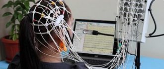
A device equipped with wires that transmit and receive radio signals is placed on the head. The equipment is quite noisy and tires with constant clicks and whistles. Therefore, the patient’s ears are protected with earplugs. After this, the table slides into the device, and the specialist sits down at the computer, in which the transmitted data is analyzed and processed.
The technique takes pictures, the quality of which depends on the characteristics of a particular MRI scanner. The thinner the visual slices the equipment makes, the more accurate the final images will be. The duration of diagnosis is 20-30 minutes, and when contrast is used - up to an hour.
After MRI diagnostics, you can immediately return to your normal life. No side effects subsequently or during the MRI examination occur, with the exception of an extremely rare allergy to gadolinium salts.
Finished photographs are handed out printed or recorded on a magnetic medium - disk or flash card. It can be sent to email with SMS notification.
- Standard - done without the introduction of contrasting solutions, but at the same time provides a sufficient amount of information.
- With contrast, before which drugs containing gadolinium salts are injected into the vein - gadopentetic and gadoteric acids, Omniscan, Magnevist, etc. These solutions penetrate into the bloodstream and, once in the rays of the MRI scanner, highlight the resulting “picture”. At the same time, the changed areas become better visible, which simplifies decoding. The technique is most often used to detect vascular abnormalities, multiple sclerosis and tumor formations. The dose of contrast agent is selected individually, taking into account weight.
- MR angiography – is performed to assess the condition of blood vessels in atherosclerosis, aneurysms, blood clots and pre-stroke conditions. It is done with gadolinium contrast to show blood flow problems in detail.
- MR imaging of the pituitary gland, an appendage that is an endocrine gland. The pituitary gland secretes hormones responsible for reproductive function, tissue metabolism and the regulation of human growth. An examination is prescribed if an adenoma is suspected - a benign tumor that causes migraine-like pain, hormonal imbalances, gigantism, infertility, obesity and sexual dysfunction. The same method is used to identify malignant pituitary formations that have similar symptoms and are accompanied by a pronounced deterioration in health.
How many MRIs of head vessels are performed?
To say how long the examination lasts, it is necessary to clarify an important factor - the quality of the resulting image.
If the time allocated for MRI diagnostics of the brain is reduced, the images will be obtained with insufficient information content. Duration is a significant indicator that influences the result.
There is no exact value that determines the time period required to obtain information when studying a particular site. The determining factor is the task set before starting the diagnosis, the type of organ/defect (tumor, edema, aneurysm). Visualization of a separate zone lasts approximately half an hour. A comprehensive MRI scan can last up to an hour. In certain situations, the procedure is made shorter.
If there is an urgent need for an emergency examination, MRI of the brain is performed promptly. It is not advisable to reduce the scanning duration.
When conducting an angiographic magnetic resonance examination, a coil is installed at the site where the vessels are scanned. During contrast-enhanced tomography, the drug is administered intravenously. The session involves complete immobility of the patient, the examination lasts 30-40 minutes.
Indications for general anesthesia for MRI
Intravenous or inhalational sedation is only necessary for patients who are unable to hold their body still for long periods of time. Main indications for general anesthesia:
- Claustrophobia is the fear of closed spaces. Such patients, while inside the device, experience panic, which negatively affects their health and makes MRI diagnostics impossible.
- Mental disorders accompanied by unpredictability of behavior and high excitability.
- Uncontrollable involuntary head movements (swaying, shaking, tics).
- Epilepsy and other types of convulsive readiness and seizures - anesthesia is given only intravenously due to the risk of provoking a seizure attack.
- Early childhood. Small children cannot lie still for a long time in an MRI scanner, so light mask anesthesia is indicated for them.
- Severe pain syndrome, in which prolonged stay in one position causes discomfort, cramps, pain and spasms.
Contraindications for use
There are relative and absolute contraindications. In the first case, conducting an examination is undesirable, but its help is resorted to in case of vital necessity. If the patient has absolute contraindications, then the use of diagnostics is strictly prohibited. Absolute contraindications include cases when the patient has:
- neuro- or pacemaker;
- electronic or ferromagnetic implant in the middle ear;
- a prosthesis or cochlear implant in the inner ear;
- metal implants, ferromagnetic fragments;
- insulin pump;
- heart valve prosthesis;
- Ilizarov apparatus.
Relative contraindications include the following:
- a clip that was installed due to removal of the gallbladder;
- tremors and conditions when a person is unable to hold his breath for a long time;
- pregnancy period;
- fixed braces and dentures, as well as stents and vena cava filters;
- heart failure;
- claustrophobia;
- coronary artery bypass grafting;
- pain that does not allow a person to remain motionless for a long time.
Indications for magnetic resonance imaging of the brain
- Neoplasms or their metastases. Diagnosis is prescribed for persistent migraine-like pain, sudden loss of vision and hearing, auditory, olfactory and visual hallucinations, attacks of confusion, sudden reading and writing disorders, often accompanying cancer pathologies.
- Epilepsy and other diseases manifested by fainting, confusion and convulsions.
- Suspicion of single or multiple cystic cavities filled with fluid, bloody or other contents.
- Possible presence of parasites (cysticercus and echinococci) carried through the vascular bed with the bloodstream into the head.
- Inflammations – meningitis, encephalitis, arachnoiditis, myelitis. Lesions caused by infections - measles, herpes, tuberculosis, toxoplasmosis, tick-borne encephalitis.
- Rehabilitation after stroke, traumatic brain injury and surgery. Using magnetic resonance diagnostics, the doctor evaluates the effectiveness of the treatment and predicts long-term results.
- The likelihood of developing multiple sclerosis, Alzheimer's disease and other degenerative processes.
- Children are examined for congenital pathologies and hydrocephalus.
With all these diseases, life and health directly depend on a timely diagnosis. Therefore, at the slightest suspicion of brain disorders in yourself or your child, you need to come to the clinic and be examined.
Preparation for the procedure
Preparation for an MRI of the brain directly depends on whether the study will be performed with or without a contrast agent. When conducting a study using contrast, approximately 5 hours before the start of the diagnosis, the patient should refuse to consume food and water. Before placing the patient in the device, he must remove all existing metal objects (wristwatches, jewelry, etc.).
If the patient has allergic reactions to certain medications, chronic diseases, or suffers from claustrophobia, this must be reported to the doctor before diagnosis.
When the patient is a child, they should not consume food or any liquids for approximately 3 hours before the MRI. If the study will be carried out with the use of anesthesia or the administration of contrast, then food and liquid intake should be limited the day before the procedure. Before the examination, the child must be examined by an anesthesiologist to determine the likelihood of allergic reactions to the contrast used in diagnosis.
MRI of the brain is carried out in several stages:
- The patient must remove metal objects and accessories, as well as all clothing.
- The patient is placed on a moving table in the supine position.
- The contrast agent is administered intravenously manually or using a special catheter.
- If it is difficult for a person to remain calm and motionless for a long time, he is given a sedative.
- To prevent the patient from accidentally moving his legs or arms, they are fixed to the table with belts before the examination.
- After this, the doctor leaves the room and observes the process from the next room. Next, the table is set in motion and directed into the tomograph capsule.
- During the examination, the patient hears slight crackling and mechanical sounds characteristic of the operation of the tomograph. The patient may feel a slight tingling sensation at the site of contrast injection.
The procedure continues for an hour. During this time, the person should remain motionless, which is necessary to obtain the most accurate results.
Since it is much more difficult for children to remain motionless for a long time, the examination is usually carried out under medical anesthesia. The child is given Propofol, children over 5 years of age are given sedatives. During the diagnostic process, the child can be shown cartoons or examined in an open tomograph (only the baby’s head is placed in the capsule, the parents can be nearby).
Immediately before diagnosis, the child should be taken to the toilet, metal objects should be removed from him and special medical clothing should be put on. It is recommended that you first let your child listen to how the device works.
What the results show
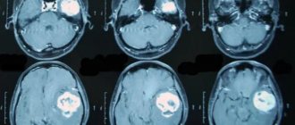
MRI studies, especially those performed with contrast, reveal numerous pathological processes. In the sections, compactions, cystic cavities, and hematomas (collections of blood) are visible in detail. Scars, parasites and their cysts, foci of degeneration, sclerosis and inflammation are identified.
Vascular changes are diagnosed, manifested by impaired patency, narrowing or dilation of blood vessels, the appearance of aneurysms (protrusion of walls) and thrombosis.
The degree of tissue damage in traumatic brain injuries, hemorrhagic and ischemic strokes is determined. The affected areas look lighter and are visible even if they are small in size and have sparse neurological symptoms.
Congenital defects are determined - underdevelopment and hypertrophy of the organ, small and incorrectly located gyri, cysts, holoprosencephaly - lack of division into hemispheres. Hydrocephalus is detected - accumulation of fluid in the ventricles, which with this anomaly are greatly enlarged.
Pathological areas and neoplasms appear as dark or lightish spots of various sizes and shapes, standing out against a grayish background. Oncological seals, especially malignant ones, have blurred, uneven edges and surrounding areas of necrosis.
MR diagnostics is recommended periodically for everyone who has been treated for oncological pathologies of any location. It detects metastases, which usually accompany cancer recurrence.
Contraindications
- Installed pacemakers and other electronic devices that are disrupted by the surrounding electromagnetic field.
- Fixed dentures with metal elements present in the mouth, crowns containing metal, braces and other orthodontic structures. The metal they contain is heated by a magnet and deteriorates, damaging surrounding tissue.
- Tattoos applied to the skin, made with metal-containing ink. Due to the heat caused by the electromagnets, burns may occur in these areas. In the absence of information about the pigment. used when applying a tattoo, it is better not to risk it and do a CT scan, ultrasound or x-ray. Examination is also prohibited for any metal piercing that cannot be removed.
- MRI examinations with contrast are not performed during pregnancy or if contrast agents are intolerant. Such an examination is not prescribed for severe kidney pathologies that make it difficult to eliminate gadolinium.
Magnetic resonance imaging is a safe and highly informative procedure that detects pathologies in the early stages. Therefore, in case of migraine-like phenomena, coordination problems, a sharp decrease in hearing and vision, fainting, or progressive memory deterioration, you must definitely go to the clinic and be examined. The price of MRI diagnostics is low and quite affordable for Muscovites and residents of Moscow Region.
Get an MRI of the brain >>>