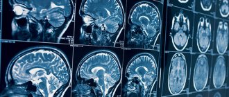
Echoencephalography is a diagnostic neurophysiological study of the structure of the brain, aimed at identifying its pathological processes and changes. The method is non-invasive and requires the use of ultrasonic waves. Thanks to it, it is possible to determine intracranial pressure and identify volumetric processes due to which structures are displaced.
At the same time, it does not allow one to accurately determine the type of detected formations, therefore it is accompanied by computed tomography or magnetic resonance imaging. These are more accurate ways to make an accurate diagnosis.
Echo-EG of the brain involves scanning and calculations by a doctor. They are necessary in order to determine the parameters of the internal volume of the skull and the presence of displacements of brain structures, to identify an increase in intracranial pressure after injuries or due to neoplasms.
You can undergo an Echo-ECG in Moscow at the CELT multidisciplinary clinic. We have everything necessary to obtain all the necessary information and correctly interpret research data.
Echoencephalography of the brain (Echo-EG) - 1,200 rubles.
15 minutes
(duration of procedure)
Outpatient
Advantages of echoencephalography of the brain in CELT
We employ specialists with many years of experience, allowing them to carry out diagnostic studies as accurately as possible and obtain all the necessary data. They have modern equipment in their arsenal that allows them to get a complete picture. Thus, Echo-EG of the head in our clinic allows:
- identify neoplasms in the early stages of their development;
- conduct a qualitative study of the midline structures and periosteal space;
- determine ICP indicators and the nature of pulsations;
- minimize the human factor and increase diagnostic accuracy.
You can find out the price of Echo-EG in our clinic in the corresponding section of our website or by calling us!
Indications for ultrasound echoencephalography
The study is carried out as part of primary neurological diagnosis when the patient complains of:
- headache;
- constant feeling of nausea;
- frequent loss of consciousness;
- feeling of pressure on the eyes;
- incessant noise in the head;
- disturbances in concentration and memory.
The indication for this is an increase in ICP or the need to measure it, suspicion of the presence of neoplasms, foreign objects, cysts and hematomas in the cranial cavity if computed tomography and magnetic resonance imaging are not available. The study is carried out if necessary:
- confirm the presence of diseases such as epilepsy, brain damage of a degenerative, vascular or inflammatory nature;
- identify the patient’s simulation of hearing and visual impairments;
- assess the brain functioning of comatose patients;
- determine the cause of sleep disturbances and abnormal surges in blood pressure;
- determine the efficiency of functioning of blood vessels and tissues.
Indications for Echo-EG
A therapist, neurologist or other specialist can refer you for echoencephalography for the purpose of primary diagnosis in cases of brain diseases or suspicion of them. One of the important reasons for the procedure is emergency cases (including TBI) that threaten a person’s life. The examination allows you to detect the following changes and disorders:
- volumetric brain lesions;
- foci of hemorrhage, intracranial hematomas;
- abscesses, purulent inflammation of tissues;
- indirect signs of intracranial hypertension;
- indirect signs of hydrocephalus.
Using echo-EG of the head, it is possible to assess the dynamics of treatment. Thus, the following symptoms and conditions may be the basis for the procedure:
- previous injuries and bruises, suspected concussion, hematomas, etc.;
- attacks of nausea not caused by pathology of the digestive system;
- dizziness, repeated fainting;
- noise in ears;
- decreased performance;
- paresthesia, tingling in the upper or lower extremities;
- sleep disorders: drowsiness, intermittent, superficial sleep, nightmares, etc.;
- frequent headaches;
- neurological reactions: enuresis, stuttering, tics, automatisms, etc.
The procedure can be prescribed for already identified diseases as a tool for monitoring the effectiveness of treatment. It can be repeated the required number of times without restrictions, unlike radiographic diagnostic methods. Echo-EG of the head can be performed after surgical interventions. In this case, it is important to wait for the skin to heal and then begin the study.
Echoencephalography can be the first diagnostic step and precede studies such as CT and MRI. In other cases, Echo-EG replaces these methods and serves as the only diagnostic method, for example, if there are contraindications to other examination tools.
Contraindications to echoencephalography in M-mode
Echoencephalography of the brain in children and adults is a safe procedure. It does not require the use of painkillers (or any other pharmacological drugs) and has no physiological contraindications. However, it should be postponed during acute respiratory diseases accompanied by a runny nose and/or cough. It is not used if the patient has open wounds or suffers from psychological disorders in the areas where the sensors need to be installed. The latter can cause hysterics due to the unfamiliarity of the situation.
Echo-encephalography (Echo-EG)
Echoencephalography (Echo-EG) is a non-invasive ultrasound diagnostic method that allows you to identify the presence of pathological processes and changes in the structure of the brain.
Echo-EG research is performed on modern computerized equipment. High quality expert-class equipment allows you to:
- examine the midline structures of the brain;
- assess the strength and nature of median pulsations in real time to assess the degree of intracranial pressure;
- accumulate and display the received data automatically to reduce the influence of the human factor and increase the reliability of diagnosis.
Why do an Echo-EG examination?
Echoencephalography is most often prescribed by a neurologist for primary diagnosis in cases of suspected brain diseases in adults and children. The results of Echo-EG allow:
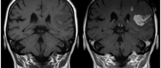
- identify volumetric lesions of brain tissue;
- identify foci of hemorrhage if intracranial hematomas are suspected;
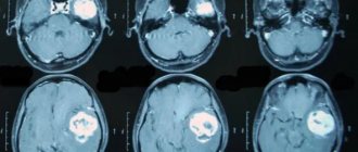
- identify the size and location of tumors, cystic formations and foreign bodies;
- identify purulent accumulations if a brain abscess is suspected;
- perform control measurements of the degree of intracranial pressure;
- assess the degree of hydrocephalus (dropsy) of the brain.
Indications for Echo-EG
Echoencephalography is prescribed for adults and children with the following symptoms and complaints:
- with traumatic brain injuries and suspicions of them;
- attacks of nausea not associated with food intake;
- dizziness and tinnitus;
- deterioration of memory and performance, attention deficit;
- loss of consciousness and loss of coordination of movements;
- feeling of lack of air;
- sleep disturbance and frequent headaches;
- neurotic reactions such as tics, enuresis, stuttering, etc.
Most often, echoencephalography is used as a preliminary diagnostic method before magnetic resonance or computed tomography of the brain. In some cases, Echo-EG can completely replace these methods, for example, due to the presence of contraindications or due to the serious condition of the subject.
Contraindications
Echoencephalography is an absolutely safe diagnostic method with no contraindications. The only limitation is open wounds on the head in the areas where the sensors are installed.
Preparation and performance of echoencephalography
Echo-EG does not require preparation. You can maintain your usual daily routine and diet.
Echoencephalography of the brain is most often performed in the supine position. During the examination, the head must be kept motionless, therefore, when conducting Echo-EG in young children, parental assistance may be required. A contact gel is applied to the skin to prevent interference, and sensors are installed. Depending on the type of study, the doctor sequentially or smoothly moves the sensors to different areas of the head. In the first case, the diagnostic result is displayed in the form of a graph, in the second - in the form of a two-dimensional image of a section of the head. The whole procedure lasts no more than 10-15 minutes.
Results of ECHO-EG of the brain
The results of echoencephalography (doctor's report, diagram and/or image) are given to the patient 15-20 minutes after the study. Depending on the data received, you may be prescribed additional examinations, as well as consultations with doctors of other specialties.
| Cost of ECHO-EG | 1,700 rubles |
Preparation for the procedure and its implementation
One- and two-dimensional echoencephalography does not require the patient to prepare independently. All he needs to do is visit the diagnostic room at the appointed time. Before starting the examination, the diagnostician will ask him to remove from his head any accessories that may interfere.
During the process, the doctor installs an ultrasound sensor in certain areas of the head. To ensure high-quality contact, a contact gel is first applied to them. This prevents air from entering and causing interference. The sensor is placed:
- above the ear canals;
- near the outer edges of the eyebrow arches (at the temples);
- behind the ears at a distance of four to five centimeters.
By sending ultrasound signals, the sensor catches them in reflected form and transmits them to the device, which displays them on the monitor in the form of a graphic image similar to the teeth of an electrocardiogram.
When carrying out the procedure in a two-dimensional format, the sensor is not rearranged sequentially - it is moved along the surface of the head, providing an image of the slice in a horizontal plane along the line of its movement. An image of the formations located at this level is displayed on the screen. The duration of the procedure is no more than 15 minutes.
Order of conduct
No special preparation is required for echoencephalography. You can lead your usual lifestyle, there are no restrictions on diet or medication.
The procedure is performed in a lying or sitting position. It is important to keep the head still, so when examining the child, parents can be present in person and help the baby not move. A special gel is applied to the scalp to improve the conductivity of the ultrasound signal, then the doctor installs the sensors.
Depending on the tasks, a specialist can install one sensor or use two located on both sides of the head. The result of this diagnostic method is a graphic image that shows whether the midline structures of the brain are displaced or whether there is an expansion of the cerebrospinal fluid spaces.
Decoding echoencephalography of the brain
The results of the diagnostic study are interpreted by a neurologist. As already mentioned, the norm provides for the same distance from the central signal to both sides. The permissible deviations are no more than two millimeters for adults and three for children.
During volumetric processes in the GM substance, a shift in the central signal occurs, changes in the shape and duration of responses. Thus, the following pathological conditions can be identified:
- intracranial neoplasms of benign and malignant etiology;
- tuberculomas;
- hematomas localized in the intracranial space;
- cerebral infarctions;
- focal accumulations of pus in the GM substance;
- syphilitic granulomas.
Their localization is determined by median deviations. The distance to the central signal on the side of the formation is greater than on the healthy side, although in certain inflammatory diseases and stroke the opposite may be true. A similar phenomenon can be triggered by a decrease in the volume of one hemisphere during recovery processes, when scarring or resorption is observed.
The doctor determines the presence of a problem based on echo-EG readings:
- with tumors of malignant etiology, a serious shift is observed;
- infarction states of the brain are characterized by small transient changes;
- in case of traumatic injuries, displacements of no more than three millimeters are observed due to swelling;
- hydrocephalus is characterized by expansions of the central signal from five minimeters and the appearance of a large number of lateral ultrasound signals;
- suspicion of hemorrhage arises with the most serious asymmetry.
It is important for patients to understand that the diagnostic accuracy of this method directly depends on the qualifications and experience of the specialist, as well as on the characteristics of the device involved in the process.
Make an appointment with our specialists by filling out and sending us an application online or by calling us by phone! Trust the diagnostics to professionals!
Indications for testing
Doctors refer for ECHO of the head for the following pathologies:
- Neoplasms are malignant and benign.
- Tuberculosis, ischemia, cerebral infarction.
- Encephalopathy.
- Swelling, abscess.
- Shaking paralysis.
- Tumor of the anterior pituitary gland.
Two-dimensional echoEG is necessary for:
- Traumatic brain and neck injuries.
- Migraines.
- Noise, ringing in the ears.
- Head spinning.
- Headache attacks.
- Shell shock.
- Acute cerebrovascular accident.
- Vascular or hypertensive encephalopathy.
- Vertebro-basilar syndrome.
- Increased intracranial pressure.
- Necrotic processes in the brain.
- Vegetative-vascular dystonia.
- Poor circulation due to damage to the vertebral artery.