The brain is one of the most important organs, the central computer of the body. His research was always fraught with difficulties and precautions. However, with the advent and implementation of magnetic resonance imaging, diagnosing brain pathologies has become faster and easier. MRI is a non-invasive test. It is based on the use of high-frequency pulses, magnetic fields, a computer system and special software that allow you to obtain the most detailed image of the brain. “Treatment and Diagnostic Center on Vernadsky” offers MRI at a cost that is one of the lowest in the capital.
Types of MRI examinations
Magnetic resonance imaging of the head is performed with or without a contrast agent. In the first case, the subject is injected with special substances (usually based on gadolinium salts), which make it possible to create a contrast between different tissues. In some cases, the contrast agent may cause allergic reactions. The type of study (with or without contrast) is determined by the specialist.
Contraindications
The diagnostic method is contraindicated for pregnant women, especially in the first trimester, because The effect of drugs on embryogenesis has not been fully studied. MRI with contrast is not recommended during lactation. If the patient is breastfeeding, the study is prescribed in case of emergency. Breastfeeding should be stopped temporarily (24-48 hours).
MRI with contrast is dangerous for patients with chronic renal failure. This is due to the difficulty in removing drugs from the body. The only proven complication of the study is nephrogenic systemic fibrosis. A side effect may occur in patients with impaired renal function when high doses of gadolinium are administered.
Doctors do not recommend conducting a contrast study if:
- decompensated diabetes mellitus;
- liver cirrhosis;
- severe forms of thyrotoxicosis.
Indications for magnetic resonance imaging
- Pathologies and diseases of cerebral vessels.
- Head injuries and bruises, which are accompanied by internal bleeding.
- Speech impairment and hearing loss.
- Tumors of the brain, cerebellopontine angle.
- Paroxysmal states.
- Infectious diseases of the central nervous system (abscess, meningitis, HIV infection).
- Pituitary adenoma.
- Abnormal development of the blood vessels of the head (thrombosis, aneurysms).
- Epilepsy.
- Systematic headaches of unknown origin.
- Sinusitis.
- Multiple sclerosis.
- Neurodegenerative diseases.
- Pathology of the base of the skull.
Contraindications to magnetic resonance imaging of the brain
Relative:
- severe claustrophobia;
- the presence of non-ferromagnetic implants, insulin pumps, heart valve prostheses;
- pregnancy up to 12 weeks;
- drug or alcohol intoxication;
- presence of tattoos containing metallic compounds in the ink;
- heart failure.
Absolute:
- the presence of metallic foreign objects in the patient’s body;
- hematopoietic anemia (with magnetic resonance imaging with contrast);
- use of electronic devices such as pacemakers, etc.
MRI for a sore head
What are the capabilities of MRI diagnostics for identifying pathologies of the brain and other head structures? We are talking about this with Vadim Vyacheslavovich Cherkasov, a radiologist at the Expert Clinic Omsk.
– Tell me, Vadim Vyacheslavovich, is MRI the best option for brain studies?
– MRI is better suited than all other methods for diagnosing the brain. If MSCT - multislice computed tomography - is effective for visualizing bones or, for example, kidney stones, then for soft tissue structures of the brain the best option is, yes, magnetic resonance diagnostics.
More information about the capabilities of computed tomography can be found here
– What does the doctor see when an MRI of the head is done?
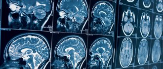
– This type of research makes it possible to detect abnormalities in brain development and visualize vascular changes in the structure of the brain. An MRI will show some volumetric changes, including oncological processes, changes in the pituitary gland. We may see signs of infectious diseases, intracranial hypertension (increased intracranial pressure). Infectious or inflammatory processes of the meninges and the brain itself are successfully diagnosed - for example, meningitis, encephalitis.
In acute pathologies in the head - ischemic or hemorrhagic stroke - we see changes characteristic of these processes.
“In simple terms, in an ischemic stroke, damage to brain tissue occurs due to blockage of a blood vessel, and in a hemorrhagic stroke, it occurs due to its rupture and hemorrhage.” Quote from the material “How to prevent a brain catastrophe?”
Post-traumatic hematomas are clearly visible both inside the brain and in the skull as a whole. This, by the way, is a very dangerous situation: when we find such hematomas, we call an ambulance right here, in our center, and the person is hospitalized (of course, with his consent).
– What types of MRI of the head are there?
– Studies of the arteries of the brain are carried out - it is clear where and which vessel is narrowed, you can measure its diameter, see the abnormal arrangement of the vessels. For neurologists, this is important information in terms of choosing further treatment tactics. Magnetic resonance venography is used to examine the venous system of the brain.
You can read more about MRI of cerebral vessels here
MRI can also clearly visualize the eye orbits - retinal detachment, clouding of the lens, that is, cataracts, nerve atrophy, space-occupying formations in the orbital area. The condition of the chiasma - the crosshairs of the optic nerves - is assessed; we see the asymmetry of the extraocular muscles and their pathological changes.
You can look at the ear canals. New growths in the internal auditory canals are clearly visualized.
MRI is also used in the study of the maxillary, frontal, sphenoid sinuses, and cells of the ethmoidal labyrinth. They can identify inflammatory processes of the mucous membranes (sinusitis, frontal sinusitis, ethmoiditis, sphenoiditis), polyps and cysts. Injuries may cause fractures of the walls of the sinuses - sometimes they are also visible.
What will an MRI of the sinuses show? Read our article
After craniotomy, osteoplastic defects are clearly visible. Some meningiomas (tumors of the meninges) can displace the brain - this displacement, and the meningiomas themselves, can also be seen.
Demyelinating diseases are best diagnosed using MRI: various forms of multiple sclerosis (including atypical ones), acute disseminated encephalomyelitis. When diagnosed early, these dangerous diseases can be treated quite successfully. The earlier they are detected, the more effective the treatment. Here it is important to carry out studies over time: we do the first one, then repeat it after some time, the doctor looks and describes what changes have occurred in the brain during therapy and, if necessary, makes some adjustments to the treatment.
Malignant brain formations, metastases, and suppurative processes are also visible - for example, a brain abscess. Neurodegenerative diseases - Alzheimer's, Parkinson's, age-related changes - are successfully diagnosed. As a rule, in cases of studying neurodegenerative processes, targeted studies are used - the cutting step is reduced to 2-3 millimeters, which allows a more detailed examination of the brain structures of interest. To obtain more complete information, in some cases, MRI studies use a hypoallergenic contrast agent.
Read more about why contrast is needed in MRI here
Based on the data obtained using MRI diagnostics, we can guide the attending physician regarding the condition of the patient’s brain, so that the doctor can draw up the most effective plan for further treatment.
– How often can an MRI of the head be done?
– In addition to good visualization of soft tissue structures, the magnetic resonance imaging method has another big advantage: it does not involve radiation exposure. That is, X-rays are not used here. And the impact of the electromagnetic field during our research is completely harmless to humans. Therefore, you can do an MRI at least every day. Provided that the person does not have any metal structures or metal fragments in his body, and does not have a pacemaker, insulin pump or inner ear prostheses installed.
When is it possible and when not to do magnetic resonance imaging? Yuri Podlevskikh, executive director of Clinic Expert Orenburg, answers these and other questions
– If a person feels dizzy from time to time or constantly, will an MRI help determine the cause?
– Your head may feel dizzy for various reasons. Let's say, due to a violation of the spinal axis - that is, due to manifestations of osteochondrosis. In humans, the vertebral arteries pass through the cervical region, which perform a very important function for supplying blood to the brain. To understand whether this is the reason, it will be necessary to do an MRI not only of the head, but also of the cervical spine with MR angiography and, perhaps, then examine the entire spine.
Dizziness may be accompanied by headaches. Clinicians are not always able to find the cause of headaches. In this situation, a research method such as neurovascular conflict can help. Sometimes the vessels pass next to the nerves, and one of them can put pressure on the nerve, causing pain. The situation is quite common; many people suffer from headaches. After our conclusion, the doctor chooses treatment tactics.
The root of the problem may lie in cancer, changes in high blood pressure. All this is best visualized with magnetic resonance imaging. But taking an MRI of the head does not mean that a final diagnosis has been made: in order to obtain complete information about the problem, a comprehensive examination should be carried out, using other diagnostic methods.
You can sign up for a head MRI here
ATTENTION: the service is not available in all cities
Interviewed by Igor Chichinov
The editors recommend:
Why undergo an MRI of the brain when nothing hurts?
Will an MRI show meningitis?
What to do when the head is cast iron?
For reference:
Cherkasov Vadim Vyacheslavovich
Graduate of the Faculty of Medicine of Omsk State Medical University (2015). Internship in the specialty "Traumatology and Orthopedics" - 2021. CHUDPO "Institute for Advanced Training of Medical Personnel" in the specialty "Radiology" (Voronezh) - 2021. Currently a radiologist at the "Expert Clinic" Omsk. Receives at the address: Omsk, st. Liza Chaikina, 7, bldg. 1.
Preparing for an MRI of the brain
Usually, there is no need to do any complicated steps before the examination. Sometimes the specialist may ask the patient to refrain from eating and drinking before the procedure. The person being examined must remove all metal jewelry. In many centers, the patient is given a loose shirt, in some they are allowed to remain in their own clothes, provided that they are loose and free of metal parts. Before a brain MRI, women should inform their doctor about the possibility of pregnancy. The patient is also obliged to inform the doctor about recent operations and diseases, allergies and the presence of metal implants. If the person being examined suffers from claustrophobia or is very worried before the procedure, he may ask the doctor for a sedative.
How does radioactive ionizing radiation affect the human body?
Radioactive radiation triggers the production of free radicals. Their excess with a low antioxidant (protective) status of the body leads to the destruction of cellular components, including the destruction and shortening of telomeres - the terminal sections of DNA molecules. Lipids and membrane proteins are also subject to oxidation.
Normally, the human body easily tolerates diagnostic measures and recovers on its own - nothing additional is needed. Following the oxidative processes caused by free radicals, restoration begins, and the body’s resources are sufficient for this.
At the end of the 20th and beginning of the 21st centuries, the telomerase enzyme was discovered (active in germ cells, stem cells and cancer cells). For its discovery, E. Black-Burn, K. Greider and J. Szostak were awarded the Nobel Prize in 2009. Telomerase is responsible for the “lengthening” of telomeres, which means that their destruction cannot be considered irreversible. However, scientists have also noticed another pattern: cancer and the growth of an oncological tumor are possible when DNA molecules are significantly shortened and damaged, while the telomerase enzyme remains in an active state. This is a kind of “failure” of the genetic program, which leads to dangerous consequences.
In general, the average healthy adult body is able to recover from radiation exposure equal to 50-100 mSv per year. With greater systematic exposure to radiation, radiation sickness develops.
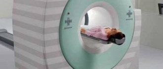
How is an MRI of the brain performed?
- An MRI machine is a cylinder with holes at both ends. The patient sits on a movable table that is placed inside the device during the MRI procedure.
- The length of the tunnel depends on the specific type of apparatus. Some devices only partially surround the table. Tomographs can also be open on the sides. They are used for patients with severe claustrophobia. In closed-type devices, the table slides in completely.
- During the examination, the patient is secured with belts. The less it moves, the more accurate the diagnostic results.
- During magnetic resonance imaging of the brain, devices with wires that receive and send radio waves are placed around the head.
- Patients are often given earplugs. This helps protect your hearing from the loud noise that comes from the MRI machine when it is in operation.
- The table is placed inside the tomograph, and the specialist moves to an adjacent office, which houses a computer system that processes the data. The device takes a series of pictures. Each one takes a few minutes to shoot. As a rule, an MRI of the brain without contrast takes about 20–30 minutes, with a contrast agent – approximately 45–50 minutes.
- An MRI machine takes layer-by-layer images of tissue. During a head scan, data is collected from approximately 20 levels, each 4–5 mm thick. The higher the magnetic field strength of the tomograph and the thinner the sections, the more accurate the research result.
Prices for magnetic resonance imaging of the brain in Moscow
Various factors influence how much an MRI costs in each case. The price may change depending on whether the specialist will use a contrast agent, whether the results will need to be recorded on disk, etc. The approximate cost of magnetic resonance imaging is indicated on the page.
You can make an appointment with a doctor at the Diagnostic and Treatment Center on Vernadsky. To do this, call us by phone at any time convenient for you. You can also leave an online application through a special form presented on the website.
Side effects
Gadolinium salts used in contrast-enhanced diagnostics rarely cause side effects or allergic reactions. Before the study, the patient must make sure that there is no individual intolerance to the components of the substance.
If allergic reactions occur after the study, the doctor should stop them with antihistamines. Reactions to contrast are usually minor and do not pose a potential threat to the patient's life.
Transient side effects of contrast-enhanced MRI include mild dizziness, headache, shortness of breath, feeling hot, and dry mouth. They disappear on their own after a day. If you experience severe nausea, uncontrollable vomiting, signs of angioedema, chills or convulsions, you should immediately consult a doctor or call an ambulance.