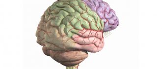
A characteristic feature of the human brain is the incredible size of the cortex and complex folding. The cortex is the most developed area of the brain, responsible for non-reflexive activity (memory, perception, cognition, thinking, etc.).
The formation of cortical-subcortical structures occurs during embryonic development, providing the possibility of placing the cortex in a limited volume of the cranium. Convolutions (giri) and grooves (sulci) make up its folded surface. Pathological changes in the size or folds of the cortex lead to severe mental disability and intractable epilepsy. Consequently, cortical expansion and folding are considered key processes in brain evolution.
Fissures and convolutions: formation and functions
The grooves and gyri in neuroanatomy that give the brain its wrinkled appearance serve two critical functions. They help increase the surface area of the cortex, which allows more neurons to pack into it and enhance the brain's ability to process information. The sulci and convolutions of the brain form divisions, creating boundaries between the lobes of the brain, dividing it into two hemispheres.
Main grooves:
- The interhemispheric fissure is a deep groove in the center of the brain that contains the corpus callosum.
- The Sylvian fissure (lateral sulcus) separates the parietal and frontal lobes.
- Roland's fissure (central sulcus), separating the fusiform gyrus and the hippocampal gyrus on the inferior surface of the temporal lobes.
- Parieto-occipital - separates the parietal and occipital lobes.
- The calcarine fissure (spur-like groove or prominent fissure) is located in the occipital lobes and divides the visual cortex.
The main convolutions of the brain:
- The angular gyrus of the parietal lobe helps in processing auditory and visual recognition.
- Broca's gyrus (Broca's center) is an area of the brain located in the left frontal lobe in most people that controls functions related to speech production.
- The cingulate gyrus, an arched fold located above the corpus callosum, is a component of the limbic system and processes sensory input regarding emotions and regulates aggressive behavior.
- The fusiform gyrus is located in the temporal and occipital lobes and consists of lateral and medial parts. It is thought to play a role in word and face recognition.
- The hippocampal gyrus lies on the inner surface of the temporal lobe, which borders the hippocampus. Plays an important role for memory.
- The lingual gyrus in the occipital lobe is involved in visual processing. It is limited by the collateral groove and calcarine fissure. In front it is in contact with the pararpopampal gyrus, and together they form the medial part of the fusiform gyrus.
As the embryo develops, gyri and sulci form with the appearance of depressions on the surface of the cortex. Not all gyri develop at the same time. The primary form is formed starting from the 10th week of pregnancy (in humans), then secondary and tertiary forms develop. The most prominent groove is the lateral one. It is followed by the central one, separating the motor cortex (precentral gyrus) from the somatosensory cortex (postcentral gyrus). Most of the cortical sulci and gyri of the brain, the anatomy of which begins to take shape between 24 and 38 weeks of pregnancy, continue to grow and develop after the newborn is born.
The early state of the brain has a strong influence on the final level of gyrification. In particular, there is an inverse relationship between cortical thickness and gyrification. Brain regions with low thickness values have a higher level of gyrification. The opposite is also true: brain regions with a high thickness value (for example, thickening of the hippocampal gyrus cortex) have a low level of gyrification.
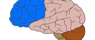
Functions of the hemispheres
The right and left hemispheres of humans perform different functions. The left hemisphere contains the centers of oral and written language. Here the processes of analysis and synthesis of information are carried out, generalizations are made and decisions are made. Verbal and logical thinking provided by the left hemisphere allows one to cognize the essence of an object and go beyond the limits of the individual world. On its basis human knowledge is formed. It is passed down from generation to generation by recording verbal or symbolic signals.
The right hemisphere carries out imaginative thinking. Operating with images of objects in the external world, it can create unprecedented, fantastic combinations from them. And this is the basis of creativity and making unusual decisions. The right hemisphere is extremely important for musical and artistic creativity. It is known that the most outstanding artists, poets, and musicians are people with a predominance of right-hemisphere thinking.
Despite the functional asymmetry, the brain works as a single whole. By summarizing information, it ensures adequate behavior, thinking, consciousness, memory, labor and creative activity of a person.
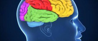
Lobes of the brain and their functions
Each hemisphere is divided into four lobes: frontal, parietal, temporal and occipital. Most brain functions rely on different regions throughout the brain working together, but each lobe performs the bulk of relatively specific functions.
The frontal lobe is located in the most anterior region of the cerebral cortex, separated from the parietal lobe by the central sulcus, and from the temporal lobe by the lateral sulcus. The region typically contains the most important executive functions for a person, including emotion regulation, planning, reasoning, and problem solving.
The parietal lobe is responsible for the integration of sensory information, including contact, temperature, pressure, and pain. Because of the processing that occurs in the parietal lobe, it is possible to distinguish between the touch of two objects at nearby points (rather than as a single object). This process is called two-point.
The temporal lobe also contains areas involved in sensory processing, especially important for hearing, language recognition, and memory formation. The primary auditory cortex receives audio information through the ears and secondary areas and processes the data so that a person understands what he hears (words, laughing, crying, etc.). The medial (closer to the center of the brain) part contains the hippocampus, an area important for memory, learning, and perception of emotions. Certain areas of the temporal lobe process complex visual information, including faces and scenes.
Function creates the center
According to Ivan Petrovich Pavlov: “Function creates the center!” In early childhood, the boundaries of the cortical centers are diffuse and less differentiated, and only as life experience is gained, a gradual concentration of functional zones occurs, and therefore in children of the first years of life, focal cortical symptoms are weakly expressed and general cerebral symptoms often predominate.
4. Significant differences in the localization of simpler and more complex functions. The simpler the function, the more accurately it is localized. Conversely, the most complex functions are determined by the integrative activity of the entire brain, therefore the concept of “cortical center” (section of the cerebral cortex, fields of the cerebral cortex, areas of the cerebral cortex, parts of the cerebral cortex) is in most cases relative and conditional. Simple cortical functions include sensory function, motor function, visual function, auditory function, vestibular function, olfactory function, and gustatory function. Complex cortical functions include speech, writing, reading, counting, praxis, gnosis, thinking, and memory.
Cellular mechanisms leading to expansion and folding of the cerebral cortex
The structure of the human brain distinguishes it from other mammals, and for this reason may explain its unique mental abilities compared to other animals. The number of folds in the cortex may correlate with some specific cognitive, sensory, and motor abilities. Although there is no clear explanation of how the unique division of the human brain into sulci and convolutions occurs. Today there is progress in understanding the extremely complex processes in the brain, the cortex of which is built with so many grooves and convolutions. Even though all cells have the same DNA, different neural stem cells are formed. It is their work with various properties that creates the basic structure of the brain, consisting of neurons and glial cells.
Telencephalic neuroepithelium
Brain growth occurs through two types of stem cells—neural stem cells and neural progenitors. Both of these forms form neurons, which become permanent in the brain, as well as intermediate cells that create the building material for building the brain. Four different types of stem cells determine the structure of the cortex.
During early embryonic development, expansion of the rostral domain of the neural tube leads to the appearance of two telencephalic vesicles. The dorsal half of these vesicles is molecularly defined as the primordium of the cerebral cortex. At this stage, the cortical primordium consists exclusively of a monolayer of neuroepithelial progenitor cells. They are highly polarized and attached to each other by tight junctions at the level of the apical domain (inner surface of the telencephalic vesicle) and move the cell nucleus between the apical (apical) and basal (lower) side of the neuroepithelium in coordination with the cell cycle.
- basal-directed movement during G1 phase;
- basal position during S phase;
- apically directed movement during G2 phase;
- mitosis on the apical surface.
The cycling movement is known as interkinetic nuclear migration and is completely asynchronous between neuroepithelial cells, giving the neuroepithelium a pseudostratified appearance. Cells undergo only symmetrical self-aggressive divisions, with each division generating two daughter cells, hence exponentially increasing their number. Because they are the fundamental progenitor cells of the cerebral cortex, the size of their association determines the number of derived neurogenic progenitor cells and the final number of cortical neurons, and therefore it has a fundamental influence on the size of the mature cerebral cortex. An increase in quantity leads to an expansion of surface area and the formation of neuroepithelium.
Spread and neurogenesis
Immediately before the onset of neurogenesis, neuroepithelial progenitor cells begin to lose tight junctions and acquire features typical of glial cells (including expression of brain lipid-binding protein, vimentin, and Pax6), thereby becoming apical radial glial cells (ARGCs). They also undergo interkinetic nuclear migration, divide at the apical surface of the developing cortex, and at this early stage also undergo self-reinforcing divisions.
However, they gradually begin to divide asymmetrically to generate one similar cell plus another cell. These new cells accumulate in the basal part of the cortical primordium, while the cell bodies of the ARGC remain on the apical side, forming the ventricular zone (VZ). With the accumulation of cells above the GC, the ARGK process is prolonged, remaining attached to the basal plate, and is now called radial glia. Asymmetric ARGK divisions generate one ARGK plus one neuron or one intermediate progenitor cell. Intermediate progenitor cells (secondary progenitor cells without apical-basal polarity) do not undergo interkinetic nuclear migration, divide in a layer located in the ventricular zone, the subventricular zone (SVZ), and all express a transcription factor (Tbr2).
However, due to the fact that each neuron itself consumes during mitosis, their relative quantity compared to ARGK is quite low. Intermediate progenitor cells in the cerebral cortex generate the majority of cortical excitatory neurons. As neurogenesis progresses, the need for ARGK expansion/renewal decreases and the production of neurons increases. In addition to the expanded ventricular zone, the subventricular zone thickens, populated in abundance by basal precursors, especially in the later stages of neurogenesis. As a result, the SVZ splits into internal and external parts. The outer portion contains a wide variety of progenitor cell types with high developmental potential, which is a key factor for the expansion and formation of the cortex.
Localization of functions and symptoms
Carrying out topical diagnostics, a reflexologist, neurologist, neuropathologist, microneuropathologist, pediatric neurologist, adult neurologist determines not only the localization of damage to the cortical centers, but also the localization of symptoms. Simple cortical functions are associated with the projection plates of the cortex (fifth and fourth), which have a direct connection with the periphery and are the cortical sections of the analyzers. Complex cortical functions are associated with the associative layers of the cortex (second and third). The last layers are connected by horizontal fibers to other areas of the cerebral cortex within the same hemisphere and do not have direct access to the periphery. Commissural connections between the hemispheres passing through the corpus callosum are also of great importance in ensuring complex cortical functions.
Simple cortical functions are usually represented in both hemispheres of the brain. Complex cortical functions often have asymmetric representation in the right or left hemisphere of the brain. So, what are the fields, areas, areas, types of cerebral cortex, divisions, analyzers, parts of the cerebral cortex?
Memory, memory function
Various areas are involved in the implementation of the memory function. The frontal lobes provide active, purposeful mnestic activity. The posterior gnostic sections of the cortex are associated with particular forms of memory - visual, auditory, tactile-kinesthetic. The speech zones of the cortex carry out the process of encoding incoming information into verbal logical-grammatical systems and verbal systems. The mediobasal regions of the temporal lobe, in particular the hippocampus, translate current impressions into long-term memory. The reticular formation ensures optimal tone of the cortex, charging it with energy.
Thinking, thinking function
The thinking function is the result of the integrative activity of the entire brain, especially the frontal lobes, which are involved in organizing the purposeful conscious activity of a person, man, woman. Programming, regulation and control take place. Moreover, in right-handed people, the left hemisphere is the basis of predominantly abstract verbal thinking, and the right hemisphere is associated mainly with concrete figurative thinking.
The development of cortical functions begins in the first months of a child’s life and reaches its perfection by the age of 20.
Praxis, praxis analyzer, praxis center
Praxis is the ability to perform purposeful motor acts. Praxis is formed during human life, starting from infancy, and is ensured by a complex functional system of the brain involving the cortical fields of the parietal lobe (inferior parietal lobe) and the frontal lobe, especially the left hemisphere in right-handed people. For normal praxis, the preservation of the kinesthetic and kinetic basis of movements, visual-spatial orientation, programming processes and control of purposeful actions is necessary. The defeat of the praxic system at one level or another is manifested by such a type of pathology as apraxia. The term "praxis" comes from the Greek word "praxis", which means "action". Apraxia is a violation of purposeful action in the absence of muscle paralysis and the preservation of its elementary movements.