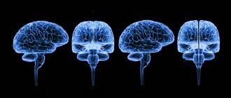
Anyone at least once in their life has heard caustic expressions about the convolutions in the head and their relationship with intelligence, but few people know that, contrary to popular belief, a frequent and far from original phrase is that “the number of convolutions in a person’s brain is the same in him.” mind” is completely unfounded. So is the number of brain convolutions an indicator of any characteristics of the human body, and is there a certain “ideal” number of them? Is there any difference between the normal number of grooves in the brain of a woman and a man? This article will provide answers to these questions.
Brain convolutions: what are they and why are they formed
The human brain is the most complex organ. It consists of more than one hundred billion neurons. This is not surprising, because this organ is the main control center that controls all processes in our body; it gives self-awareness, which makes a person a person, an individual.
Retaining all this number of elements in a limited space, the surface of the brain, called the cerebral cortex, is naturally covered with countless grooves. Such anatomy is a consequence of the body’s adaptation to “crowding,” that is, the limited space of the skull.
The mechanism of formation of convolutions can be easily illustrated as follows: it is easier to push a square leaf into a small round box by crumpling it. In this case, the lump into which the once square sheet has turned becomes a set of grooves similar to those found in the brain when the organ is compactly located in the cranium.
Contrary to popular belief, the number of grooves on a person’s gray matter can neither increase nor decrease, regardless of what activity he engages in throughout his life. The structure of the brain, externally similar to the kernels of a walnut, is formed in humans while still in the embryonic state. Thus, the smooth surface of the gray matter begins to be streaked with grooves at the twentieth week of pregnancy, and they cease to appear in the child at the age of one and a half years. That is, from this very time on, the number and position of the folds have been formed completely and for the rest of life, so that talk about the fact that the convolutions can straighten over time is completely unfounded.
Interestingly, the normal weight of a newborn's brain is about 0.3 kg - this is about 1/8 of his total body weight. In a mature healthy person, the weight of the brain organ should increase five times with an average area of 2200 cm2.
Parietal lobe
The boundaries of this part of the brain are outlined by the following grooves:
- central;
- parieto-occipital;
- transverse occipital;
- central.
Behind the central sulcus is the postcentral gyrus of the brain. At the back it is bounded by a groove with the appropriate name - postcentral. In some literary publications, the latter is further divided into two parts: upper and lower.
The parietal lobe, using the interparietal sulcus, is divided into two regions, or lobules: superior and inferior. The latter contains the supramarginal and angular gyri of the cerebral hemispheres.
In the postcentral, or posterior central, gyrus there are centers that receive sensory (sensitive) information. It is worth noting that the projection of different parts of the body in the posterior central gyrus is unevenly located. So, most of this formation is occupied by the face and hand - the lower and middle third, respectively. The last third is occupied by projections of the torso and legs.
Praxis centers are located in the lower part of the parietal lobe. It implies the development of automatic movements throughout life. This includes, for example, walking, writing, tying shoelaces, etc.
What determines the number of convolutions and is it possible to count them?
According to the latest data obtained in the course of research by Brazilian scientists, the number of convolutions in a person depends on two main variables: the area of the cortex and its thickness. This discovery fits organically into the general theory, because a large area is more difficult to locate in the cranium, and it is also more difficult to form folds in a thick layer of gray matter.
Interestingly, the folds inherent in the human brain are almost not observed in other mammals. The exception is the whale, pig, dog, cat and some primates. A dolphin, for example, has significantly more convolutions than a human.
It is impossible to know exactly the number of grooves, and there is no “absolute” for this parameter. The appearance of the cerebral cortex is individual for everyone, and during an external examination it is not possible to see the total area of its cortex: approximately 2/3 of the gyri are located in deeper grooves.
However, for a person we can name the main convolutions present in the head of everyone:
- toothed;
- tape;
- occipitotemporal;
- lingual;
- parahippocampal;
- straight;
- hook of the brain.
Well, the total number is not at all impressive, but we can say with confidence how many convolutions in the human brain are guaranteed to end up in the same place in anyone’s head.
Structure
The pattern of convolutions and sulci of the brain is best seen in schematic images. The depressions dividing the cortex into two parts (hemispheres) are called primary . In addition, there are other fundamental limitations of the cortex, namely:
- Sylvian fissure (lateral, lateral): separates the temporal and frontal cortex.
- Roland's fossa (central): separates the parietal from the frontal.
- Parieto-occipital fossa: separates the occipital and parietal lobes of the brain.
- The cingulate recess, which passes into the hippocampal recess: separates the surface of the olfactory brain from other parts.
These structures also have another name: first-order sulci of the brain.
Each part of the telencephalon contains several convolutions, separated by secondary cavities. Tertiary depressions develop purely individually: their presence depends on the personal characteristics of a person and his mental abilities. The third type of notches gives individual relief to the folds.
Superolateral part of the hemisphere
This area of the telencephalon is limited by three sulci: the lateral, part of the occipital and central. The lateral cavity originates from the lateral fossa. Developing slightly upward and backward, the formation ends on the superolateral surface.
The central sulcus begins at the upper edge of one of the hemispheres. From its middle it goes backward and partially forward. In front of this notch is the frontal lobe of the brain, and behind it is the parietal cortex.
The end of the occipital region serves as the edge of the parietal region. This groove does not have a clear boundary, so the separation is carried out artificially.
Medial surface of the brain
This part of the hemispheres has permanent deep grooves. When talking about the formations of the medial surface, first of all, as a rule, one thinks of the groove of the corpus callosum (1). Above this groove there is a belt cavity (2), forming a knee and subsequently a branch. Also in this area is the hippocampal sulcus (3) or seahorse sulcus. Closer to the occipital lobe is the collateral groove (4). On the territory of the posterior part of the median surface there is a calcarine groove (5).
Between the first two formations is the encircling gyrus. And the hippocampal and collateral groove limits the gyrus belonging to the temporal cortex of the hemisphere.
Furrows and convolutions of the lower surface of the cortex
This part of the brain is distributed in different parts of the cortex - temporal, occipital and frontal. The lower surface includes the following grooves:
- Olfactory (1)
- Orbital (2)
- Straight (3)
- Inferior temporal (4)
This area of the hemisphere does not have prominent gyri, however, one should still be noted - this is the lingular gyrus (5).
Does the number of convolutions affect the level of intelligence?
Today it has been scientifically proven that the number of convolutions, as well as the mass of the brain, cannot in any way affect the mental development of a person. And even if you read the works of ancient Greek philosophers from morning to night, you will not gain as many gyri as you do grams of weight. This is logical, because the human convolutions in the form in which they remain throughout life are formed during the period of intrauterine development, and the weight of the brain depends on the body composition.
Some scientists and ordinary citizens who donate their bodies to science after death have allowed repeated studies to be conducted that have established that physiological differences between the brains of ordinary people and scientists do not correlate with the intelligence demonstrated during life.
Frontal lobe
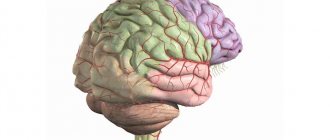
The frontal part of the cerebral hemispheres is located in front of all other structures of the brain. Posteriorly, this area is limited from the parietal lobe by the central sulcus, and laterally, by the lateral sulcus, from the temporal region.
In front of the central sulcus is the precentral gyrus of the brain. The latter, in turn, is limited from other formations of the frontal lobe cortex by means of the precentral recess.
The precentral gyrus, together with the adjacent posterior parts of the frontal lobe, plays an important role. These structures are necessary for the implementation of voluntary movements, that is, those that are under the control of consciousness. In the fifth layer of the cortex of the precentral gyrus there are giant motor neurons, which are called pyramidal cells, or Betz cells. These neurons have a very long process (axon), the endings of which reach the corresponding segment of the spinal cord. This pathway is called the corticospinal pathway.
The relief of the frontal region of the brain is formed by three large convolutions:
- superior frontal;
- average;
- bottom.
These formations are delimited from one another by the superior and inferior frontal grooves.
In the posterior part of the superior frontal gyrus there is an extrapyramidal center, which is also involved in movements. This system is historically more ancient than the pyramidal one. It is necessary for accuracy and smoothness of movements, for automatic correction of motor acts that are already normal for humans.
In the posterior part of the inferior frontal gyrus there is Broca's motor center, which was already mentioned earlier in the article.
Is there a connection between a person's gender and the number of convolutions?
The long-known fact that the male brain weighs more than the female brain has given rise to many ridiculous jokes and stereotypes. However, a worthy answer to the jokers was given by scientists who found that the female brain, as opposed to the male brain, has a more complex structure with a significantly larger number of convolutions, which compensates for less weight. For the same reason, neurons in men's heads are located at a greater distance. Thus, the area of the human brain, regardless of the gender of its owner, is equal.
Studies have found that the gray matter content in men's heads is 20% less. In accordance with this, the difference in the number of convolutions or the mass of the brain does not give a single advantage to the genders: in the level of intelligence, both sexes do not differ.
The human brain is the largest element of the central nervous system, which determines the complexity of its structure. It is he who makes a person himself, gives him the miracle of consciousness. Naturally, scientists have long been interested in whether there is a connection between the appearance of the brain and the kind of personality it makes its owner. For now, we can say for sure: neither its mass, nor how many convolutions a person has in the brain, defines an individual as smart or stupid. The grooves in the gray matter are just folds of a huge organ squeezed into the human skull. Attempts to calculate their average number are pointless, because for each person this number is individual, and in structure and appearance they can be both deep and barely visible to the eye, which makes the counting process impossible.
Outside surface
The anatomy of the cerebral convolutions and sulci is best studied in sections. Let's start with the outer surface. It is on the outer surface of the brain that the deepest groove is located - the lateral one. It begins in the basal (lower) part of the cerebral hemispheres and moves to the outer surface. Here it branches into three more recesses: the ascending and anterior horizontal, which are shorter, and the posterior horizontal, which is much longer. The last branch has an upward direction. It is further divided into two parts: descending and ascending.
The bottom of the lateral groove is called the insula. It then continues as the transverse gyrus. The insula is divided into anterior and posterior lobes. These two formations are separated from each other by a central groove.
Temporal lobe
This structure of the brain is limited by the following grooves: the lateral one from above, a conventional line between the lateral and posterior occipital grooves at the back.
The temporal lobe, by analogy with the frontal lobe, consists of three large convolutions:
- superior temporal;
- average;
- lower
The name of the depressions corresponds to the convolutions.
On the lower surface of the temporal region of the brain, the hippocampal gyrus and the lateral occipitotemporal gyrus are also distinguished.
Wernicke's speech center is located in the temporal lobe, which was already mentioned earlier in the article. In addition, this area of the brain performs the functions of perception of taste and olfactory sensations. It provides hearing, memory, and synthesis of sounds. Specifically, the superior temporal gyrus, as well as the inner surface of the temporal region, is responsible for hearing.
Thus, the lobes and convolutions of the brain are a complex and multifaceted topic to understand. In addition to the parts discussed in the article, there is also the limbic cortex with its own relief, a structure called the insula. There is a cerebellum, which also has a cortex with its own characteristics. But the anatomy of the brain should be studied gradually, so this article provides only basic information.


