Astrocytoma is one of the most common brain tumors. The inside of the tumor often contains cysts, which can grow to large sizes and cause compression of the brain matter.
Benign astrocytomas, located in an accessible location, give a better prognosis for life expectancy than high-grade astrocytomas or benign astrocytomas, located in a location inaccessible to the surgeon and having a large tumor size. The earlier a tumor is detected, the better the prognosis for its treatment.
At the Yusupov Hospital they deal with the diagnosis and treatment of astrocytes. The hospital is equipped with innovative diagnostic equipment that allows you to receive all diagnostic services.
Brain astrocytoma: what is it?
An astrocytoma is a glial brain tumor that develops from astrocytes, neuroglial cells that look like stars or spiders. Astrocytes support the structural component of the nervous system of the brain - neurons. Astrocytes influence the movement of substances from the wall of a blood vessel to the plasma membrane of a neuron, participate in the growth of nerve cells, regulate the composition of intercellular fluid, and much more. Astrocytes in the white matter of the brain are called fibrous or fibrous. Protoplasmic astrocytes predominate in the gray matter of the brain. Astrocytes perform the function of protecting brain neurons from chemicals and injuries, providing nutrition to neurons, and participating in regulating brain blood flow.
Brain tumors cannot be called cancer, since cancer does not develop from epithelial cells, but from cells of more complex structures. A malignant brain tumor practically does not metastasize beyond the brain, but the brain can be affected by metastases from tumors located in other organs and tissues of the body. A malignant brain tumor may be no different from a benign tumor. A brain tumor does not have a clear boundary, so its complete removal is almost impossible. The difficulty of treating such tumors is that the brain has a blood-brain barrier, through which many antitumor drugs do not pass; the brain has its own immunity. A brain tumor can affect the entire brain - the tumor itself can develop in one part of the brain, and its cells can be located in different places in the brain.
Polyclonal brain tumors are a tumor within a tumor. These include primary brain tumors. The difficulty is that such a combination of tumors should be treated with different groups of drugs - one of the tumors is insensitive to drugs for treating another type of tumor. A major role in the effectiveness of treatment of a brain tumor is played not by the determination of the histological type of the tumor, but by the location and size of the tumor.
Bibliography
- B.V. Gaidar, T.E. Rameshvili, G.E. Trufanov, V.E. Parfenov, Radiation diagnostics of tumors of the brain and spinal cord - St. Petersburg: FOLIANT Publishing House LLC, 2006-336p.
- V.N. Kornienko and I.N. Pronin Diagnostic neuroradiology Moscow 2009 0-462.
- Abdullah ND, Mathews VP (1999) Contrast issues in brain tumor imaging. Neuroimaging Clin North Am 9(4):733–749
- Abul-kasim K, Thurnher MM, Mckeever P et-al. Intradural spinal tumors: current classification and MRI features. Neuroradiology. 2008;50(4):301-14.
- Agrawal V, Ludwig N, Agrawal A et al. Intraosseous intracranial meningioma. AJNR Am J Neuroradiol. 2007;28(2):314-5
Causes
There is currently no data on the reasons for the development of tumors from astrocyte cells. There is an opinion that some negative factors can serve as a trigger for tumor development:
- radioactive exposure. Radiation often causes the development of malignant brain tumors in patients. The risk of developing astrocytoma increases in patients who have undergone radiotherapy;
- long-term exposure to toxic chemicals. Working in hazardous work can cause the development of brain tumors.
- oncogenic viruses;
- hereditary predisposition;
- injuries;
- patient's age. Some types of tumors primarily affect children, others are more common in young people between 20 and 30 years of age, and a third type of tumor affects older people more.
When studying the causes of astrocytomas, two types of damaged genes were identified.
Preventive actions
Prevention of the growth of astrocytomas includes protecting the body from radiation exposure, excess ultraviolet radiation and other negative factors, including the consumption of foods and drinks with potentially dangerous chemical components. It is also important to pay attention to working conditions. It is recommended to limit contact with chemicals, objects and equipment that provide a high radiation dose to the body.
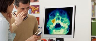
Regular preventive examinations and screening studies will allow timely diagnosis of brain cancer processes. The best methods of prevention are timely seeking medical help and regular diagnosis. Today, every person can undergo all the necessary studies and receive detailed information about the state of their health. If an astrocytoma is detected, it is better to remove it immediately. This will prevent the spread of the cancer process to healthy tissue and increase the patient’s life expectancy.
Symptoms of astrocytoma development
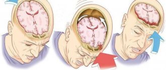
Symptoms of tumor development depend on its location and size. Depending on its location, it can impair coordination of movement (tumor in the cerebellum) and cause disturbances in speech, vision, and memory. Tumor growth in the left hemisphere can cause paralysis on the right side of the body. A patient with a brain tumor suffers from a headache, sensitivity is impaired, weakness appears, blood pressure surges, and tachycardia. When the hypothalamus or pituitary gland is damaged, endocrine disorders develop. Symptoms of the disease depend on the location of the tumor in a specific part of the brain responsible for certain functions.
Primary
When astrocytoma is localized in the frontal lobe of the brain, patients experience the appearance of psychopathological symptoms: a feeling of euphoria, decreased criticism of their illness, aggressiveness, emotional indifference, the psyche can completely collapse. If the corpus callosum or the medial surface of the frontal lobes is damaged, patients experience memory and thinking impairment. When Broca's area is damaged in the frontal lobe of the dominant hemisphere, motor speech disorders develop. Patients with tumors in the posterior regions develop paresis and paralysis in the upper and lower extremities.
In case of damage to the temporal lobe, patients may experience the appearance of hallucinations: auditory, gustatory, visual, which over time are replaced by generalized epileptic seizures. Often the development of auditory agnosia - a person does not recognize previously known sounds, voices, melodies. An astrocytoma located in the temporal region threatens to dislocate and herniate into the foramen magnum, resulting in almost inevitable death. When the tumor is localized in the temporal and frontal lobes, patients often experience epileptic seizures.
When a tumor affects the parietal lobe of the brain, sensory disturbances, astereognosis, and apraxia in the opposite limb occur (with apraxia, purposeful actions are impaired in a person). Patients experience the development of focal epileptic seizures. If the lower parts of the left parietal lobe are damaged, right-handers experience impairment in speech, counting, and writing.
The least commonly diagnosed astrocytomas are those in the occipital lobe of the brain. Patients with these tumors develop visual hallucinations, photopsia, and hemianopsia (loss of half the visual field of each eye).
Secondary
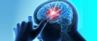
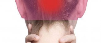
One of the main signs of cerebral astrocytoma is the presence of paroxysmal or aching diffuse pain in the head. The headache has no clear localization and is caused by intracranial hypertension. In the early stages of the disease, the pain is paroxysmal and aching, but over time it becomes constant, which is associated with the progression of the tumor.
In patients with cerebral astrocytoma, intracranial pressure increases as a result of compression of the cerebrospinal fluid ducts and venous vessels. Headaches, vomiting, persistent hiccups appear, cognitive functions and visual acuity decrease. Severe cases are accompanied by a person falling into a coma.
Diagnosis: types of tumor
The structure of malignant cells divides astrocytomas into two groups:
- fibrillar, gemistocytic, protoplasmic.
- piloid (pilocytic), subepidemic (glomerular), cerebellar microcystic.
Astrocytoma has several degrees of malignancy:
- first degree of malignancy - this type of benign tumor grows slowly, is small in size, is limited to healthy areas of the brain by a kind of capsule, and rarely affects the development of neurological deficits. The tumor is represented by normal astrocyte cells, which develop in the form of a nodule. A representative of such a tumor is piloid astrocytoma, pilocytic. Affects children and adolescents more often;
- second degree of malignancy - the tumor grows slowly, the cells begin to differ from normal brain cells, more common in young people aged 20 to 30 years. A representative of this degree of malignancy is fibrillar (diffuse) astrocytoma;
- third degree of malignancy - anaplastic astrocytoma. Grows rapidly, tumor cells are very different from normal brain cells, the tumor has a high level of malignancy;
- fourth degree - malignant glioblastoma, the cells do not look like normal brain cells. It affects important centers of the brain, grows quickly, and often it is impossible to remove such a tumor. It affects the cerebellum, the cerebral hemispheres, and the area of the brain responsible for the redistribution of information from the sensory organs - the thalamus.
Over time, tumor cells of the first and second degrees degenerate and turn into cells of the third and fourth degrees of malignancy. Transformation of a tumor from a benign to a malignant neoplasm most often occurs in adults. Benign brain tumors can be as life-threatening as malignant tumors. It all depends on the size of the formation and the location of the tumor.
Morphology
There are three different types of morphological structure: - cyst with a parietal node (the most common form), this form is most typical for infratentorial localization and occurs mainly in children, and solid formation and cystic-solid formation in adults.
On MRI, the cyst is ↑T2 and ↑Flair↓T1, and the solid component is → along T2 and T1 brain matter. A wall cyst usually consists of healthy cerebellar tissue, much less often tumor tissue. Calcifications occur in 11% of cases, and vasogenic edema in 5%.
On CT, the contents of the cyst are → cerebrospinal fluid, and the structures of the solid part → cerebral. Hemorrhages and necrosis are not typical.
Fig.3
Tumor cyst in the left hemisphere of the cerebellum (arrow heads in Fig. 3), the contents of the cyst have a ↑ Flair MR signal compared to unchanged cerebrospinal fluid (asterisk in Fig. 3). A parietal node on the inner side of the cyst wall (arrow in Fig. 3).
Treatment of astrocytoma
Treatment tactics for brain tumors depend on the location of the tumor, its size, and the type of tumor. A favorable prognosis in the treatment of diffuse astrocytoma exists for young patients, provided that the brain tumor is completely removed. Anaplastic astrocytoma is treated using a combined approach - surgery, radiation therapy, and polychemotherapy. The average life expectancy is about three years after surgery. The prognosis is favorable for young people who were in good health before surgery, provided that the tumor was completely removed.
Pilocytic (piloid) astrocytoma develops in children, has limited growth, characteristic localization, and morphological features. Treatment of the tumor has a favorable prognosis due to its slow growth and low malignancy. Treatment is carried out by surgery and complete removal of the tumor, which in some cases is not possible due to the location of the tumor in the hypothalamus or brain stem. Some types of piloidal astrocytomas (hypothalamic) have the ability to metastasize.
Biological behavior and follow-up
Dynamic changes in the absence of treatment are expressed in the growth of the tumor, an increase in its cystic part, which compresses and deforms the IV ventricle, reducing the communication of the ventricular and subarachnoid space, which leads to an increase in the pressure of the cerebrospinal fluid in the ventricular system, its expansion and damage to the brain substance. Dynamic changes are not fleeting, but always progressive.
Fig.7
As the tumor grows, an enlargement of the cyst is determined (Fig. 7), as well as the “mass effect” it produces with compression of the fourth ventricle and the formation of occlusive hydrocephalus with expansion of the third and lateral ventricles, as well as smoothing of the grooves.
Fig.8
Brain astrocytoma: consequences after surgery
The consequences after surgery for astrocytoma depend on the size of the tumor and its location. Benign astrocytomas, located in an accessible location, give a better prognosis for life expectancy than high-grade astrocytomas or benign astrocytomas, located in a location inaccessible to the surgeon and having a large tumor size. After removal of an astrocytoma, tumor recurrence often occurs, which occurs within two years after surgery. The earlier a tumor is detected, the better the prognosis for its treatment.
Forecast
After surgical removal of nodular forms of astrocytoma, long-term remission (more than ten years) can occur. Diffuse astrocytomas are characterized by frequent relapses, even after combined treatment.
With glioblastoma, the average life expectancy of a patient is one year, with anaplastic astrocytoma of the brain - about five years.
With other types of astrocytomas, the average life expectancy is much longer; after adequate treatment, they return to a full life and normal work activity.