Symptoms of a mild concussion include a decrease in the speed of psychomotor reactions and impaired attention. Sometimes there are mild impairments in the ability to abstract, make adequate judgments and make logical conclusions. In most cases, disorders are caused by psychogenic (mental, psychological, emotional, but not physical) factors.
Symptoms and signs
Symptoms of a concussion are quite variable and include the following:
- Headache.
- Unsystematic dizziness.
- General weakness.
- Nausea and vomiting once or twice during the first time after injury.
- Loss of consciousness for a short time at the time of injury.
- Impaired coordination of movements.
- Mood swings.
Signs of a concussion should also include an improvement in general condition on the first or second day after the injury. This is sometimes associated with such undesirable phenomena as the desire to stop treatment in a hospital setting.
Read also
Herpes zoster
Varicella zoster first enters the human body at an early age and manifests itself as chickenpox, most often in children.
Then follows the latent phase of circulation of the pathogen in the body and under certain conditions... Read more
Neuralgia
Neuralgia is a disease that is a lesion of the peripheral nerve. It manifests itself as a strong, burning, acute pain that is felt in the area of innervation of the affected (along) nerve. Kinds…
More details
Multiple sclerosis
Multiple sclerosis (MS) is a chronic disease, contrary to popular belief, completely unrelated to attention disorders and senile sclerosis. In this case, the term “sclerosis” means...
More details
Chiari malformation
What is Chiari Malformation? Chiari malformation (formerly Arnold-Chiari malformation) is a congenital defect of brain development that involves the dislocation of the cerebellar tonsils into the spinal canal through the large…
More details
Vertebral artery syndrome
The main vascular highways carrying blood to the brain pass in the neck - these are two carotid and two vertebral arteries. A decrease in blood flow through the vertebral arteries will lead to the development of acute...
More details
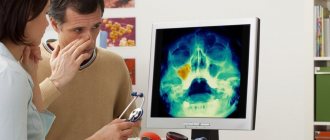
Diagnostics
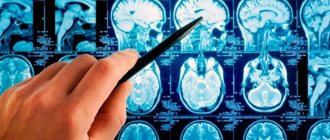
The diagnosis of concussion is rather a diagnosis of exclusion (it is necessary to exclude subarachnoid and subdural hemorrhage, brain contusion, diffuse axonal damage, etc.). To reliably make this diagnosis, it is necessary to conduct an examination (MSCT or MRI) to exclude violations of the integrity of bone structures, hemorrhages, and focal changes.
Diagnostic criteria also include the absence of severe symptoms, the presence (or absence) of mild cerebral symptoms, and diffuse organic symptoms. The fact of loss of consciousness after injury or the presence of severe nausea and one or two vomiting is also very important.
Concussion or not? Severe symptoms, pronounced neurological manifestations, the fact of prolonged loss of consciousness and repeated vomiting, amnesia after injury, even without structural changes in the brain, the fact of a violation of the integrity of the skull bones, persistent symptoms without dynamics against the background of prescribed treatment for 4-5 days, the slightest suspicion of focal lesions favor a more severe diagnosis.
Characteristics of mild concussion
Head injuries often cause disability and death. More often, traumatic brain injuries in adult patients are associated with car accidents, and in children - with outdoor games, sports, and falls from a height. Mild concussion occurs with a frequency of 80% among all traumatic injuries in the head area.
In the pathogenesis of disorders provoked by head trauma, the leading place is given to factors of sharp acceleration or braking to which the brain substance is exposed as a result of a collision of the head with a solid object. The large hemispheres are distinguished by the absence of tight fixation and mobility. At the moment of a blow to the head, they begin to rotate relative to the brain stem, which is tightly connected by nerve roots to the base of the skull.
As a result of the mobility of the cerebral hemispheres, the upper parts of the brain stem and thalamic structures are damaged, which leads to dysfunction of the reticular formation (a formation that runs along the entire axis of the brain stem). The reticular formation consists of nuclei and a branched network of neurons, abundantly equipped with axons and dendrites. The main functions of the reticular formation are activation of the cortical parts of the brain and control of the reflex activity of the spinal cord structures.
Damage to the structures of the reticular formation is clinically manifested by stupefaction or loss of consciousness. At the time of injury, a short-term increase in intracranial pressure occurs. A mild concussion of the brain is not accompanied by a change in the morphological structure of the tissue. Against the background of injury, metabolic disorders develop, which leads to dysfunction of neuronal membranes and disruption of the physiological processes in the nervous tissue.
Autopsy often shows the presence of axonal damage (tension and rupture of axons due to rapidly increasing acceleration or deceleration of the head) of a diffuse type in patients who suffered a TBI but died from other causes. The appearance of characteristic symptoms is based on microvascular damage of a reversible type, partial damage to the nervous tissue in the hippocampus and reticular formation of the thalamus. Damage to nerve tissue is also caused by the neurotoxic effect of excitatory neurotransmitters (amino acids).
Treatment
In order to answer the question of how to treat a concussion, you should turn to the manifestations, because treatment is most often symptomatic. Treatment of this pathology is carried out in a neurosurgical hospital for a period of 9 to 12 days, followed by outpatient rehabilitation if necessary. A certificate of incapacity for work is usually issued for two weeks in a hospital setting and only if necessary is extended for a longer period, especially for certain work factors that require concentration and quick psychomotor reactions. It is necessary to prescribe semi-bed rest for a period of one to two weeks.
Drug treatment of concussion is reduced to the prescription of general restorative and symptomatic drugs. For severe headaches, analgesics are prescribed (paracetamol, analgin, baralgin, etc.); for dizziness, it is sometimes advisable to prescribe betahistine, cavinton. In all cases, it is permissible to prescribe neuroprotective therapy; phenotropil, vitamin therapy, and glycine are often used. As a rule, all manifestations disappear completely within the first month after the injury.
MINOR BRAIN INJURY
Classification of mild brain injuryAccording to the domestic classification, traumatic brain injury (TBI) is divided into mild, moderate and severe. Mild TBI includes concussion and mild brain contusion [1]. While domestic and foreign authors generally agree on the understanding of severe TBI, there are differences regarding mild TBI. American authors group head trauma into severe (severe), moderate (moderate) and moderate (mild). Some American authors, in turn, divide moderate head trauma into mild (minor) and minimal (minimal) [5]. A minimal head injury corresponds to a concussion according to the Russian classification, and a mild head injury corresponds to a brain injury resembling a mild contusion according to the Russian classification. In the following, we will use the term “mild brain injury” (MTBI), since the term “mild TBI” is inappropriate in this situation, since the size and extent of brain damage are of greatest importance, while cranial trauma plays only an indirect role.
Statistics
The incidence of hospitalization for MTBI of mild head injury in the United States is 131 cases per 100,000 population and accounts for 60 to 82% of all hospitalizations due to head injury. With such a huge number of patients, LTBI has been called a “hidden epidemic.” More than $3.9 billion per year is spent on treating such patients in the United States [4]. Without having similar domestic statistics, we can only guess about the enormous economic damage caused by LTHM.
Definition of LTHM
LTBI includes two forms of TBI—concussion and mild brain contusion. 1. Concussion - SHM
(commotio cerebri, brain concussion) is a clinical syndrome caused by the action of mechanical forces on the brain and characterized by loss of consciousness for a short time after injury, without immediate and long-term evidence of structural damage to the brain.
Characterized by the absence of any residual effects, except for transient post-traumatic amnesia and other nonspecific symptoms such as dizziness, headache, etc. The diagnosis of FMS can only be made retrospectively. SHM can occur due to both acceleration-deceleration (impulsive) trauma and blows to the head. Trauma that causes head rotation is more likely to result in FMS. The mechanism of concussion is not precisely known. It is assumed that there is a functional disconnection between the brain stem and hemispheres of the brain. In this case, macroscopic and histological changes in brain tissue are not detected [2]. The definition of "loss of consciousness for a short time" can be interpreted in different ways. In the domestic classification, the time interval allotted for loss of consciousness during SHM ranges from several seconds to several tens of minutes, and for mild brain contusion - from tens of minutes to an hour. In practice, all this is quite conditional. Many Western experts consider the maximum duration of coma with BGM to be 6 hours [5]. If the patient regains consciousness before this time, the injury is interpreted as a concussion with a fairly good long-term prognosis. If the coma lasts more than 6 hours, then damage to the brain tissue is almost certain. Now all researchers are convinced that in this case we are talking about diffuse brain damage caused by acceleration-deceleration trauma, as a result of which axons in the white matter of the hemispheres and the brainstem are stretched and/or cut off. This is called diffuse axonal brain injury (DABI). It has always seemed illogical to many specialists in our country and abroad to make the same diagnosis of FMS both for boxers knocked down for a few seconds and for patients in a coma for more than an hour. Well-founded attempts to eliminate these contradictions have been made both in our country and abroad. This found a place in the classification of FMS according to severity. In the domestic literature of the 60s and 70s, one can find a division of FMS into three degrees of severity: mild, moderate and severe. At the level of modern knowledge, severe FMS is interpreted as DAP, and the remaining two degrees refer to various manifestations of FMS in its current understanding. In Western literature, some authors distinguished 3 or 4 degrees of severity of SHM. Currently, the division of FMS into degrees is of only historical interest, although much in the pathogenesis of FMS is still unclear. 2. The second manifestation of LTBI is a mild cerebral contusion
(UMCL), in which mild damage to the brain substance is microscopically determined in the form of areas of local edema, pinpoint cortical hemorrhages, possibly in combination with limited subarachnoid hemorrhage as a result of rupture of pial vessels [1]. It is almost impossible to distinguish between SHM and UGMLS by the duration of coma and post-traumatic amnesia, as well as by clinical manifestations. The classification adopted in Russia allows for the presence of linear fractures of the calvarium in UGMLS. An analogue of the UGMLS of the domestic classification is a minor head injury by American authors, which implies a condition that meets the following criteria [3]: 1) more than 12 points on the Glasgow Coma Scale (when observed in the clinic); 2) loss of consciousness and/or post-traumatic amnesia not exceeding 20 minutes; 3) hospitalization for less than 48 hours; 4) absence of clinical signs of contusion of the brainstem or cortex. Most American authors exclude patients with linear fractures of the cranial vault from this group of patients, thereby emphasizing that a skull fracture is a fundamentally more serious condition.
Clinical manifestations of LTBI
After the patient awakens, more or less adequate reactions are possible. After recovery of consciousness, but no more than 24 hours later, some confusion may persist. All patients have at least minor amnesia, usually retro- and/or anterograde. There may be friendly or unfriendly floating movements of the eyeballs, while oculomotor reflexes are preserved. The reaction of the pupils to light is preserved, but their width can spontaneously change. Spontaneous nystagmus is often found with the presence of fast phases, or it is easily caused by caloric tests. There are no motor disorders or they are transient in the form of a plastic increase in muscle tone and pathological foot signs. Subsequently, muscle hypotonia is characteristic. Some authors believe that decerebrate or decortication rigidity may occur rarely and for a very short time [2]. Vital functions usually do not change. There may be a vegetative reaction in the form of slight bradycardia or tachycardia, hypotension or hypertension.
Review of additional examination methods used for LTBI
1. Radiological research methods a) X-ray of the skull (RF) [4]. To date, RF has been a fairly reliable method for screening patients with LTBI. In cases of severe head trauma, computed tomography (CT) is preferable to radiography, which is not worth wasting time on. A CT scan must be performed if a skull fracture is detected. There is ongoing debate regarding the indications for routine RF or CT after mild head injury. All experts agree that X-ray examination is not necessary for completely adequate patients who have not lost consciousness after injury, who were clearly conscious during the examination without neurological symptoms, with the exception of those in whom a depressed fracture is suspected. One large study showed that more than 70% of patients who subsequently developed intracranial hematomas had some impairment of consciousness on admission, and the remainder had other indications for hospitalization. At the same time, the presence of a linear skull fracture in a conscious patient increases the risk of developing an intracranial hematoma by 400 times. b) Computed tomography. CT is the method of choice in the acute stage of head injury. The method makes it possible to detect skull fractures, meningeal and intracerebral hemorrhages, cerebral edema, and areas of contusion. CT scans in patients with BMS do not reveal any abnormalities in the state of the brain matter. With UGMLS, CT does not detect changes in 50% of cases. In the other half of cases, a limited area of cortical edema is detected with possible local narrowing of the subarachnoid fissures. c) Carotid angiography. Carotid angiography in the presence of CT is not used, but it can be performed when an intracranial traumatic mass lesion is suspected in the absence of CT or when a traumatic arterial aneurysm or arteriovenous fistula is suspected. d) Magnetic resonance imaging (MRI). Can be of great help in diagnosing DAP, as well as the evolution of various forms of brain injury in later phases of the process. In the acute stage, the method is less convenient than CT (skull fractures are not visible, fresh blood is poorly visualized, takes longer, etc.). In diagnosing UGMLS, MRI is a more informative method than CT.
2. Lumbar puncture (LP)
In case of brain injury, LP is contraindicated. The information content of the puncture is minimal: 1) based on the presence of blood in the cerebrospinal fluid, it is impossible to draw a conclusion about the severity and extent of brain damage (for example, with DAP and epidural hematoma, the cerebrospinal fluid may not contain blood); 2) there is no correlation between the number of red blood cells in the cerebrospinal fluid and the prevalence of traumatic subarachnoid hemorrhage, as well as the brain damage that caused it; 3) it is not always possible to judge intracranial pressure from the cerebrospinal fluid pressure measured during LP; 4) the threat to the patient’s life during and after LP is quite real due to possible dislocation of the brain stem. The only absolute indication for LP is suspicion of post-traumatic meningitis. But the disease rarely occurs in the first days after injury.
3. Electroencephalography (EEG)
EEG is rarely used in emergency neurotraumatology, in particular for diagnosing LTBI. EEG can help in severe brain injury regarding prognosis, as well as in diagnosing epileptic activity weeks and months after injury. The method allows you to make a decision on the prophylactic use of anticonvulsants. 4. Echoencephaloscopy (ECHOes)
One-dimensional echocardiography is a simple, fast, non-invasive and publicly available method for diagnosing traumatic supratentorial hematomas. Echo signals passing perpendicular to the temporal bone are assessed, and the position of the M-complex from the midline structures of the brain is revealed. The value of the method increases when used in dynamics. In case of pneumocephalus, associated, in particular, with a fracture of the base of the skull, ECHO is impossible due to the complete reflection of echo signals on the surface bordering the air. It is hardly advisable to perform echocardiography in patients with soft tissue trauma to the head without any neurological symptoms when examined adequately. At the dawn of the method’s development, as often happens, it was overestimated. Overdiagnosis concerned not so much volumetric intracranial formations, but the notorious “hypertensive-hydrocephalic syndrome” that developed after a mild head injury. Many neurologists are still confident that with the help of echoes it is possible to diagnose dilatation of the ventricular system and intracranial hypertension by dilatation of the median complex and additional echo signals. Control CT and MRI studies most often completely refute these findings.
Hospitalization for LTBI
All patients with moderate to severe TBI should, of course, be hospitalized. Patients whose head injury was not accompanied by loss of consciousness, amnesia, and who did not show any abnormalities during a neurological examination may be allowed to go home. The area of contention lies between these two extreme positions. We are talking about patients with LTBI who score 13–15 points on the Glasgow Coma Scale. Criteria for hospitalization of patients with PTHM, adopted in the United States: loss of consciousness for 10 minutes or more (or there is doubt about this); focal neurological symptoms; post-traumatic seizures; suspicion of a depressed fracture or penetrating wound; persistent change in consciousness; suspected fracture of the base of the skull and liquorrhea. The duration of hospitalization for SGM and UGMLS is 24 - 48 hours. In certain cases, it can be increased (mainly for social reasons). Recovery is considered complete 48–72 hours after injury. It must be emphasized that LTBI is an outpatient injury; patients with it are hospitalized mainly for close observation by medical personnel, and not for treatment in a specialized setting (in particular, a neurosurgical hospital). It has been proven that the duration of hospitalization and outpatient treatment does not affect the frequency and severity of post-concussion syndrome.
Treatment of LTBI
The mainstay of treatment for LTBI is rest for several days. Medications include analgesics and tranquilizers. As a rule, this is enough. If any vegetative symptoms are excessively pronounced, then additional correction may be required, for example, b-blockers for severe tachycardia and hypertension. The presence of concomitant diseases and old age contribute to symptomatic treatment. The effectiveness of nootropics and drugs that improve cerebral blood flow in LTBI has not been proven. Prophylactic use of anticonvulsants during LTHM is indicated only for patients with a history of seizures and the presence of seizure readiness on the EEG. The prescription of diuretics, xanthine derivatives (in particular, aminophylline), B vitamins, magnesium sulfate and other drugs with unproven effects for IHT can be considered nothing more than an anachronism.
Prognosis for LTBI
The mortality rate with SHM is 0. If a CT scan reveals slight swelling in the cerebral cortex (which corresponds to UGMLS), then the mortality rate is less than 2%. With brain contusions, the mortality rate is 5%. Exit from coma during SGM and UGMLS of the brain without residual effects. Headache, dizziness, and autonomic symptoms with LTBI disappear within a few days or weeks after injury, but can last for months. The development of any consequences, in particular convulsive syndrome, is not typical for LTHM.
Post-traumatic syndrome
Post-traumatic syndrome - PS (post-commotion syndrome, post-traumatic neurosis) is a common complication of brain injury, including mild ones. Occurs in 35–40% of patients who have suffered mild to severe head trauma [5]. In principle, any of the listed syndrome names is unsatisfactory, since in all cases it is implied that the symptoms are a direct result of the brain injury or the patient's psychological reaction to the injury. In reality, everything is much more complicated. Intensive psychological and physiological research has not found any criteria that could help determine the role of physiological or psychological response to brain injury in the occurrence of the syndrome. In some patients, symptoms may depend on the degree of brain damage; in others, they appear to be purely psychogenic. In practice, it is difficult to find the cause of symptoms, especially if there are no abnormalities on neurological examination. Symptoms can develop in patients who have undergone only BMS with loss of consciousness for a few seconds, but PS is more often observed in patients who have been unconscious for a more or less long time. However, there was no clear correlation between the severity of PS and the duration of coma or post-traumatic amnesia. Signs of PS: headache, dizziness, insomnia, increased irritability, decreased ability to concentrate, increased sensitivity to noise and light, various fears, anxiety, hypochondria and depression. The duration of symptoms varies from several weeks to several years. Symptoms of PS, usually characteristic of adults, are rare in children, but behavioral disorders and personality changes are more common in children.
PS diagnostics
There are no clear criteria for PS. PS can develop in mentally healthy patients with no psychological abnormalities in the premorbid period, but it is more common in individuals who had any neurotic symptoms before the injury. Factors such as high-risk work, home or financial problems, and the desire to receive material or other compensation play an important role in the development of PS and its prolongation. The role of MRI in the diagnosis of PS has not yet been determined. Symptoms of the disease may appear in patients without any changes on CT and MRI.
Prognosis for PS
The outcome of a PS is never known in advance. More often than not, you can expect progressive improvement. The duration of symptoms does not depend on the severity of the brain injury.
Treatment of PS
Adequate treatment in the acute period of injury is a very important factor in the prevention of PS. For suspicious patients, the idea of receiving even a mild brain injury is closely connected with the fear of acquiring all sorts of unimaginable complications and consequences, such as mental illness or brain tumors. Often, medical personnel play an important role in the development of PS, aggravating the fear of suffering the consequences of a brain injury with stories about how close the patient was to the brink of death, or frightening them with late complications of a brain injury, and most importantly, by keeping the patient from activating within a reasonable time frame. After several weeks or months, patients may not notice any improvement in their symptoms, which further neuroticizes them, supporting the idea that the consequences of the trauma with which they were frightened in the hospital have developed. A decrease in the patient's economic or social status further intensifies and prolongs symptoms. PS is also aggravated by the untimely activation of law enforcement agencies, which constantly returns the patient to a stressful situation with their interrogations. The basis of treatment is psychotherapy. Physiotherapeutic procedures and antidepressants may also be used.
Literature:
1. Konovalov A. N., Likhterman L. B., Potapov A. A. Classification of traumatic brain injury. Sat. scientific works of the Institute of Chemistry. - M., 1992. - P. 28-29. 2. Plum F., Pozdner J.B. Diagnosis of stupor and coma: Transl. from English - M.: Medicine, 1986. - P. 181-184. 3. Mandel S, Sataloff RT, Schapiro SR (editors). Minor head trauma. Springer-Verlag 1993;8-44. 4. Masters SJ, McClean PM, Arcanese MS, et al. Scull X-ray examinations after head injury: Recommendations by a multidisciplinary panel and validation study. N Engl J Med 1987;316:84-91. 5. Rowland LP. Merritt`s textbook of neurology. - 9th ed.: Williams and Wilkins 1995;418-423,437-438.
First aid for a concussion
Help for a mild concussion is to alleviate the condition of the victim and prevent complications. First of all, you should call an ambulance, since most often not every person can correctly assess the condition.
Important! It is strictly forbidden to leave the victim unattended.
After a brain injury, nausea may occur, which may be accompanied by vomiting. To prevent a person from choking on vomit, his condition should be closely monitored. Convulsions also occur, the state of health may sharply deteriorate, and the victim will need to be brought to consciousness. This is why a person should not be left alone.
Many people do not know what to do if they have a mild concussion. The sequence of actions for providing first aid for a concussion is as follows:
- Lay the victim on his side or back. But a person may not realize what happened and claim that everything is fine with him and try to leave, which is prohibited from doing with a mild concussion. In this case, the victim should be persuaded to wait for an ambulance.
- If there is loss of consciousness, you need to make sure that breathing and heartbeat are present.
- Check your pulse.
- If there are other injuries, it is necessary to treat the wounds with an antiseptic. This will help prevent infection.
- Apply cold to the bruises.
- Ask witnesses about the condition and details of what happened.
If the victim is conscious and answers all questions, you should put a pillow under his head. This is necessary so that the upper body is on a slight elevation. In cases where the victim is indoors, it is recommended to turn off the bright lights. If there is a threat to life, it is necessary to provide resuscitation measures, consisting of artificial respiration and cardiac massage.
If a child has a concussion, it is necessary to take him to a neurologist. It is important that the baby remains conscious during the first hour. His physical activity should be limited in order to avoid negative consequences. Many parents try to give medications for a mild concussion, which is strictly prohibited. This can only harm the baby.
How to treat?
With a mild form of concussion, it is necessary to be under constant supervision of doctors such as an ophthalmologist, neurologist, neurosurgeon and traumatologist. A patient with a concussion is prescribed strict bed rest for two or three weeks. It is advisable to spend this time in a hospital to be under constant medical supervision.
What to take and how to treat a mild concussion? To relieve pain, the patient is prescribed analgesics - Baralgin, Ketorol, Sedalgin. Mainly used as sedatives for concussions are tinctures of motherwort and valerian, as well as tranquilizers - Phenazepam, Relanium.
Effective remedies - Bellataminal, Bellaspon, Cinnarizine - are what treat dizziness. In order to relieve the patient's general tension, magnesium sulfate is prescribed. Diuretics, in particular Diacarb, help prevent cerebral edema. Often, for concussions, doctors prescribe the use of vascular drugs and nootropics - Cavinton, Trental, Piracetam, Nootropil, as well as B vitamins.
Attention! If the patient is not in a hospital setting, but at home, then it is important to provide him with complete rest and remove all irritants - in no case should you watch TV, work or play on the computer, or listen to loud music. With the permission of a doctor, herbal medicine can be used.
What is a mild concussion, how does it manifest and how is it treated?
A concussion is one of the most common types of traumatic brain injury. There are many reasons for the occurrence, most often these are sports, household and impacts received as a result of an accident. The signs of a mild concussion in children and adults have some differences. In case of injury, it is important to provide first aid to the person, as the condition may worsen.
Important! If left untreated, a concussion can cause serious consequences.
Complications
If left untreated, post-concussion syndrome develops in case of injury. This is a set of symptoms for a mild concussion that occurs several months after a blow or bruise. Among the complications observed:
- Migraine-like headache.
- Dizziness.
- Sensitivity to light and loud, sudden sounds.
- Tinnitus.
- Double or blurred vision.
- Sleep disorders.
- Unstable psycho-emotional state.
- Impaired concentration.
- Difficulties in learning new information.
- Forgetfulness.
There is no special treatment in this case, but based on many years of research, the effectiveness of anti-migraine medications has been proven. Antidepressants and psychotherapy are also used.
When diagnosing a mild concussion, the attending physician will decide what to take based on the condition and individual characteristics of the patient’s body. Self-medication can cause serious complications in the future. That is why, if you suspect an injury, you should contact a specialist.
What is a concussion
Concussion is a mild type of TBI (traumatic brain injury), which is characterized by temporary impairment of cerebral functions and morphological abnormalities of the brain. In some cases, there is an unconscious state from several seconds to half an hour. A longer stay of the victim in an unconscious state indicates damage to cerebral tissue.
In medicine, there are three degrees of injury. The first is considered the easiest and may not be noticed immediately after a bruise or blow. Concussion occurs in patients of any age, including children over 2 years of age.
Causes of concussion
A mild concussion is the result of direct or indirect mechanical impact. When a bruise or blow occurs, the brain is suddenly displaced, causing damage to the synoptic apparatus and movement of tissue fluid. As a result, characteristic symptoms arise.
The main causes of concussion are:
- Road accident. A large number of injuries of this type occur after road traffic accidents. A mild concussion occurs due to a blow or sudden change in the position of the head and neck.
- Domestic injuries. Minor head impacts on furniture.
- Sports. Most often, concussions occur in people involved in martial arts, alpine skiing, and acrobatics.
- Production. In factories and various enterprises there is a fairly high risk of head injury.
A slight concussion can also be obtained through criminal means. This category includes injuries after fights or beatings.