Find out more about diseases starting with the letter “O”: Fainting (syncope); Occlusive hydrocephalus; Occlusion of the carotid arteries; Olivopontocerebellar degenerations; Optico-chiasmatic arachnoiditis; Neuromyelitis optica; Brain tumors; Cerebellar tumors; Tumors of peripheral nerves; Spinal cord tumors; Brain stem tumors; Central nervous system tumors; Tumor of the cauda equina; Tumor of the pineal gland; Oromandibular dystonia; Osteocondritis of the spine; Brain swelling; Carbon monoxide poisoning; Ophthalmoplegic migraine.
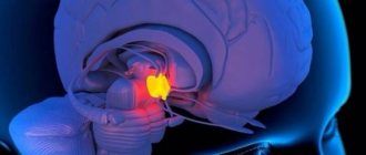
A cerebellar tumor is a pathological formation in the cerebellum, benign or malignant in nature. It may have an independent origin or be a secondary disease of a metastatic nature. The diverse clinical picture is divided into 3 groups - cerebral, cerebellar and brainstem symptoms.
Establishing a conclusion about a tumor is based on data from magnetic resonance scanning of brain structures. The diagnosis is confirmed after receiving the results of a histological examination of the formation material. Treatment of a cerebellar tumor involves surgery with absolute removal of the lesion, restoration of the flow of cerebrospinal fluid, and elimination of compression of the brain stem.
general information
Of the total number of diseases with brain tumors, cerebellar tumors account for approximately 30%. A detailed histological study makes it possible to determine more than 100 morphological types of cerebellar formations. The results of numerous studies by neurologists, neurosurgeons, and oncologists confirm that in the vast majority of cases—70%—glioma develops.
The possibility of developing pathology does not have limited age criteria. However, medulloblastoma is diagnosed more often in children, hemangioblastoma, astrocytoma - in middle-aged people, in older patients - glioblastoma, metastatic neoplasms. The risk of developing cerebellar cancer is higher in Caucasian men.
Forecast
If cerebellar cancer is detected, the prognosis depends on the stage at which treatment was performed and the size of the tumor.
But even with therapy in the early stages, life expectancy does not exceed five years. This is due to the fact that the tumor affects important parts of the brain, rapidly spreads to neighboring structures and causes complications.
In later stages, surgical intervention is usually not performed, as the tumor begins to disintegrate and becomes inoperable. In this case, maintenance therapy is prescribed. Life expectancy is no more than 1 year.
A cancerous tumor affecting the tissue of the cerebellum occurs as a result of exposure to a number of negative factors. But experts have not established the exact causes of the development of the pathology, which does not allow determining preventive measures.
Treatment of pathology is carried out comprehensively, using not only surgery, but also chemotherapy. But even with this approach it is not possible to achieve a favorable approach. Removing the tumor can only prolong the patient’s life.
Pathogenesis and causes of disease development
Factors promoting the development and growth of tumor cells are not known. According to medical research, it has been determined that with the primary disease of Recklinghausen's neurofibromatosis, 10 of the patients develop a hereditary tumor of the cerebellum. It is generally accepted that tumor provocateurs can be exposure to radiation, oncogenic viruses (herpes, human papillomas, some adenoviruses), and the effect of carcinogenic chemical compounds on humans. The risk of developing malignant tumors increases in patients undergoing immunosuppressive treatment (for example, HIV-infected patients).
The mechanisms of neoplasm development occur in the following directions:
- Damage to the cerebellar substance occurs due to compression by the tumor with further death. This process is accompanied by focal cerebellar symptoms.
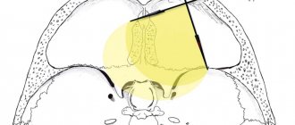
- The longer the tumor develops, the more it fills the fourth ventricle and compresses the brain stem, which is accompanied by dysfunction of the cranial nerves and brain stem clinical signs.
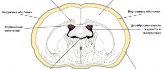
- Increasing hydrocephalus of the brain is accompanied by a general cerebral clinical picture and an increase in intracranial pressure. The cerebellar tonsils descend and become pinched in the posterior cranial fossa. The medulla oblongata is located between the occipital bone and the cerebellum. These processes cause bulbar disorders, complications from the cardiovascular system and breathing.
Methods for preventing cerebellar tumors
Specialists from Israeli cancer centers emphasize that without knowing the exact mechanisms of pathology, it is not easy to recommend preventive measures. In addition, doctors suggest that the pathogenesis of the disease involves factors such as the negative effects of ionizing radiation, a decrease in the body's immune defense, as well as inherited predisposition.
In order to prevent the development of the disease, n
It is necessary to adhere to a number of recommendations:
- take care of your health;
- visit the doctor periodically and strictly follow his instructions;
- live an active life;
- rest periodically;
- avoid unnecessary stress;
- devote time to sports;
- eat rationally;
- do not practice self-treatment.
Classification
Cerebellar tumors, like all tumors, regardless of location, are divided into malignant and benign. Medulloblastoma is a tumor characterized by rapid growth and invasion of the subarachnoid membranes. In children, it is the most commonly diagnosed type of cerebellar malignancy.
The second place is occupied by cerebellar sarcoma, according to general data on prevalence. Locally spreading hemangioblastomas and astrocytomas with infiltrative growth are the most common benign tumors of the cerebellum. In most cases, they have a cystic modification and consist of a small node and a cystic cavity of significant size.
With any form of tumor, the brain stem is subject to compression due to the small lumen of the posterior cranial fossa. This is risky in any case, so the division of neoplasms into benign and malignant is very arbitrary.
Based on the source of development, tumor processes of the cerebellum are divided into 2 significant groups - primary and secondary neoplasms. The primary tumor is formed from the cells of the cerebellum, as a result of persistent replacement of cells of its substance with pathological cells (the process of metaplasia starts).
Primary tumor foci can be benign or malignant. Metastatic origin is characteristic of secondary cerebellar neoplasms. They can develop in breast cancer, thyroid cancer, malignant tumors of the gastrointestinal tract and lungs. Secondary tumors invariably have a malignant nature.
Predictions for the development of cancer of cerebellar tumors for different stages
The results of treatment of cerebellar pathologies are significantly determined by their scale, distribution patterns, and level of malignancy. If the tumors are benign and completely eliminated, the prognosis is favorable. If the necessary treatment does not occur with progress in the growth of the formation, the patient dies from compression of the stem structures responsible for respiratory processes and cardiovascular processes. If a benign tumor is not completely eliminated, the pathology recurs and after a couple of years another operation will be needed. As for malignant tumors, they are much less favorable. Survival after surgery in combination with adjuvant therapy is usually about 1-5 years.
Symptoms
The picture of the disease in cerebellar tumors consists of general cerebral, cerebellar symptoms, and phenomena of damage to the brain stem. Very often, all three groups of symptoms appear simultaneously.
Sometimes the onset of the disease is manifested by symptoms of one type. General cerebral phenomena occur first if the tumor is localized in the cerebellar vermis. The process taking place at this time directly in the tissues of the cerebellum may not produce any symptoms and remain in the compensation stage for a long time. Symptoms of cranial nerve damage or indications of brainstem compression may also be the first manifestations of the disease.
Tumor diseases of the cerebellum have the same general cerebral symptoms as tumors of the cerebral hemispheres. Patients report constant headaches, sometimes of a paroxysmal, progressive nature. The headache comes in the morning, is widespread, and is less often centralized in the back of the head. The feeling of nausea comes regardless of food. At the peak of the headache, vomiting, dizziness, stunned state, and drowsiness are possible.
Patients complain of fatigue. Sometimes hallucinations of the olfactory, auditory or light type develop. The more the tumor blocks the vessels through which the cerebrospinal fluid passes, the more the clinical picture increases. The patient is forced to take a position that alleviates his condition - tilt his head forward or backward, knee-elbow position with his head down.
Attacks of nausea and vomiting come more often. If the patient suddenly changes the position of the head, sudden blocking and the onset of a hypertensive-hydrocephalic crisis are possible.
Focal (cerebellar) signs depend on the location of the tumor process. Cerebellar ataxia is the main clinical factor. If the process is located in the cerebellar vermis, instability and gait disturbances are observed. When walking, the patient stumbles, staggers, and spreads his legs wide. To maintain balance, the patient balances with his hands. The patient may skid when turning.
Involuntary movements of the eyeballs are observed - nystagmus. Cerebellar speech disorders develop. It has an intermittent and chanting character (divided into syllables). When the cerebellar hemispheres are involved in the tumor process, disorders of coordination and proportionality of movements are noted on the affected side. The patient cannot perform finger-nose, knee-heel tests. There are changes in handwriting to a more sweeping, larger one, as well as dysmetria and intention tremor.
A cerebellar neoplasm can grow from one hemisphere of the cerebellum to the other, from the hemisphere to the vermis and vice versa. Impaired coordination and confusion of symptoms of damage to the cerebellar structures are manifested in a variety of clinical pictures.
Damage to the brain stem has its own signs of compression, as well as dysfunction of individual cranial nerves. Patients experience trigeminal neuralgia, neuritis of the facial nerve, hearing impairment, pathologies of taste perception, speech impairment, and complete loss of sensitivity of the soft palate.
Vomiting not associated with headache is a typical manifestation of brainstem lesions. It is caused by irritation of the receptors in the posterior fossa of the skull and can appear due to sudden movements or changes in body position. Long-term compression of the brainstem provokes motor restlessness, irregular heartbeat, double vision, increasing nystagmus, oculomotor disorders - gaze paresis, divergent strabismus, drooping eyelid, mydriasis.
Possible autonomic disorders, tonic convulsions, arrhythmia. Impairment of respiratory activity is possible until breathing stops completely, which can lead to the death of the patient.
Diagnostics
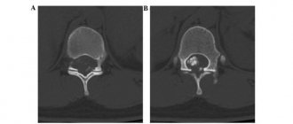
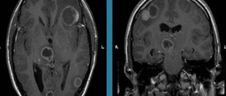
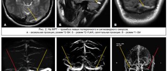
An examination by a neurologist does not always make it possible to determine the presence of a cerebellar tumor, since specific manifestations may be absent. Identification of cerebral symptoms is the reason why the patient is referred to an ophthalmologist for consultation and ophthalmoscopy. Hydrocephalus is indicated by congestion of the optic nerves. A CT or MRI of the brain is required. The examination results provide complete information about the location of the tumor and its size.
It is necessary to differentiate cerebellar tumors from cerebellar cysts, aneurysms of cerebral vessels, brain abscesses, and stroke.
Etiology
Medulloblastomas belong to the group of embryonal neuroepithelial tumors. It is believed that medulloblastomas develop from cells of the outer granular layer of the cerebellum and the posterior cerebellar velum. There are 2 types of medulloblastomas: “classical” type medulloblastomas and dysmoplastic medulloblastomas.
Medulloblastomas disseminate through the cerebrospinal fluid system, producing metastatic nodes in the subarachnoid space of the brain and spinal cord, in the walls of the ventricles, in the area of the chiasm, and in the basal parts of the brain. It is extremely rare that metastases occur extracranially - in the bone marrow, lungs, and liver.
Treatment
Surgical treatment is the main treatment for cerebellar tumors. The question of the need and extent of the operation is considered by a neurosurgeon. Radical removal of the tumor is considered the most effective. A contraindication to surgery is considered to be tumor growth into the brain structures, into the fourth ventricle. In such a situation, the circulation of cerebrospinal fluid is restored and the possible volume of tumor formation is removed. It is advisable to perform shunt manipulations, ventricular puncture, external ventricular drainage, depending on the symptoms.
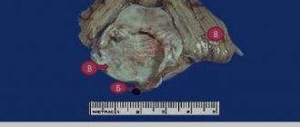
The material obtained during the operation is subject to histological examination to determine the degree of malignancy. Based on the results, further treatment tactics, chemotherapy and radiation treatment are determined. If necessary, painkillers, diuretics, sedatives, and antiemetics are prescribed.
The presence of a cerebellar tumor can be detected using MRI. You can find a clinic that allows you to undergo tomography using modern equipment on our website. Here you can find hundreds of clinics with high ratings and affordable prices. Making an appointment through the website gives you the opportunity to receive a discount on the examination.
Treatment strategy
Treatment of patients with medulloblastomas currently consists of three stages: surgical removal of the tumor and restoration of normal cerebrospinal fluid circulation, radiation therapy and chemotherapy.
Complex therapy begins with tumor removal. With timely comprehensive treatment, the 50-year survival rate of patients with medulloblastomas is 70-80%. The progress of surgical technology (use of a microscope, ultrasonic suction), anesthesiology and resuscitation has significantly affected the reduction of surgical mortality, as well as increasing the radicality of tumor removal.