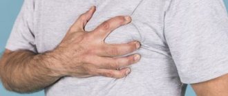
Severity of injury
In order for the provision of assistance to the victim to really have the desired effect, experts recommend conducting at least a cursory initial examination before starting pre-medical procedures. This will allow you to determine the approximate severity of the head injury.
The easiest way to cope is with a mild degree of the disease, which is more typical of accidental household injuries such as hitting your head on the edge of a cabinet. The deviation is not accompanied by any heavy bleeding and does not bring any particular discomfort during recovery. Typically, such mild bruises are limited to the formation of a small swelling, which is popularly called a “bump.”
It goes away on its own, but sometimes, to speed up the process, it is allowed to apply cold immediately after the blow. This will somewhat reduce pain and also limit the spread of swelling.
The situation with damage to the integrity of the skin is a little more complicated. The category includes not only extensive wounds that occur when a large area of skin along with hair is combed. Even ordinary abrasions provoke an outflow of blood. But only in the first case the patient has to deal with a large amount of blood, and in the second case only capillary bleeding is stopped.
If emergency measures are taken, it will be possible to block capillary breaks to stop bleeding within 5-10 minutes.
Three other categories are considered separately:
- jaw damage;
- traumatic brain injury;
- combined options.
Complex pathologies are more suitable for describing polytrauma, when, in addition to deformation of the head, destabilization of the spine or any other part of the body is simultaneously recorded. This format is most often found in road accidents, fights and industrial injuries, when there is damage to the chest and limbs.
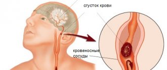
A particularly serious development of events is a related deviation in the issue of the integrity of the neck systems. We are talking about the rupture of nerve endings and large vessels designed to nourish the brain.
Almost always, a head blow is accompanied by minor, but still skin injuries. In addition to the immediate top layer, the natural integrity of the fatty tissue is disrupted. Because of this, blood flow through the affected vessels is obstructed. The clinical picture ends with the collection of blood at the site of localization, which externally transforms into a hematoma of various sizes.
Depending on the severity and duration of the blow, the color of the hematoma changes. If initially it turns purple due to fresh blood stains under the skin, then later the tone changes. The transformation is due to the launch of the process of hemoglobin breakdown and increased activity of leukocytes. Externally, such manifestations are expressed in a change in color to blue-green with yellowness.
The swampy tint is provided by biliverdin, and bilirubin is responsible for imparting yellowness.
If there is strong external pressure during a mechanical injury, it is often accompanied by destabilization of the tendon helmet. Doctors recognize such a deviation due to a hematoma, which seems to be spreading across the face.
When a victim falls on their face, they have to deal with soft tissue damage. There is one pitfall here that ordinary people do not know about. Some people unknowingly confuse the outer rim of a hemorrhage with a depressed cranial fracture.
Principles of providing first aid for severe TBI
When providing assistance to a victim with a traumatic brain injury, you must adhere to the following rules:
- put the person on a horizontal surface and ensure peace;
- the head and cervical region should be fixed. A homemade roller made from clothing or any dressing material is suitable for this. This point is especially important if it is necessary to move the victim.
- to prevent swelling, apply cold (ice, snow, a bottle of water) to the injury site;
- Cover the open wound with clean material. If there is a foreign object in the wound, do not try to pull it out under any circumstances. Cover it with tampons on all sides and bandage it using the crosswise technique.
- if you notice blood from the ear, nose or mouth, cover with a sterile bandage and turn the person to the side where the liquid is flowing;
- monitor the victim’s condition: check pulse, breathing;
- transportation is carried out only in cases of emergency. Definitely with neck and head fixation.
- in the presence of vomiting and any brain damage, pain relief cannot be administered until the ambulance arrives;
- if breathing stops, begin resuscitation measures (indirect cardiac massage and artificial respiration).
Thus , before the medical team arrives, you must provide the victim with rest, apply cold, immobilize the head and neck, and monitor the general condition. It is prohibited to give medications to the victim. Given the seriousness of traumatic brain injuries, hospitalization or evaluation by an appropriate specialist is necessary to prevent unwanted health problems.
Specific symptoms
All symptoms accompanying any type of head injury are expressed differently depending on the specific location of the lesion. If we are talking about the facial part, when the usual integrity of the skin is broken and deep tissues are affected, then the first dangerous sign is pain. The syndrome is reinforced by hemorrhage. After a while, a bruise appears on the face.
If serious damage to the skull is recorded, the victim is faced with the same phenomena that can be observed with a cerebral contusion:
- nose bleed;
- dizziness;
- headache;
- vomit.
In such a scenario, medical care should be provided immediately, before irreversible processes begin.
With occipital lesions, patients often complain of the inability to focus their eyes and other vision problems. This is explained by the anatomical features of the human body, because it is in the back of the head that the center responsible for visual functions is located.
In this situation, concussions and general weakness are not uncommon, which often becomes the basis for frequent fainting.
An eye injury is marked by a circular hemorrhage. But not everyone knows how to distinguish it from the so-called glasses effect. The latter is suitable to describe the condition of a cranial fracture.
Those whose jaws are injured will need emergency medical care. Here, the first step is to check the chewing function. If, during a light test, it turns out that a person is not able to make the usual chewing movement, then this indicates a fracture.
The condition of children who have been victims of head injuries should be checked especially carefully. In children, all types of presented deviations may not manifest themselves at first, but in adolescence or even adulthood they may result in serious deviations in brain activity.
ARTIFICIAL RESPIRATION
ARTIFICIAL RESPIRATION is an emergency first aid measure for drowning, suffocation, electric shock, heat and sunstroke. This is carried out until the victim’s breathing is restored.
THE MECHANISM OF ARTIFICIAL RESPIRATION is as follows:
- place the victim on a horizontal surface;
- clear the victim’s mouth and throat of saliva, mucus, soil and other foreign objects; if the jaws are tightly clenched, move them apart;
- tilt the victim’s head back, placing one hand on the forehead and the other on the back of the head;
- take a deep breath, bend over to the victim, seal the area of his mouth with your lips and exhale. The exhalation should last about 1 second and help lift the victim’s chest. In this case, the victim’s nostrils should be closed and his mouth covered with gauze or a handkerchief for reasons of hygiene;
— frequency of artificial respiration – 16-18 times per minute;
- periodically empty the victim’s stomach of air by pressing on the epigastric region.
Clinical features of head injuries
All head injuries can be divided into several broad categories according to a standard principle, as is typical for sorting injuries to the abdomen or chest.
Schematically, the classification is divided into three divisions:
- closed;
- open;
- penetrating.
The first point implies the absence of destabilization of the aponeurosis. Instead, there are bruises of various locations and sizes, as well as other versions of soft tissue rupture.
With open analogues, the emphasis is on damage to the aponeurosis. But the worst situation is with penetrating wounds, when in addition to destruction of the integrity of the surface and subcutaneous tissue with vessels and nerve roots, damage to the dura mater is noted. It is these variations that harm the patient the most.
In addition to the typical classification by mechanism of injury, the medical classification has another structured sorting. It is based on clinical manifestations, including:
- brain concussion;
- bruises;
- compression by intracranial hematomas.
Classic concussion occurs in approximately 80% of all recorded cases of damage to the head of the body. It is characterized by the absence of macrostructural pathology, which is explained by the presence of damage only at the cellular level. Physiologically, a concussion can be designated a functionally reversible pathology.
Specific manifestations of this condition are called:
- being unconscious for up to several minutes;
- nausea;
- vomiting;
- amnesia;
- dizziness;
- spreading headache;
- increased sweating;
- double vision.
At the same time, vital functions remain normal. But neurological abnormalities, even in minor manifestations, are still diagnosed. They cover:
- asymmetry of tendon reflexes;
- small-scale nystagmus.
All of the above goes away over the next week with fairly effective therapy. But in order to ensure that there are no more significant consequences, the victim will be required to undergo additional examination. This includes the need to provide images obtained during a CT scan or magnetic resonance therapy.
No less often, patients are found with intracranial hematomas, which, as they grow, begin to compress still healthy tissue. Bruises form under or above the dura mater. The cause of the disease is depressed fractures of the cranial bones.
Despite the fact that their clinical manifestations are similar to those of a brain contusion, they still have their own distinctive features. The main sign of this is the so-called “light gap”.
Doctors call this the time interval between waking up after fainting and a sharp plunge into a coma. At this time, the person feels relief, but in fact, cerebral edema begins to rapidly progress.
FAINTING
Fainting is a sudden short-term loss of consciousness, accompanied by weakening of the heart and breathing. It occurs with rapidly developing anemia of the brain and lasts from a few seconds to 5-10 minutes or more.
SIGNS. Fainting is expressed in a sudden onset of lightheadedness, dizziness, weakness and loss of consciousness.
Fainting is accompanied by paleness and coldness of the skin. Breathing is slow, shallow, weak and rare pulse (up to 40-50 beats per minute).
FIRST AID. First of all, it is necessary to lay the victim on his back so that his head is slightly lowered and his legs are raised. To make breathing easier, free your neck and chest from constricting clothing. Cover the victim warmly and place a heating pad at his feet. Rub the patient's temples with ammonia and bring a cotton swab soaked in ammonia to his nose, and sprinkle his face with cold water. In case of prolonged fainting, artificial respiration is indicated. After regaining consciousness, give him hot coffee.
Brain contusions
Lesions such as bruises of the brain itself are considered separately. They are divided into three degrees of severity, which are distinguished by gross macrostructural damage to the brain matter.
This manifests itself as hemorrhage and destruction.
There is no bruise without subarachnoid hemorrhage. Fractures of the skull bones are frequent accompaniments of the presented injuries. They are accompanied by significant edema with swelling of the brain substance.
If the patient, after direct external influence on the skull, remained unconscious for no more than twenty minutes, then this indicates a mild version of the development of events. After waking up, victims begin to complain of endless dizziness, which is complemented by nausea and vomiting.
Such patients have retrograde or anterograde amnesia. Against the background of stable vital functions, disruption of the usual activity of the cardiovascular system looks more depressing. We are talking about bradycardia or hypertension.
On the neurological side, experts note pyramidal insufficiency, clonic nystagmus and mild anisocoria.
With moderate severity, disconnection from reality can last about two hours. Almost immediately after waking up, a person begins to experience repeated vomiting. Additionally, pronounced amnesia manifests itself, and even some deviations in mental health.
Vital disturbances are expressed in prolonged bradycardia, which is supported by hypertension along with tachypnea. But the airways remain normal, which reduces the risks of more serious diseases.
The neurological status may include all those signs that are characteristic of a mild course of the disease, and may also be diluted by the asymmetry of tendon reflexes with muscle tone.
In some cases, meningeal symptoms and pathological traces make themselves felt. Among other things, focal types of destabilization such as pupillary, oculomotor, speech disorders, as well as paresis of the limbs are occasionally possible.
Severe degree is characterized by a long duration. A person may remain in a coma for several weeks, while his vital functions are seriously impaired, posing an increased threat to life.
The most pronounced alarming symptoms are brainstem symptoms, which include floating movements of the eyeballs, destabilization of the rhythm and frequency of breathing. Paresis of the limbs, convulsive seizures, and pathological foot signs are diagnosed.
The longer the coma lasts, the less favorable the prognosis for later life becomes. The reason for this is the large affected areas. Including disruption of the bone structures of the head, as well as massive subarachnoid hemorrhages.
If you do not see a doctor in time after an injury, the consequences for a person can be quite unpleasant: decreased or loss of memory, impaired attention, difficulty doing household chores, emotional distress, etc.
Bleeding after open traumatic brain injury and other open head wounds
In case of injury accompanied by heavy bleeding with pulsation, hemostasis should be performed immediately. With frontal head injuries, fractures of the bones that form the upper jaw and frontal, maxillary sinuses and nose may occur with rupture of blood vessels and nerves . With open fractures of the lower jaw, even larger vessels can also be damaged with bleeding into the oral cavity. In this case, if a person is unconscious after a traumatic brain injury, blood can either be swallowed or enter the bronchi.
- If you can see the direction of the vessel on the head, apply pressure to the damaged blood vessel, pressing it to the underlying bone with your fingers. If bleeding from the head is accompanied by pulsation, press the carotid artery of the corresponding side with your fingers for 10 minutes. with breaks. Some blood loss during these breaks is inevitable, but the body can handle it. At the same time, prolonged shutdown of the external carotid artery is unfavorable. In this situation, at least a minimal understanding of the location of the vessels is required, but leaving the situation as it is will simply lead to the rapid death of the victim.
- Tie the limb over the wound with a rubber tube, bandage, or cloth. Place a sheet on the bandage with the exact time of manipulation. (Of course, this applies specifically to the limbs).
- Sterile dressing. The wound is tightly bandaged to reduce blood loss. If blood comes out of the wound in spurts and is scarlet in color, insert a sterile swab into the wound and press on top. Use a sterile bandage from your car first aid kit as a tampon.
- In all cases where the victim is unconscious, but retains breathing and cardiac activity, it is necessary to place him in a lateral stable position to prevent swallowing and the flow of possible vomitus, blood and saliva into the lungs.
- The principles of first aid in peacetime and in combat conditions differ significantly. In a city with a developed ambulance service, the main advice is to call a medical team in a timely manner and cover the victim with warm clothes. But what should you do if, say, an accident occurs somewhere on the evening highway and the injured person fades away before your eyes? What rescue methods should you use if you are not a doctor? It seems that the principles of first aid from military field medicine will be more correct here. Let us remind you that each serviceman in the army is given an individual package with instructions and a reliance is placed on common sense and restraint. And for what purpose then is each car equipped with a first aid kit with a tourniquet, sterile wipes and bandages.
In case of traumatic brain injury with loss of consciousness, and therefore concussion, it is necessary to ensure absolute immobility of the head and body. This also applies to any area where injury may occur. Doctors recommend that the body be placed horizontally. If the injury occurred in winter, first place warm clothes or car seat covers under the body and cover the person on top. In severe frost, if help is delayed, you will have to transport the victim to a warm car in the back seat of the car. A coat or jacket can serve as an improvised stretcher, which can be slipped under the body, and a rubber mat will give them sufficient rigidity so that the head does not sag.
What not to do when providing first aid for a suspected concussion.
- Do not slap the person on the cheeks to try to bring them to consciousness.
- Don't try to get him to his feet.
- Do not use iodine or rinse a head wound.
- Do not panic. Everything has already happened, it is not your fault for what happened, and it is necessary to strictly follow the sequence of actions - ensure blood circulation and hemostasis, breathing, do not cause additional injuries and do not freeze the person in winter. Until the doctors arrive, you have no right to do anything else and no one demands this. For more serious manipulations, training is required.
Should you seek medical help if you have symptoms of a concussion?
In the event of a traffic accident, fall, head injury, severe headache, confusion in speech or any of the other symptoms listed above, consult a healthcare professional immediately.
Which doctor should we see?
In case of a head injury, you will need to consult a neurologist, orthopedist, traumatologist, or maxillofacial surgeon . Any injury to the skull with loss of consciousness is considered by surgeons as a concussion. To diagnose head injuries in dental surgery and general surgery, the most common imaging studies are used: X-ray (R), computed tomography (CT), nuclear magnetic resonance imaging (MRI). For any head injury, the degree of injury must be assessed using the well-known Glasgow scale. Nowadays, the diagnosis of surgical trauma is carried out quickly and with great accuracy.
Glasgow Coma Scale
The Glasgow Scale is a neurological scale designed to objectively assess a patient's mental status. The scale was first published in 1974 by professors of neurosurgery at the Institute of Neuroscience at the University of Glasgow, Scotland. The scale consists of three tests: ocular, verbal and motor responses. Three values are considered separately, as well as their sum. The lowest possible GCS (sum) is 3 (deep coma or death) and the highest is 15 (fully awake person).
HEART MASSAGE
HEART MASSAGE is a mechanical effect on the heart after it has stopped in order to restore activity and maintain continuous blood flow until the heart resumes functioning.
SIGNS OF SUDDEN HEART STOP - loss of consciousness, severe pallor, disappearance of pulse, cessation of breathing or the appearance of rare convulsive breaths, dilated pupils.
THE MECHANISM OF EXTERNAL HEART MASSAGE is as follows: with a sharp push-like pressure on the chest, it is displaced by 3 - 5 cm, this is facilitated by relaxation of the muscles of the victim who is in a state of agony. This movement leads to compression of the heart and it can begin to perform its pumping function - it pushes blood into the aorta and pulmonary artery when compressed, and when expanded, it sucks in venous blood. When performing an external cardiac massage, the victim is placed on his back, on a flat and hard surface (floor, table, ground, etc.), and the belt and collar of his clothes are unfastened.
The person providing assistance, standing on the left side, places the palm of the hand on the lower third of the sternum, places the second palm crosswise on top and applies strong measured pressure towards the spine. Pressures are applied in the form of pushes, at least 60 per minute. When performing a massage on an adult, significant effort is required not only from the hands, but also from the entire body. In children, massage is performed with one hand, and in infants and newborns - with the tips of the index and middle fingers, with a frequency of 100-110 shocks per minute. The displacement of the sternum in children should be within 1.5-2 cm.
The effectiveness of indirect cardiac massage is ensured only in combination with artificial respiration. It is more convenient for two people to carry them out. In this case, the first one makes one blow of air into the lungs, then the second one makes five pressures on the chest. If the victim’s cardiac activity has recovered, the pulse is determined, the face has turned pink, then the cardiac massage is stopped, and artificial respiration is continued in the same rhythm until spontaneous breathing is restored. The issue of stopping measures to provide assistance to the victim is decided by a doctor called to the scene of the incident.
POISONING BY ACCIDENTAL CHEMICAL HAZARDOUS SUBSTANCES
POISONING OF PEOPLE BY ACCIDENTAL CHEMICALLY HAZARDOUS SUBSTANCES (HAS) during accidents and disasters occurs when hazardous substances enter the body through the respiratory and digestive organs, skin and mucous membranes. The nature and severity of the lesions are determined by the following main factors: the type and nature of the toxic effect, the degree of toxicity, the concentration of chemicals in the affected object (territory) and the timing of human exposure.
SIGNS.
The above factors will also determine the clinical manifestations of the lesions, which in the initial period may be:
- irritation phenomena - cough, sore and sore throat, lacrimation and pain in the eyes, chest pain, headache;
- the increase and development of phenomena from the central nervous system (CNS) - headache, dizziness, feelings of intoxication and fear, nausea, vomiting, a state of euphoria, impaired coordination of movements, drowsiness, general lethargy, apathy, etc.
FIRST AID should be provided as soon as possible and consist of:
- putting a gas mask on the victim, carrying out partial sanitary treatment of open areas of the body and clothing adjacent to open areas of the body;
- to protect the respiratory system, in the absence of a gas mask, using improvised means (a piece of cloth, a towel and other materials) moistened with a solution of baking soda;
- introduction of an antipode (antidote);
- removal (removal) of the victim from the infection zone;
- in carrying out, if necessary, artificial respiration and chest compressions in an uncontaminated area;
- providing first aid in the presence of a chemical outbreak (see section “Chemical burn”);
- transporting the victim to the nearest medical facility.
STRETCH
Sprain is damage to soft tissues (ligaments, muscles, tendons, nerves) under the influence of a force that does not violate their integrity. Most often, sprains of the ligamentous apparatus of the joints occur during incorrect, sudden and sharp movements that go beyond the normal range of motion of a given joint (when turning the foot, lateral turns of the leg with a fixed foot, etc.). In more severe cases, a tear or complete rupture of the ligaments and joint capsule may occur.
SIGNS: the appearance of sudden severe pain, swelling, impaired movement in the joints, hemorrhage into the soft tissues. When you feel the stretched area, pain appears.
FIRST AID involves providing rest to the victim, tightly bandaging the damaged joint, ensuring its mobility and reducing hemorrhage. Then you need to consult a traumatologist.
first aid for head injuryconsultation on life safety on the topic
FIRST AID
IN CASE OF HEAD INJURY
DAMAGE TO SOFT TISSUE OF THE HEAD
A bruise is a mechanical damage to soft tissues without violating their integrity. The main signs of bruises are pain, swelling, and discoloration of the skin. Since there are especially many vessels in the soft tissues of the head, swelling can be very large with bruises.
First aid consists of applying a pressure bandage and applying cold (ice packs, cold water bottles, snow wrapped in oilcloth, etc.).
REMEMBER! PATIENTS WITH HEAD CONTUSES NEED A MEDICAL EXAMINATION TO EXCLUDE SKULL FRACTURES AND CONCUSSION.
Wounds of soft tissues of the head. Their characteristic features are very high bleeding and detachment of soft tissues with the formation of flaps of skin (so-called scalped wounds).
First aid consists of applying a pressure bandage with a sterile bandage (or a clean, ironed cloth if possible).
In case of arterial bleeding (blood flows out in a scarlet pulsating stream), a pressure bandage is ineffective. If the stream comes from a wound on the scalp, you can apply a tourniquet (medical rubber or improvised material), passing it horizontally across the forehead and over the ears (Fig.).
Rice. Application of a tourniquet when
stream bleeding from
scalp.
In case of large blood loss with pallor and dizziness, the patient should be placed horizontally on a stretcher (special or improvised) and transported to the hospital.
Particular attention should be paid to the moment when, when a flap of soft tissue is detached, it comes off completely. The torn flap should be thoroughly washed with boiled water (or better, of course, with a sterile solution of sodium chloride), wrapped in a clean (sterile) cloth and sent along with the victim (this flap will then be used by traumatologists to close the defect in the same victim).
CLOSED INJURIES OF THE SKULL AND BRAIN
For a person providing first aid for a closed fracture of the outer part of the skull, the so-called vault, in the absence of skin damage, it is sometimes very difficult to determine whether there is a bone fracture or just a severe bruise of soft tissues. This can only be done accurately enough by a doctor using an x-ray.
At the slightest suspicion of a fracture of the skull bones, assistance should be provided as with an obvious fracture - place the victim on a stretcher without a pillow, put cold on the head and transport him to the hospital.
Those victims who have a fracture of the skull vault combined with brain damage (concussion, contusion, contusion with compression) deserve special attention. They must be given assistance in full accordance with the severity of their condition, including artificial respiration.
A basal skull fracture is a very severe injury to the central part of the skull. It occurs more often when falling from a height on the head or legs and, as a rule, is accompanied by damage or bruise to the brain. With a fracture of the base of the skull, soft tissue contusions are not detected. A characteristic sign of this is bleeding (or leakage of clear cerebrospinal fluid) from the nose and ears. Early symptoms of this fracture may include facial asymmetry (with compression and damage to the facial nerve), a sharp decrease in pulse (up to 35-30 beats per minute) due to increased intracranial pressure. After 18-24 hours, signs that are very typical for a fracture of the base of the skull appear: extensive, bruising on the eyelids of both eyes in the form of “spectacles” or behind both ears.
First aid. The victim is given a cold pack on his head and transported on a stretcher to the hospital. If the patient is unconscious, then, as is known, his tongue may become stuck and suffocation may occur. Therefore, for transportation you can choose one of the options:
1) lying horizontally on the stomach - then the tongue does not sink, and when vomiting, the masses from the stomach flow freely out and do not enter the respiratory tract. However, it is possible to transport a patient in this way only with sufficient control over him - after all, his face is turned downward;
2) lying horizontally on your back with fixation of the tongue with a puncture (stepping 2 cm from its tip with a safe pin, attach the tongue to a braid or strip of bandage tied around the neck) or a pressure bandage, pressing the tongue to the lower jaw.
When lying on your back, you must carefully monitor the patient and, if you have the urge to vomit, turn his head to the side so that the vomit does not enter the respiratory tract. It must be emphasized that patients with a fracture of the base of the skull, especially if it is accompanied by brain damage and blackouts, need to be very carefully transferred to a stretcher and gentle transportation. If there is no stretcher at hand, you need to build one from available materials (Fig.).
ATTENTION! THOSE CARRYING STRETCHERS SHOULD WALK AT WALKING WALK: THIS WILL PROVIDE GREATER PEACE FOR THE VICTIM.
BRAIN DAMAGE
They occur as a result of a fracture of the vault or base of the skull, and are also observed as an isolated injury during a fall or blow, when the bones of the head are not damaged.
Brain concussion. Its signs are: short-term loss of consciousness, dizziness, nausea, vomiting. Often the victim does not remember the events preceding the injury. Upon examination, the patient is weak, lies motionless and does not perceive his surroundings well.
First aid. Cold on the head and transportation on a stretcher in a prone position.
Brain contusion without compression. Due to the violation of the integrity of the blood vessels, the crushed brain matter is fed with blood: a “bruise zone” is created. With a mild injury, loss of consciousness lasts from several minutes to an hour, and with a moderate injury, up to 3-4 hours. The situation of the patient with a severe injury is especially alarming—consciousness is lost for many hours, and motor agitation develops.
First aid is the same as for a fracture of the base of the skull. Requires rapid transport to hospital.
Brain contusion with compression. Compression of the brain during trauma can occur due to the accumulation of blood in the cranial cavity (rupture of blood vessels), due to the introduction of bone fragments into the brain, or, finally, as a result of swelling of the brain substance. Victims with such compression are usually in serious condition from the moment of injury, but in some cases there is a so-called bright period when the patient is conscious for several hours, but then his condition sharply worsens; In both cases, a crisis can occur every minute, since increasing compression of the brain can lead to respiratory and cardiac arrest at any moment. Particularly reliable and ominous signs of a severe brain contusion with compression are the dilation of one pupil in the patient (this means that the brain is more compressed on this side) and the occurrence of convulsions.
First aid is organized in the same way as for fractures of the base of the skull. However, there are a few special notes to make:
Transporting such a victim to the hospital should be as fast and as gentle as possible.
At any time during transportation, the rescuer must be prepared to begin artificial respiration and chest compressions.
If convulsions occur that cover the entire body, you should insert the handle of a spoon wrapped in a scarf (or any cloth) between the patient’s teeth, otherwise he may bite his tongue.
REMINDER! AN UNCONSCIOUS VICTIM USUALLY HAS A STOPPED TONGUE! CHOKING MUST BE PREVENTED!
OPEN CRANIO-BRAIN INJURIES
The most severe are penetrating wounds of the skull, when a wounding weapon (bullet, knife or other object) violates not only the integrity of the skin and bones of the skull, but also destroys the substance of the brain: compression of the brain occurs due to its swelling, as well as due to the accumulation of blood.
First aid is the same as for closed skull injuries with compression. However, you need to additionally apply a sterile bandage (or ironed fabric) to the wound and make sure that the edges of the wound do not touch the stretcher. To do this, you can place a low roll of clothing under your head (the ends of the roll must be tied to make a ring).
DAMAGE
MAXILLOFACIAL AREA
A facial bruise is characterized by swelling and tenderness at the site of injury, which requires the application of cold and a pressure bandage. It is very important to distinguish a facial bruise from a jaw fracture.
Fracture of the lower jaw occurs most often. It should be remembered that it differs from a bruise in that with a fracture the patient cannot speak, chew or swallow. The mouth is usually half-open and saliva flows abundantly from it - up to 1.2-1.5 liters per day. In addition, the victim's tongue (even though he is conscious) may become stuck (danger of suffocation!). It is not uncommon to find malocclusion and tooth fractures.
A fracture of the upper jaw does not occur as often as the lower jaw. It often occurs as an additional injury following a fracture of the base of the skull. Characteristics of such a fracture are considered to be severe pain when palpating the injury site and a sudden change in the face (disfigurement) due to rapidly increasing hemorrhage in the tissue.
First aid. First of all, you need to prevent suffocation from falling tongue. The tongue must be pulled forward and fixed. After this, remove blood and vomit from the oropharynx with a finger wrapped in some kind of cloth.
With jaw fractures there is very strong jet (arterial) bleeding. As a rule, it cannot be stopped with a pressure bandage. Therefore, the rescuer needs to know the points on the face, by pressing which you can press down the arterial vessels and stop the flow of blood. There are two such points: one slightly in front of the tragus of the auricle (the artery at this point must be pressed against the zygomatic bone, the second - on the lower jaw slightly in front of the anterior edge of the masticatory muscle. If, by pressing hard with your finger on these points, it is not possible to stop the bleeding, you will have to doctor or until delivery to the hospital, press the carotid artery on this side (Fig.). Such patients must be transported in a prone position so that blood does not enter the windpipe. If the bleeding is severe and the patient is dizzy, “flies” fly in front of eyes, and he suddenly turns pale, you need to slightly raise the foot end of the stretcher to increase blood flow to the head and avoid shock.
NOSE DAMAGE.
The nose is often subject to injury, and there may be fractures of the nasal bones, which are always accompanied by bleeding.
REMEMBER! BLEEDING FROM THE NOSE MAY BE A SIGN OF A FRACTURE OF THE BASE OF THE SKULL. This is usually accompanied by loss of consciousness. You can’t put such a sick person in prison!
On the contrary, in case of fractures of the nasal bones, the victim should not be laid horizontally, as blood may enter the respiratory tract. After sitting the patient down, you need to put cold on his nose and slightly tilt his head forward to reduce the pressure in the blood vessels. Usually the bleeding stops after 3-5 minutes. If this does not happen, only a medical professional can stop the bleeding by performing anterior or posterior nasal packing. In any case, if a fracture of the nasal bones is suspected, the victim should be taken to an emergency room or hospital.
EYE DAMAGE
Eye injuries occur very often. They account for about 30% of all human injuries. Eye damage has dangerous consequences and often leads to disability.
Foreign bodies (specks) quite often get behind the eyelids and, in contact with the surface of the eyeball (cornea), cause severe pain and lacrimation. When you blink, the tear washes away the speck and the pain goes away. However, this does not always happen.
REMEMBER! IF A FOREIGN BODY ENTERS, DO NOT RUB YOUR EYE, as the speck can penetrate into the eyeball and cause inflammation!
If there is no foreign body on the lower eyelid, you need to look behind the lower eyelid first. You need to pull the eyelid down with your finger, inspect it and, having found a speck, carefully remove it with a wet cotton wool wound on a match, or the tip of a handkerchief.
If there is no foreign body on the lower eyelid, you need to pull the upper eyelid by the eyelashes and pull it back and forth. If the pain stops, it means the speck has come out; if it doesn’t stop, you need to turn out the upper eyelid. To do this, take the upper eyelid with the index finger and thumb of your right hand and pull it slightly anteriorly and downward. At the same time, the fingers of the left hand should be placed on the patient’s head so that the thumb fits comfortably under the eyebrow on the upper eyelid. Then you need to sharply pull the eyelid forward and upward with your right hand, as if wrapping it around the thumb of your left hand. At this moment, with the finger of your left hand, on which the front part of the eyelid is “wrapped”, you need to move the back part of it downwards - the eyelid should turn out! Immediately you need to press the eyelashes of the inverted eyelid to the eyebrow with the thumb of your left hand and hold the eyelid. The speck from the upper eyelid is removed with a wet cotton swab in the same way as from the lower eyelid.
Wounds to the eyelids (especially through them) can lead to facial disfigurement, especially if the wound is directed perpendicularly or at an angle to the edge of the eyelid. It is very important not to contaminate the wound, as the infection can spread to the entire orbit and further into the cranial cavity. Therefore, if the eyelids are wounded, the skin around the circumference (carefully!) should be lubricated with a 1% alcohol solution of brilliant green (“brilliant green”). Do not wash the wound of the eyelid, tear off or cut off hanging fragments.
If the eyelid is completely torn off, you should not throw it away, but you should wrap it in a clean (preferably sterile) cloth and take it with you to the doctor. If you have a first aid kit at hand, the patient should be given 0.5-1 g of sulfadimethoxine or an antibiotic tablet orally. A bandage is applied to the eye (Fig.) and the victim is sent to the hospital (and, if available, directly to the eye
trauma center).
If an injury or bruise to the eyeball occurs, the patient should be taken to a specialist as soon as possible, since the victim is at risk of blindness in this eye. Therefore, if, after bruising the eye or being injured by a sharp object, vision has sharply decreased, the eye has turned red, pain has appeared, the shape of the pupil has changed, and even more so if a wound to the eyeball is visible, it is necessary to drip a 30% solution of sodium sulfacyl (albucid) into the eye, and if it is not available, drink cold, freshly brewed tea, apply a sterile bandage to the eye without pressure, and quickly transport the victim to an ophthalmologist.
A chemical burn to the eye can lead to blindness, especially if the substance has alkaline properties. If a liquid chemical, such as glue, alcohol, or household chemicals gets into your eye, you MUST FIRST RINSE YOUR EYE WITH WATER. To do this, a lump of clean cotton wool should be immersed in water and, without squeezing, rub it many times from the temple to the nose along the palpebral fissure. This will wash away the chemical. One note: you need to apply the cotton wool, barely touching the eyelids; the patient should not squeeze the eyelids tightly.
It is important to emphasize that if solid chemicals (for example, lime, potassium permanganate, etc.) get into the eye, before rinsing the eye with water, it is necessary to remove all solid particles from the surface of the eyeball and from the inner surface of the eyelids (otherwise they form highly concentrated solution of a chemical substance). After washing the eye, the victim should be immediately referred to a doctor who specializes in eye diseases.
BLEEDING
Bleeding is the effusion of blood from damaged blood vessels. It is one of the frequent and dangerous consequences of wounds, injuries and burns. Depending on the type of damaged vessel, there are: arterial, capillary and venous bleeding.
ARTERIAL BLEEDING occurs when the arteries are damaged and is the most dangerous.
SIGNS: scarlet blood flows from the wound in a strong pulsating stream.
FIRST AID is aimed at stopping bleeding, which can be done by elevating the bleeding area, applying a pressure bandage, maximally bending the limb in the joint and squeezing the vessels passing in this area, finger pressure, and applying a tourniquet. The vessel is pressed above the wound, at certain anatomical points, where muscle mass is less pronounced; the vessel passes superficially and can be pressed against the underlying bone. It is better to press not with one, but with several fingers of one or both hands.
When bleeding in the temple area, the artery is pressed in front of the earlobe, at the cheekbone.
When bleeding in the cheek area, the vessels should be pressed to the edge of the lower jaw, in front of the masticatory muscle.
When bleeding from wounds of the face, tongue, or scalp, the carotid artery is pressed against the transverse process of the cervical vertebra, along the anterior edge of the sternocleidomastoid muscle, at its middle.
When bleeding in the shoulder area, the subclavian artery is pressed under the collarbone to the rib; The axillary artery is pressed in the axilla against the head of the humerus.
If there is bleeding in the area of the forearm and elbow, press the brachial artery at the inner edge of the biceps brachii muscle to the humerus.
When bleeding in the groin area, press the abdominal aorta with a fist below and to the left of the navel to the spine.
When bleeding in the thigh area, pressure is applied to the horizontal branch of the pubic bone at a point located below the inguinal ligament.
Finger pressure to temporarily stop bleeding is rarely used, only as an emergency. The most reliable way to temporarily stop severe arterial bleeding in the upper and lower extremities is to apply a hemostatic tourniquet or twist, i.e. circular tugging of a limb. There are several types of hemostatic tourniquets. In the absence of a tourniquet, any available material can be used (rubber tube, trouser belt, scarf, rope, etc.).
The procedure for applying a hemostatic tourniquet:
1. A tourniquet is applied when large arteries of the extremities are damaged above the wound so that it completely compresses the artery.
2. Apply a tourniquet with the limb elevated, placing soft tissue (bandage, clothing, etc.) under it, and make several turns until the bleeding stops completely. The coils should lie close to one another so that folds of clothing do not fall between them. The ends of the tourniquet are securely fixed (tied or fastened with a chain and hook). A properly tightened tourniquet should stop the bleeding and the disappearance of the peripheral pulse.
3. A note indicating the time of application of the tourniquet must be attached to the tourniquet.
4. The tourniquet is applied for no more than 1.5-2 hours, and in the cold season the length of stay of the tourniquet is reduced to 1 hour.
5. If it is absolutely necessary to keep the tourniquet on the limb for a longer period of time, loosen it for 5-10 minutes (until the blood supply to the limb is restored), during which time finger pressure is applied to the damaged vessel. This manipulation can be repeated several times, but each time reducing the length of time between manipulations by 1.5-2 times compared to the previous one. The tourniquet should lie so that it is visible. The victim with a tourniquet applied is immediately sent to a medical facility to completely stop the bleeding.
VENOUS BLEEDING occurs when the walls of the veins are damaged.
SIGNS: dark blood flows from the wound in a slow, continuous stream.
FIRST AID is to stop the bleeding, for which it is enough to elevate the limb, bend it at the joint as much as possible, or apply a pressure bandage. This position is given to the limb only after applying a pressure bandage. In case of severe venous bleeding, they resort to pressing the vessel. The damaged vessel is pressed against the bone below the wound. This method is convenient because it can be performed immediately and does not require any equipment.
CAPILLARY BLEEDING is a consequence of damage to the smallest blood vessels (capillaries).
SIGNS: the entire wound surface is bleeding.
FIRST AID involves applying a pressure bandage. A bandage (gauze) is applied to the bleeding area; you can use a clean handkerchief or bleached cloth.
THERMAL BURN
THERMAL BURN is a type of injury that occurs when body tissue is exposed to high temperature. Depending on the nature of the agent that caused the burn, the latter can be obtained from exposure to light radiation, flame, boiling water, steam, hot air, or electric current.
Burns can be of a wide variety of locations (face, hands, torso, limbs) and occupy different areas. According to the depth of damage, burns are divided into 4 degrees: I degree is characterized by hyperemia and swelling of the skin, accompanied by burning pain; Stage II – formation of blisters filled with a clear yellowish liquid; IIIa degree – spread of necrosis to the epidermis; IIIb – necrosis of all layers of skin; IV degree – necrosis of not only the skin, but also the underlying tissues.
FIRST AID is:
- cessation of the action of the traumatic agent. To do this, you need to throw off the burning clothes, knock down the person running in burning clothes, pour water on him, cover him with snow, cover the burning area of clothing with an overcoat, coat, blanket, tarpaulin, etc.;
- extinguishing burning clothing or incendiary mixture. When extinguishing napalm, damp earth, clay, and sand are used; Napalm can only be extinguished with water by immersing the victim in water;
- prevention of shock: administration (giving) of painkillers;
- removing (cutting off) affected clothing from affected areas of the body;
- applying an aseptic dressing to the burned surfaces (using a bandage, an individual dressing bag, a clean towel, a sheet, a handkerchief, etc.);
- immediate referral to a medical facility.
The effectiveness of self- and mutual aid depends on how quickly the victim or the people around him can navigate the situation and use first aid skills and means.
RESUSCITATIVE MANAGEMENT in the affected area is reduced to closed heart massage, ensuring airway patency, artificial respiration from mouth to mouth or mouth to nose. If resuscitation by these methods is ineffective, it is stopped.