Competition “Bio/Mol/Text”-2020/2021
This work was published in the “Own Work” category of the “Bio/Mol/Text” competition - 2020/2021.
The nomination partner is the Russian Science Foundation.
The general partner of the competition is the annual biotechnology conference BiotechClub, organized by the international innovative biotechnology company BIOCAD.
The sponsor of the competition is SkyGen: a leading distributor of life science products on the Russian market.
Competition sponsor: the largest supplier of equipment, reagents and consumables for biological research and production.
"Book" sponsor of the competition - "Alpina Non-Fiction"
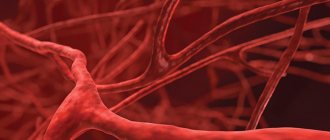
More than 5 million people in the world suffer from various forms of paralysis, the main causes of which are stroke (34%) and spinal cord injury (24%).
Stroke is currently one of the main causes of disability in the population. In Russia, more than 450,000 strokes are registered annually, and 70–80% of stroke survivors become disabled, with approximately 20–30% of them requiring constant outside care.
Over the past 70 years, the number of patients with spinal cord injury has increased 200 times, and in Russia more than 8,000 people suffer such injuries every year. Most often, this leads to the patient’s inability to move independently and provide for their basic needs. As a result of using a wheelchair, physical activity decreases, which provokes the development of a number of diseases: heart disease, osteoporosis, bedsores. Therefore, there is an active search for alternative methods of restoring the ability to move. One of the newest developments in this direction is the neural interface.
Neurointerface (also known as brain-computer interface, BCI) is a system that allows you to transmit brain signals directly to an external device (this can be a wheelchair, exoskeleton, computer, etc.), in fact, control the “power of thought” (Fig. 1) .
In “Biomolecule” you can read in more detail about the history of the development of neurocomputer technologies, as well as about Elon Musk’s modern Neuralink project [1], [2].
Figure 1. BCI operation diagram.
adapted from materials from the Tritriwulansari website
Contraindications to TES therapy
The procedure cannot be performed in the following cases:
- age up to 5 years;
- skin damage where contacts are made;
- presence of pacemakers;
- hypertensive crises;
- epilepsy;
- hyperthyroidism.
Physiotherapeutic equipment. Photo Gallery
Methods for recording brain signals
The first link in the BCI operation scheme is receiving a signal from the brain. The following methods are used for this:
- electroencephalography (EEG), which records electrical signals from the brain;
- magnetoencephalography (MEG), which records magnetic fields resulting from electrical activity in the brain;
- near-infrared spectrometry (NIRS), which measures the saturation of blood with hemoglobin (the more active a group of neurons, the more it uses oxygen carried by oxyhemoglobin);
- functional magnetic resonance imaging (fMRI), based on measuring blood flow to different areas of the brain (the more active a group of neurons, the greater the blood flow to it).
Nowadays, EEG is most often used in BCIs to obtain information about the electrical activity of the brain, since it has a high temporal resolution (electrodes allow one to read the immediate activity of individual parts of the brain), is relatively cheap, portable and does not pose a risk to users. EEG-based BCIs consist of a set of sensors that detect EEG signals from different areas of the brain. However, the quality of EEG signals is degraded because the signal travels through the scalp, skull, and many other layers, which creates noise.
To reduce noise and improve recording quality, they resort to invasive methods - implanting a set of microelectrodes inside the skull [3]. This implies significant health risks, which is why they are rarely used in experimental practice. There are two invasive approaches to BCI research: electrocorticography (ECoG), in which electrodes are placed on the surface of the cerebral cortex, and intracortical recording of neural activity, in which sensors are implanted into the cortex (Fig. 2). Such solutions are currently used extremely rarely, only in exceptional cases: either when the patient is already undergoing brain surgery, or when this is the only chance to regain the ability to interact with the outside world.
Figure 2. Electrode layout for EEG, ECoG and intracortical microelectrodes.
adapted from [3]
2.How does it work?
Intracerebral electrodes
for chronic brain stimulation, electromagnetic pulses are received from
a generator
, which is located subcutaneously and is adjusted to a specific program using
a remote control.
It should be noted that electrical brain stimulation is not a treatment. This is symptomatic care for patients
who are doomed (often for life) to experience functional muscle disorders that significantly reduce the quality of life. Neurostimulation of the brain is most justified and indicated for patients with Parkinson's disease, dystonia, essential tremor, epilepsy, and various movement disorders.
Visit our Neurosurgery page
Sensorimotor rhythm and motor cortex
As we have already said, the purpose of a BCI is to capture the user's intention by recording their brain activity. When recording brain activity using EEG, we get a graphical representation of a complex oscillatory electrical process, in which we can identify a number of specific rhythms that differ in amplitude and frequency: alpha, beta, delta, mu and others. Now we are interested in the mu rhythm, since it is on its basis that the neural interfaces used in neurorehabilitation of movements work.
The mu rhythm, or sensorimotor rhythm (SMR), has a frequency of 8–13 Hz and is recorded over the motor area of the cerebral cortex, located in the posterior part of the precentral gyrus (Fig. 3). Suppression of the mu rhythm occurs when a person performs a movement or imagines performing a movement—this is called event-related desynchronization (ERD). This occurs because neurons that previously fired synchronously acquire individual firing patterns that are different from each other. In this case, a person can train in imagining movements, and over time, the suppression of the mu rhythm becomes more and more pronounced, which is used when learning to control a BCI.
The motor cortex is characterized by a topical organization. This means that each section of the cortex corresponds to a specific area of the body that it controls. Figure 3 shows Penfield's homunculus, whose body parts are proportional to the brain areas in which they are represented. As can be seen from the figure, the representations of the upper and lower extremities are located far enough from each other, making it possible for the neural interface to separately recognize the movements of the arms and legs.
Figure 3. Somatosensory and motor homunculus.
adapted from materials from the BioNinja website
Note that the representation of the lower extremities in the motor cortex is much smaller than that of the upper extremities. This is easily explained by the presence of fine motor skills of the hands: the brain needs to control many individual muscles of the fingers. The legs, on the other hand, have fewer muscles to control and are larger. In addition, it is clear that the representation of the lower extremities falls into the interhemispheric fissure, which makes it difficult to recognize EEG signals generated by imagining the movements of different groups of leg muscles. Therefore, the use of BCIs for the legs causes certain difficulties, and most of the existing scientific works on neurorehabilitation using BCIs are devoted specifically to the upper extremities, since it is easier to work with their imagination. In the laboratory of movement physiology of the Institute of Physiology named after. I.P. Pavlov RAS, where the author works, conducts research aimed at studying the processes of rehabilitation of the lower extremities, as well as the possibility of using transcutaneous electrical stimulation of the spinal cord (TESCS) and special practices that help increase the efficiency of BCI control [4].
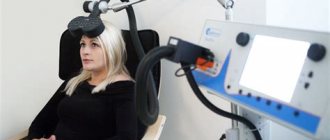
Design and operation of a beckoning stimulator
Transcranial magnetic stimulator Neuro MS/D
1 - main unit; 2 - cooling module; 3 — replenishment module; 4 — bracket for the inductor; 5 - software
TMS device
consists of three main parts: a system of high voltage (more than 3.5 kV) and high current capacitors, a coil (coil) and a control unit. 1718 There are additional technical elements that ensure a comfortable procedure and proper operation of the magnetic stimulator: a cooling module for the device, a handle (bracket) for fixing the coil, software.
- The main block is the basis of the system. Its front panel contains controls and indicators that reflect the operating parameters of the stimulator. Can be connected to a computer via a USB cable. The main unit can operate at frequencies up to 30 Hz; maximum induction is carried out at a frequency of up to 5–7 Hz;
- The cooling module is a liquid cooling system. With the help of the cooling module, the session is carried out much faster, without the need for breaks to exchange or cool the coil during the session or between patients. The coolant does not fill the inductor, but moves along the winding, removing thermal energy from the place of its formation;
Cooling module operation
- Replenishment module - an additional power supply increases the maximum stimulation frequency to 100 Hz, and the maximum induction frequency to 20-25 Hz. Using the replenishment module makes it possible to conduct theta-burst stimulation (TBS), in which the session is significantly shorter than conventional TMS;
- Bracket for the inductor – allows you to hold the inductor in the same position relative to the patient’s head throughout the entire session;
- Software – “neuro-MS.NET” program. Using a computer, the software provides control of the patient database, management of courses and sessions, conducting stimulation according to existing programs and creating your own programs for TMS.
Types of inductors:
Ring inductor –
has a coil diameter of 150 mm. Used for peripheral muscle stimulation and cortical bilateral stimulation. Suitable for peripheral stimulation in urology and coloproctology;
Ring inductor
Inductor – figure eight –
has a coil diameter of 100 mm. Compared to a ring inductor, it provides stimulation of deeper tissues.
Figure-of-eight inductor
Distribution of the magnetic field of the field in a ring inductor and a figure-of-eight inductor
Angular figure-of-eight inductor
– has a coil diameter of 100 mm. It has an anatomical shape that matches the shape of the head. Used for deep cortical stimulation.
Angular figure-eight inductor
Double conical inductor
– has a coil diameter of 125 mm. Provides the deepest stimulation. Suitable for stimulating the cortical representations of the muscles of the lower limbs and pelvic floor, cerebellum and DMPFC.
Double conical inductor
Each inductor has a device readiness indicator on the side surface (lights up green) and a magnetic stimulus supply button.1920
The principle of operation of a magnetic stimulator
consists in generating a current by a system of capacitors, transmitted to the coils, with the subsequent formation of a magnetic field in them (up to 2.2 Tesla). As a result, the nerve tissues form their own electrical impulse, passing through the pathways from the stimulated cortical structures to the tissues and muscles that have representation in them.21
It is worth noting that magnetic stimulators, depending on the purpose of TMS, are capable of generating different types of stimuli: 222324
- Monophasic stimulus
- the current in the inductor with this stimulus flows in one direction, increasing according to a sinusoidal law and decreasing exponentially;
Monophasic stimulus
- Biphasic stimulus
- the current in the inductor with this stimulus is characterized by one period of a damped sinusoid;
Biphasic stimulus
- Biphasic burst stimulus
- stimulation in which a series of biphasic stimuli with a high frequency (up to 100 Hz) and decreasing amplitude is issued;
Burst stimulus
- Paired monophasic stimulus
- two stimuli with a specified inter-stimulus interval and amplitude set independently for each stimulus.
Paired monophasic stimulus
Thus, today a number of magnetic stimulators have been created that meet different requirements and purposes of TMS.25
Line of magnetic stimulators from the Neurosoft company
In addition to magnetic stimulators from the Neurosoft company, stimulators from the MagVenture company are widely used: MagPro R100, MagPro R30, MagPro x100.262728 Magnetic stimulators of this line differ in terms of maximum stimulation frequency and pulse shapes, depending on the purposes of TMS. The principle of operation and structure of the devices is the same as in the description above. These devices operate without specific software based on a built-in computer, which has everything necessary to track indicators and store results during TMS.
How to Imagine Movement Effectively
The following features of movement imagination are known that increase its effectiveness:
- Kinesthetic (paying attention to sensations from muscles and joints) representation, rather than visual [5];
- First person presentation rather than third person [6];
- Imagination of movements after an actual action [7];
- Using feedback (when a person is shown how well he is doing a task) [8]. Feedback in the form of virtual reality showed high efficiency: when imagining leg movements, the avatar controlled by the subject goes forward, and when imagining stops, it stops. The subject’s task is to go forward and stop at certain points in virtual space [9–12];
- Simultaneous viewing of a video in which the corresponding movement is performed helps to enhance desynchronization of the mu rhythm due to the work of mirror neurons [13];
- The use of meditative practices, in particular, mindfulness meditation [14].
In addition, we have shown that the effectiveness of motor imagery depends on a person’s personal characteristics [15].
For the experiment, 44 people with a dominant right hand were recruited. All of them were tested using the Cattell questionnaire, which identifies 16 basic individual characteristics. Next, the subjects controlled the BCI based on the imagination of hand movements. It turned out that when imagining the movements of the right hand, expressive, sensitive extroverts were more successful, while when imagining the movements of the left hand, practical, reserved, skeptical and not very sociable people were more successful.
We hypothesize that this can be explained by different levels of dopamine in the right and left hemispheres, as well as differences in the way information about movements is encoded [16]. You can read more about this in the article published by the author and colleagues in the journal “Proceedings of the Academy of Sciences” [15]. Knowledge of the personal psychological parameters of a BCI user can help in the development of individual training and preparation methods before operating neural interfaces.
Why is it necessary to imagine movements and work with neural interfaces? How can this help people with movement disorders? Let's look at these questions using the example of the two most common causes of movement disorders - stroke and spinal cord injury.
Indications and contraindications, side effects
Carrying out TMS with therapeutic and restorative
The purpose is carried out for the following pathology:[/efn_note]31 32
- consequences of traumatic brain and spinal trauma;
- violation of cerebral and spinal circulation, accompanied by motor disorders;
- demyelinating diseases (multiple sclerosis, Guillain-Barré polyneuropathy);
- neurodegenerative diseases (amyotrophic lateral sclerosis, Alzheimer's disease, Parkinson's disease);
- congenital pathology of the central nervous system (cerebral palsy, consequences of perinatal hypoxia);
- mental disorders (depression, obsessive-compulsive disorder, schizophrenia);
- developmental disorders of the central nervous system in children (autism spectrum disorders, aphasia, dysarthria, alalia, etc.).
TMS for diagnostic purposes
shown to record the following indicators:
- conductivity of central and peripheral neural pathways;
- cortical excitability and plasticity of motor areas of the brain;
- evoked motor response;
- radicular delay time.
Contraindications
for TMS is: 33
- The presence of magnetic implants (pumps for the delivery of medicines, metal structures, a pacemaker and electrodes for deep brain stimulation are acceptable);
- Convulsive syndromes and seizures;
- Vascular aneurysms and brain tumors;
- Acute infectious diseases;
- Negative consequences of previously performed TMS procedures (headaches, nausea, vomiting, dizziness and other adverse reactions);
- Severe heart pathology;
- Pregnancy;
- Age up to 3 years.
Side effects
and their prevalence under different TMS protocols are presented in the table.
| Undesirable effect | Single-pulse TMS | Stimulation with paired stimuli | Low frequency rTMS | High frequency rTMS | Theta burst stimulation |
| Epileptic seizures | rarely | not described | rare (3 cases described) | possible | 1 case described |
| Development of hypomanic state | No | No | rarely | possible by stimulating the prefrontal cortex | not described |
| Syncope | possible (not related to biological effects of stimulation) | ||||
| Head/neck pain, discomfort at the stimulation site | possible | not described | 20-40% | 20-40% | possible |
| Transient hearing changes | possible | not described | possible | possible | not described |
| Transient changes in cognitive function | not described | not described | possible (insignificant) | possible (insignificant) | Transient impairment of working memory described |
| Other biological effects | not described | not described | transient changes in serum hormone levels | transient changes in serum hormone levels | not described |
EFFECTIVENESS AND LIMITATIONS
The effectiveness of therapeutic TMS for various pathologies is shown in the table: 34
| Disease/condition | Magnetic stimulation protocol | Level of evidence |
| Movement disorders after stroke | Low-frequency stimulation of the M1 zone of the hemisphere contralateral to the lesion in the acute and subacute periods Low-frequency stimulation of the M1 zone of the hemisphere contralateral to the lesion in the chronic period | C B |
| Broca's aphasia | Low frequency stimulation of the right inferior frontal gyrus High frequency stimulation of the left inferior frontal gyrus | More research needed |
| Wernicke's aphasia | Low frequency stimulation of the right inferior frontal | More research needed |
| Neglect syndrome | cTBS mode of the left (contralateral) inferior parietal cortex Low-frequency stimulation (contralateral) of the left inferior parietal cortex | C C |
| BP | High-frequency stimulation of M1 zones of both hemispheres | WITH |
| Dystonia | Low frequency stimulation of PMC, M1 or S1 | More research needed |
| Neuropathic pain | High frequency stimulation of M1 contralateral to the side of pain | A |
| Migraine | High-frequency stimulation of the left M1 or DLPFC Single stimuli of the occipital cortex during the onset of a migraine attack | More research needed |
| CRPS type 1 | High frequency stimulation of M1 contralateral to the side of pain | WITH |
| Fibromyalgia | High frequency stimulation of the left M1 or DLPFC or low frequency stimulation of the right DLPFC | More research needed |
| Epilepsy | Low frequency stimulation of epileptic focus | WITH |
| Tinnitus | Low-frequency stimulation of the temporoparietal cortex contralateral to tinnitus | WITH |
| Spinal spasticity | High frequency stimulation of M1 zones | WITH |
| Depression | High frequency stimulation of the left DLPFC Low frequency stimulation of the right DLPFC | A B |
| Anxiety | High frequency stimulation of the right DLPFC for post-stress anxiety disorder | WITH |
Note
: Effect: A - definite, B - probable, C - possible.
The limitations of the TMS method are associated with the small number of studies in this area, the novelty of most of the data obtained with an insufficient level of evidence, as well as the lack of official clinical recommendations regarding the use of TMS in the diagnosis and treatment of diseases.
Footnotes
- Magnetic stimulation in the diagnosis and treatment of diseases of the nervous system. Guide for doctors. Ed. Nikitina S.S., Kurenkova A.L. Publishing house M.: SASHKO; 2003.
- Wassermann EM et al. Safety and side-effects of transcranial magnetic stimulation and repetitive transcranial magnetic stimulation //Handbook of transcranial magnetic stimulation. London: Arnold. - 2002. - p. 39-49.
- Barker AT, Jalinous R., Freeston IL
Non-invasive magnetic stimulation of human motor cortex // The Lancet: journal. - 1985. - Vol. 325, no. 8437. - P. 1106-1107. - Bickford RG et al. Magnetic stimulation of human peripheral nerve and brain: response enhancement by combined magnetoelectrical technique. // Neurosurgery: journal. - 1987. - Vol. 20, no. 1. - P. 110-116.
- George MS et al. Daily repetitive transcranial magnetic stimulation (rTMS) improves mood in depression. // Neuroreport: magazine. - 1995. - Vol. 6, no. 14. - P. 1853-1856.
- Pascual-Leone A. et al. Akinesia in Parkinson's disease. I. Shortening of simple reaction time with focal, single pulse transcranial magnetic stimulation// Neurology: journal. - 1994. - Vol. 44, no. 5. - P. 884-884.
- Pascual-Leone A. et al. Akinesia in Parkinson's disease. II. Effects of subthreshold repetitive transcranial motor cortex stimulation// Neurology: journal. - 1994. - Vol. 44, no. 5. - P. 892-892. DOI: https://doi.org/10.1212/WNL.44.5.892
- Jose Luis Rodriguez-Martin, José Manuel Barbanoj, V Pérez M Sacristan. Transcranial magnetic stimulation for the treatment of obsessive-compulsive disorder // Cohrane library. - John Wiley & Sons, Inc., 2003. - April 22. — doi:10.1002/14651858.CD003387
- Brain Stimulation Approved for Obsessive-Compulsive Disorder by Rebecca Voelker, MSJ in JAMA. Published September 18, 2021 – DOI: 10.1001/jama.2018.13301
- Stepanchenko A.V., Mamedov T.R., Sharov M.N., Savushkin A.I., Krymshaukhalova S.Ya. // Magnetic stimulation in the treatment of exacerbation of trigeminal neuralgia // Pain: magazine - No. 3 (4) - pp. 40-45 // Medical Encyclopedia Publishing House (Moscow) / 2004.
- L.M. Oberman, P.G. Enticott, M.F. Casanova, A. Rotenberg, A. Pascual-Leone, J.T. McCracken Transcranial Magnetic Stimulation in Autism Spectrum Disorder: Challenges, Promise, and Roadmap for Future Research // Autism research: official journal of the International Society for Autism Research - 2021 Feb;9(2):184-203. doi: 10.1002/aur.1567. Epub 2015 Nov 4.
- Voitenkov V.B., Mally J., Skripnichenko N.V., Klimkin A.V. Transcranial magnetic stimulation as a diagnostic and therapeutic technique // Neurological Journal 2015, vol. 20, No. 5 – pp. 4-13 // OJSC Publishing House: Medicine (Moscow), 2015
- Gimranov R. F. Transcranial magnetic stimulation. - M.: “Allana”, 2002. - 164 p. ISBN 5-86656-115-1
- Magnetic stimulation in the diagnosis and treatment of diseases of the nervous system. Guide for doctors. Ed. Nikitina S.S., Kurenkova A.L. Publishing house M.: SASHKO; 2003.
- Voitenkov V.B., Mally J., Skripnichenko N.V., Klimkin A.V. Transcranial magnetic stimulation as a diagnostic and therapeutic technique // Neurological Journal 2015, vol. 20, No. 5 – pp. 4-13 // OJSC Publishing House: Medicine (Moscow), 2015.
- Gimranov R. F. Transcranial magnetic stimulation. - M.: “Allana”, 2002. - 164 p. ISBN 5-86656-115-1
- A.A. Sorochinsky Transcranial magnetic stimulation // Journal: News of the Southern Federal University. Technical Sciences – 2010 – No. 9 – pp. 207-210.
- https://kandel.com.br/equipamentos/emt/neuro-msd/
- https://kandel.com.br/equipamentos/emt/neuro-msd/
- https://neurosoft.com/ru/catalog/tms/neuro-msd-therapeutic#delivery
- A.A. Sorochinsky Transcranial magnetic stimulation // Journal: News of the Southern Federal University. Technical Sciences – 2010 – No. 9 – pp. 207-210.
- Sommer M, Alfaro A, Rummel M, Speck S, Lang N, Tings T, et al. Half sine, monophasic and biphasic transcranial magnetic stimulation of the human motor cortex. Clin Neurophysiol 2006, 117:838-44.
- Valls-Solé J, Pascual-Leone A, Wassermann EM, Hallett M. Human motor evoked responses to paired transcranial magnetic stimuli. Electroencephalogr Clin Neurophysiol 1992 85: 355–64.
- Huang YZ, Edwards MJ, Rounis E, Bhatia KP, Rothwell JC. Theta burst stimulation of the human motor cortex. Neuron 2005, 45: 201-6.
- https://neurosoft.com/ru/catalog/tms/neuro-msd-therapeutic#delivery
- https://www.magventure.com/tms-research/products-overview/research-stimulators/stimulators/magpro-r100-4
- https://www.magventure.com/tms-research/products-overview/research-stimulators/stimulators/magpro-x100-w-magoption
- https://www.magventure.com/us/tms-research/products-overview/research-stimulators/stimulators/magpro-30
- Voitenkov V.B., Mally J., Skripnichenko N.V., Klimkin A.V. Transcranial magnetic stimulation as a diagnostic and therapeutic technique // Neurological Journal 2015, vol. 20, No. 5 – pp. 4-13 // OJSC Publishing House: Medicine (Moscow), 2015.
- A.V. Chervyakov, A.G. Poydasheva, Yu.E. Korzhova, N.A. Suponeva, L.A. Chernikova, M.A. Piradov // Rhythmic transcranial magnetic stimulation in neurology and psychiatry // Journal of Neurology and Psychiatry (Moscow), No. 12, 2015 – pp. 7-18 // doi: 10.17116/jnevro20151151127-18
- A.V. Chervyakov, A.G. Poydasheva, Yu.E. Korzhova, N.A. Suponeva, L.A. Chernikova, M.A. Piradov // Rhythmic transcranial magnetic stimulation in neurology and psychiatry // Journal of Neurology and Psychiatry (Moscow), No. 12, 2015 – pp. 7-18 // doi: 10.17116/jnevro20151151127-18
- A.A. Sorochinsky Transcranial magnetic stimulation // Journal: News of the Southern Federal University. Technical Sciences – 2010 – No. 9 – pp. 207-210.
- ON THE. Suponeva, I.S. Bakulin, A.G. Poydasheva, M.A. Piradov // Safety of transcranial magnetic stimulation: review of scientific recommendations and new data // Journal: Neuromuscular DISEASES, vol. 7, 2' 2021 – pp. 21-36 // DOI: 10.17650/2222-8721-2017-7- 2-21-36
- A.V. Chervyakov, A.G. Poydasheva, Yu.E. Korzhova, N.A. Suponeva, L.A. Chernikova, M.A. Piradov // Rhythmic transcranial magnetic stimulation in neurology and psychiatry // Journal of Neurology and Psychiatry (Moscow), No. 12, 2015 – pp. 7-18 // doi: 10.17116/jnevro20151151127-18
Mechanisms of neuroplasticity
During a stroke, an acute disruption of the blood supply to the brain occurs (either as a result of blockage of a vessel by a blood clot - ischemic stroke, or as a result of hemorrhage - hemorrhagic). Since, along with the blood, everything that they need for life ceases to flow to the neurons, the areas of the brain where blood circulation has stopped die off. And if these are areas responsible for motor activity - for example, the motor cortex, then the patient experiences hemiparesis, decreased muscle strength on one side of the body, or hemiplegia, complete paralysis of half the body.
The restoration of motor function is carried out mainly due to the mechanisms of neuroplasticity - the ability of the brain to change under the influence of experience: to establish new connections between neurons, destroy old and unnecessary ones, and restore those lost after damage. These processes involve not only neurons, but also neuroglial cells, as well as the vascular system [17]. The activity of synapses and their number also change [18]. To activate these mechanisms, motor rehabilitation is used in medicine. However, in patients with paralysis or a high degree of paresis, the implementation of real movements is impossible, so they resort to training with a BCI based on the imagination of movements. When imagining movements, the same areas of the brain are activated that are also involved in preparing a real action and in its execution, as a result of which such neurorehabilitation becomes real [19].
Thanks to such rehabilitation training, neurons around the damaged area undergo restructuring: the volume of gray matter in the motor zone of the brain increases, and neighboring areas take on lost functions [20]. The motor areas of the undamaged hemisphere are also involved in this process.
The effectiveness of these exercises can be increased through the use of biofeedback - visual or tactile - when the patient sees on a monitor screen how well he is completing a task (imagining the movement of a limb), or when he feels vibration from a special device when he successfully completes a task.
There are also systems that provide motor feedback: for example, when a person imagines moving his right leg, setting it in motion with a special mechanism. The Biokin system (Kosima LLC), developed under the leadership of Yu.P. Gerasimenko, works on this principle. (Institute of Physiology named after I.P. Pavlov RAS) (Fig. 4) [21]. It includes feedback, functional electrical stimulation (FES) and transcutaneous electrical stimulation of the spinal cord (TESCS), which makes it a highly effective tool in the field of neurorehabilitation of the lower extremities [22].
Figure 4. Biokine. A complex for neurorehabilitation of the lower extremities, based on the use of a BCI with feedback, FES (functional electrical stimulation) and tESCS (transcutaneous electrical stimulation of the spinal cord).
Biokin website
Such systems make it possible to close the sensorimotor loop: from the efferent (outgoing) signal of motor activity sent by the brain to the afferent (incoming) signal of sensory feedback (Fig. 5) [23].
Figure 5. Neuroplasticity induced by motor imagery-based BCI use. When the motor areas of the cortex are damaged, real movement becomes impossible, so to activate the processes of neuroplasticity, only the possibility of imagining movements remains. The use of a BCI with visual and tactile feedback enhances these processes.
adapted from [23]
This rehabilitation mechanism can be explained by the concept of Hebbian plasticity: with the simultaneous activation of two neurons connected to each other, their synaptic interaction increases, which leads to more reliable contact between them (Fig. 6). If we assume that signal transmission from the motor cortex to the limb muscles has been disrupted due to stroke or injury, then simultaneous activation of the sensory and motor cortices may enhance previously inactive connections between neurons through plasticity and thus lead to restoration of motor function of the limbs [ 24].
Figure 6. Hebbian plasticity mechanism. Strengthening the synaptic interaction between two neurons occurs due to repeated stimulation of the postsynaptic cell by the presynaptic cell.
adapted from ""
Figure 7. Formation of new neural connections in the area of spinal cord injury (SCI).
adapted from [25]
When restoring motor function after spinal cord injury, the same mechanisms of neuroplasticity are involved. With such damage, some of the nerve fibers, including motor ones, are interrupted, which causes paralysis of the limbs, while some retain their integrity. Due to this, during neurorehabilitation, it is possible to activate neuroplasticity processes: intact fibers form synaptic connections with motor neurons (motoneurons), which, in turn, transmit a signal to the muscles (Fig. 7) [25].
To increase the effectiveness of neurorehabilitation using BCIs, functional electrical muscle stimulation (FES) is often additionally used. It provides contraction of a muscle at the moment when the user imagines a movement involving this muscle (Fig. [26]. This leads to increased neuroplasticity via the Hebbian mechanism: there is a simultaneous activation of the motor areas of the brain, which transmit signals to the motor neurons of the spinal cord, and sensory neurons, activated by a muscle contracting under the influence of FES, which closes the sensorimotor loop.
Figure 8. IMC-FES system. When imagining movements, a signal from the motor cortex is processed by a computer (PC) and transmitted to a functional electrical stimulation (FES) device, which causes contraction of the corresponding muscle. The signal from the muscle is then transmitted to the sensory cortex, providing feedback.
adapted from [26]
Advantages of transcranial electrical stimulation at Medicenter
- Professionalism . We employ experienced physiotherapists with many years of experience.
- Modern equipment . We use modern physiotherapeutic devices for electrical stimulation.
- Safety . The electrical stimulation procedure is safe for patients.
- Saving on medicines . During the treatment process, electrotherapy can replace some medications.
- Saving time . Patients are accepted by appointment.
Electrostimulation of the spinal cord
In recent years, electrical stimulation (ESS) has shown great effectiveness in neurorehabilitation after spinal cord injury. The spinal cord has two thickenings: in the neck and lumbar region, which corresponds to the place where the roots of motor neurons of the upper and lower extremities exit from them. In the lumbar enlargement of the spinal cord there are specialized neural networks that ensure the automatic process of stepping (stepping movement generators, GSM). In other words, if you apply electrodes that deliver a current of a certain amplitude and frequency to the dura mater of the spinal cord at the site of the lumbar enlargement, you can induce involuntary walking movements even in people with paraplegia [27]. However, this method requires surgery, so there is a risk of developing postoperative complications.
Currently, transcutaneous electrical stimulation of the spinal cord (TESCS) is considered the safest and most painless. In video 1 (Edgerton Lab, University of California) you can see how involuntary stepping movements of the legs are caused when the patient is in a relaxed position, with his legs suspended on a swing frame [28].
Video 1. Involuntary walking during transcutaneous electrical stimulation of the spinal cord.
Edgerton Lab, University of California
When using TECS, the question of the correct location of the stimulating electrodes arises. If, when installing invasive electrodes during surgery, the segments and roots of the spinal cord are clearly visible, then when installing cutaneous electrodes, it may be difficult to find the desired area. This problem is solved by applying single impulses to the electrode and recording reflex muscle responses - after all, each segment of the spinal cord corresponds to strictly defined muscle groups.
There is also the problem of insufficient amplitude of the impulses sent - due to degenerative processes in the case of spinal cord damage, a large amplitude of stimulation is required to obtain the desired response. However, this can result in burns. Our laboratory has created an optimal device for non-invasive electrical stimulation of the spinal cord [29].
In addition, a system was developed that detects the phases of the walking cycle online and stimulates the spinal cord according to these phases [30]. While walking, different muscles tense at different moments, and joints bend at certain angles, which can be recorded with special devices - accelerometers and gyroscopes. Both legs move in a coordinated manner, and based on the position of one leg, the position of the other can be predicted. The principle of operation of the system is as follows: for a patient with hemiparesis, motion sensors are applied to the healthy leg, which transmit a signal to the TECS device. It, in turn, stimulates at certain points in time groups of spinal cord motor neurons responsible for the movement of the flexor and extensor muscles of the leg, which helps normalize walking and restore movement of the affected limb.
3.Preparation and performance of the operation
Each disorder is caused by disturbances in a specific area of the brain, so the installation of electrodes is carried out after a thorough diagnosis, which includes:
- electroencephalogram;
- MRI;
- CT;
- general diagnosis of the body's condition.
The procedure for installing electrodes and generator is carried out under local anesthesia
, since the brain itself has no pain receptors and only skin anesthesia is required. During surgical procedures, the patient is in full contact with the surgeon, which allows him to assess the condition and progress of the operation.
After operation
measures are taken to prevent infection of the affected areas, including a course of antibiotics. Discharge from the hospital occurs on days 3-5. After two weeks, a second visit to the neurosurgeon is necessary.
About our clinic Chistye Prudy metro station Medintercom page!
Advances in modern neurorehabilitation
The largest study in the field of neurorehabilitation using a BCI based on motor imagery is the work of Donati et al., published in Nature in 2021 [31]. This study involved eight people with paraplegia caused by spinal cord injury. A special rehabilitation system was developed for them, including six stages of increasing complexity, and about 255 (!) sessions were conducted with each patient during the year.
The first stage included a deep immersion in a virtual reality environment, during which the subject controlled the movement of his avatar (computer character), imagining the movement of the lower limbs in a sitting position. Then the patient did the same, only in a standing position, leaning on a special table. During the third stage, training took place on a treadmill: the subject walked using a device that supports body weight (Lokomat). At the fourth stage, the legs moved in the air, and not on the treadmill. In the fifth stage, the patient trained on a treadmill using a robotic limb support system controlled by a BCI. And at the final stage, the subject walked in an exoskeleton controlled by a BCI: the exoskeleton took a step when the person imagined the movement of the corresponding leg. During all trainings, subjects received tactile feedback - a vibration that was applied to the forearm when a virtual or robotic leg on the same side touched the ground. You can see the experiment diagram in Figure 9, and the rehabilitation process itself in Video 2.
Figure 9. Scheme of the experiment, which includes six stages: 1 - BCI + virtual reality (VR) in a sitting position; 2 — BCI + VR in a standing position; 3 — walking on a treadmill while supporting body weight; 4 - movement of legs in the air; 5 — walking on a treadmill using a robotic system controlled by a BCI; 6 — walking in an exoskeleton controlled by a BCI. Designations: EEG - electroencephalography; EMG - electromyography, which records muscle activity; Tact. - tactile feedback.
[31]
Video 2. The process of conducting the experiment.
[31]
After 12 months of training using this system, all eight patients had improved tactile sensation scores and regained free control of key muscles of the lower extremities. As a result, noticeable progress was seen in their ability to walk. Many patients were able to walk with the help of assistive devices. In addition, all patients showed a significant increase in emotional stability and assessment of quality of life, as well as a decrease in the level of depression and an increase in self-esteem. The condition of the skin and the function of the digestive system have improved, which is apparently associated with the normalization of the activity of the sympathetic and parasympathetic systems. The fact is that along the spine there are nodes of the autonomic nervous system, which regulates the functioning of the internal organs. They are damaged when the spinal cord is injured, which causes disruption of the digestive system, which in turn affects the condition of the skin through the release of signaling molecules, including pro-inflammatory ones [32], [33].
Neurological recovery was associated with plasticity mechanisms at both the spinal cord and sensorimotor cortical levels. Cortical and spinal plasticity alters neural connections in the surviving region of the spinal cord through motor and sensory connections (Figure 10).
Figure 10. Plasticity of the spinal cord (SC) and cerebral cortex through motor (red) and sensory (blue) connections.
adapted from [31]
Don't forget to turn off the electricity
Micropolarization in doses prescribed by doctors is considered safe for humans, although, in addition to itching and burning of the scalp, in 17% of cases patients complained of headaches. But these are official experiments, and at home TES fans are going all-in. Firstly, wanting a quick and noticeable result, some people increase the current recommended by scientists. Second, they leave the device on for longer periods of time and sit with the electrodes on during notes and lectures in hopes of better retention. Third, homemade devices may produce incorrect currents. The negative impact on the brain of experimenters under such conditions is unknown. On forums, people complain about pain in the arm, increased anxiety instead of decreasing it, increased irritability, heaviness and unpleasant tension in the head.
Another problem with “brain hackers” is an overly optimistic interpretation of research results. If scientists write that TES in some cases improves learning abilities or creativity, then in the eyes of a person who wants miracles, this turns into direct evidence that electrical discharges will make him a great mathematician. A keen experimenter is unlikely to think that it is extremely difficult to objectively measure creativity, and the improvement of cognitive abilities is assessed by scientists as success in very specific tests that are not always applicable to reality.