Parts of the brain
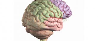
As noted above, the structure of the brain is truly complex. To simplify its study, depending on the functions performed and the characteristics of intrauterine development, the brain is divided into the following parts:
- the forebrain (telecephalon), which consists of the cerebral hemispheres;
- the diencephalon (diencephalon), which includes the thalamus and surrounding structures;
- the midbrain (mesencephalon), consisting of the quadrigeminal cord and cerebral peduncles;
- the hindbrain (metencephalon), which includes the pons and cerebellum;
- medulla oblongata (myelencephalon).
You may be interested in: Venous and arterial blood: features, description and differences
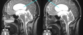
Brain malformations: polymicrogyria, agyria and pachygyria
These anomalies arise as a result of a disrupted process of formation of the brain or its individual structures during the prenatal period, and represent abnormal changes in the structure of brain structures.
There are no exact data on the prevalence of congenital brain defects, but polymicrogyria is considered the most common.
More details about each malformation:
- Polymicrogyria is a rare pathology of the development of the cerebral cortex, in which a significant number of small and underdeveloped convolutions are formed on the surface of the cerebral hemispheres. It is often combined with other genetic pathologies and abnormalities in the development of the cerebral cortex. Most often, pathology develops in the area of the Sylvian fissure (60% of cases), but can occur in any part of the cerebral cortex. Pathology can be diffuse, multifocal and focal. It can also be divided into unilateral (40% of cases) and bilateral (60% of cases), symmetrical and asymmetrical.
- Agyria (lissencephaly) is an anomaly in the formation of the cerebral cortex, in which the gyri are underdeveloped, weakly expressed, or absent, and the architecture of the cortex is disrupted. The appearance of the child's brain is identical to the appearance of the fetal brain at 3-4 months of the prenatal period. Agyria is a severe form of lissencephaly. It can be either an idiopathic disease or accompany other pathologies (Miller-Diecker syndrome, Norman-Roberts syndrome, Walker-Warburg syndrome, Fukuyama congenital muscular dystrophy).
- Pachygyria is a rare developmental anomaly of the central nervous system, in which a small number of wide and flat convolutions are formed in the cerebral cortex. With pachygyria, the main gyri are enlarged, and the secondary and tertiary gyri are absent, the sulci are shortened and straightened, and the architecture of the cerebral cortex is disrupted. This pathology is considered an “incomplete”, milder form of lissencephaly.
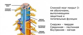
Cross section of the brain
You may be interested in: Aromatase inhibitors: purpose and list of drugs
If you conventionally cut the brain in the frontal plane, you can see that part of the brain is dark in color, and part is light. The dark part is the gray matter, which is a collection of nerve cell bodies (neurons). It is represented by the cerebellum and the cerebral cortex, which is located along the perimeter. However, there are areas of gray matter inside the brain, they are called the basal ganglia, or extrapyramidal system.
While the cortex, together with the grooves and convolutions of the brain, performs the functions of coordinating higher nervous activity (speech, writing, thinking, memory, attention, emotions), the gray matter of the extrapyramidal system is necessary for the implementation of high-precision coordinated movements.
The basal ganglia include the following structures:
- striopallidal system, which consists of the caudate nucleus and the lentiform nucleus (the putamen together with the globus pallidus);
- the limbic system, which includes the fence and the amygdala.
White matter, in turn, is a collection of processes of nerve cells that ensure the interaction of the overlying parts of the brain with the underlying ones, as well as the interaction of different neurons within the same structure.
Types and functions
The central gyrus contains the center responsible for voluntary movements; the pyramidal motor pathway begins in this part of the cerebral cortex. The area is responsible for creating complex movements and maintaining motor coordination. The pyramidal system controls the function of upright walking. The main convolutions of the brain and their functions:
- Direct gyrus of the brain. It lies in the frontal region. Receives signals from sensor networks. Interacts with limbic structures, including the hippocampus, participating in the expression of certain emotions, memory regulation, and the formation of spatial memory, which helps to navigate in space. Receiving signals from motor neurons and the autonomic system, it combines information and contributes to the continuous modulation of reactions - cognitive, behavioral, psycho-emotional.
- Angular gyrus, also known as angular gyrus. It lies in the parietal region. Borders the posterior segment of the superior temporal gyrus. The angular gyrus of the brain regulates speech function and is involved in the formation of reading skills. The control center for the visual analyzer of written images is located in this area.
- Supramarginal gyrus of the brain. Located in the parietal region. The supramarginal gyrus surrounds the posterior segment of the Sylvian fissure. In this area of the medulla there is a center of praxia - the ability to perform habitual or learned muscle contractions in a given sequence, which is associated with the regulation of complex motor activity. Controls purposeful movements.
- Fusiform gyrus. Located in the temporal region at the junction with the occipital region. Responsible for recognizing people's faces. When viewing or mentally reproducing a person's face, the neurons that form this part of the brain matter are activated.
- Lingual gyrus. It lies in the occipital region. Contains the control center for the visual analyzer - a neuroreceptor system that perceives and processes visual information. The analyzer core is similar in cellular structure in children and adults. As adults gain experience, this brain structure becomes more complex.
The cingulate gyrus is part of the cingulate region, which determines its functions. The cingulate region is part of the limbic system, belongs to the mediobasal structures, which lie in the middle part of the base, and are considered the oldest parts of the brain.
The cingulate cortex, which lies on the surface of the brain, regulates memory functions, the formation of emotions and the ability to learn. Participates in the implementation of executive functions (a set of cognitive processes that allow you to plan and reproduce actions in accordance with the goal) and control of the respiratory system.
Brain: functions
You may be interested in: “Indapamide”: instructions for use and analogues
In fact, there are a huge number of functions of the human brain, and more than one article can be written about them. In the list below, all functions are combined into separate groups:
- processing information coming from outside;
- planning and decision making;
- carrying out movements;
- emotions;
- memorization and memory;
- attention;
- speech;
- intelligence and thinking.
Signs of defeat
Disturbances in the structure of the sulci and gyri in humans are more often associated with malformations at the stage of embryogenesis, less often with TBI - if such changes are present, the likelihood of developing epilepsy and mental retardation increases. The clinical picture in the presence of malformations of the cortical relief includes convulsive and epileptic seizures, dementia, paresis and spastic paralysis. Common malformations:
- Pachygyria. Thickening, flattening, expansion of cortical convolutions.
- Lissencephaly. Underdevelopment or absence of convolutions (effect of a smooth surface of the brain).
- Polymicrogyria. Abnormal folding of the cortical layers. Lack of clear relief - in some cases the convolutions merge.
Damage to the cortical layers of the brain is accompanied by symptoms: agnosia (impaired perception - visual, auditory, tactile), amnesia (partial or complete loss of the ability to remember past experiences), aphasia (impaired formed speech), apraxia (impaired complex voluntary movements while maintaining basic motor skills) .
Damage to individual areas of the cortex is accompanied by specific symptoms. Analysis of symptoms allows us to identify the localization of the pathological focus. Damage to the gyrus recta, which lies in the cerebrum, is associated with a slowdown in the formation of a cognitive response, a disturbance in the emotional background and behavioral skills.
Research shows a connection between damage to this area of the cortex and disturbances in social perception (social role, social status, interpersonal relationships). Damage to the angular gyrus, which lies in the cerebral cortex, manifests itself as difficulties in developing writing, speaking, and reading skills.
A patient with damage to brain structures of such localization experiences difficulties in performing mathematical operations, is unable to distinguish between fingers, and does not distinguish between the right and left half of the body. Damage to the supramarginal gyrus leads to loss of the ability to make complex voluntary movements leading to a specific goal.
Damage to the fusiform gyrus leads to impaired recognition of faces and symbols. When the lingual gyrus is damaged, the processes of perception and analysis of visual stimuli are disrupted. With the help of vision, a person receives about 90% of information coming from outside. Damage to the medulla in this area is accompanied by hemianopsia - partial loss of visual fields (vision is usually preserved in the central field).
Damage to the central gyrus is accompanied by impaired motor activity and the appearance of convulsive syndrome. Damage to the cingulate cortex is associated with the development of schizophrenia and other psychopathologies. The function of social cognition and the formation of emotions is impaired in patients.
Structure of the cortex
The cerebral cortex is the center of higher nervous activity in humans. Thanks to her work, we experience emotions, have the ability to learn, memorize and remember. The cortex is precisely the structure that distinguishes humans from representatives of other species of living beings.
What makes her so special? The cortex is not just a solid mass of gray matter; its structure includes the grooves and convolutions of the brain. These are important components of this body. These formations divide the cerebral hemispheres into separate functionally significant parts.
Does the number of convolutions affect the level of intelligence?
According to the latest data obtained in the course of research by Brazilian scientists, the number of convolutions in a person depends on two main variables: the area of the cortex and its thickness. This discovery fits organically into the general theory, because a large area is more difficult to locate in the cranium, and it is also more difficult to form folds in a thick layer of gray matter.
It is impossible to know exactly the number of grooves, and there is no “absolute” for this parameter. The appearance of the cerebral cortex is individual for everyone, and during an external examination it is not possible to see the total area of its cortex: approximately 2/3 of the gyri are located in deeper grooves.
However, for a person we can name the main convolutions present in the head of everyone:
- toothed;
- tape;
- occipitotemporal;
- lingual;
- parahippocampal;
- straight;
- hook of the brain.
Today it has been scientifically proven that the number of convolutions, as well as the mass of the brain, cannot in any way affect the mental development of a person. And even if you read the works of ancient Greek philosophers from morning to night, you will not gain as many gyri as you do grams of weight. This is logical, because the human convolutions in the form in which they remain throughout life are formed during the period of intrauterine development, and the weight of the brain depends on the body composition.
Some scientists and ordinary citizens who donate their bodies to science after death have allowed repeated studies to be conducted that have established that physiological differences between the brains of ordinary people and scientists do not correlate with the intelligence demonstrated during life.
Types of furrows
Fissures are, roughly speaking, gaps in the brain that form more convex parts - convolutions. The following main grooves of the brain can be distinguished:
- primary formed - the deepest, divide the cortex into separate lobes (frontal, occipital, temporal, insular, parietal);
- secondary - less deep, they divide the brain into small convoluted parts - convolutions;
- additional (tertiary) - the most superficial, designed to give a specific shape to the gyri and to increase the surface of the cortex.
Main grooves
There are many grooves and convolutions in the brain. The most important ones are listed below:
- Sylvian fissure - the border between the frontal and temporal lobes;
- Roland's fissure - the border between the frontal and parietal lobes;
- The parieto-occipital sulcus separates the occipital and parietal regions;
- the lateral sulcus is one of the largest and deepest in the brain;
- cingulate sulcus - located on the medial plane of the brain;
- the hippocampal sulcus is a continuation of the cingulate;
- the circular sulcus borders the insula on the lower part of the brain.
Structure
The pattern of convolutions and sulci of the brain is best seen in schematic images. The depressions dividing the cortex into two parts (hemispheres) are called primary. In addition, there are other fundamental limitations of the cortex, namely:
- Sylvian fissure (lateral, lateral): separates the temporal and frontal cortex.
- Roland's fossa (central): separates the parietal from the frontal.
- Parieto-occipital fossa: separates the occipital and parietal lobes of the brain.
- The cingulate recess, which passes into the hippocampal recess: separates the surface of the olfactory brain from other parts.
These structures also have another name: first-order sulci of the brain.
Each part of the telencephalon contains several convolutions, separated by secondary cavities. Tertiary depressions develop purely individually: their presence depends on the personal characteristics of a person and his mental abilities. The third type of notches gives individual relief to the folds.
Superolateral part of the hemisphere
This area of the telencephalon is limited by three sulci: the lateral, part of the occipital and central. The lateral cavity originates from the lateral fossa. Developing slightly upward and backward, the formation ends on the superolateral surface.
The central sulcus begins at the upper edge of one of the hemispheres. From its middle it goes backward and partially forward. In front of this notch is the frontal lobe of the brain, and behind it is the parietal cortex.
The end of the occipital region serves as the edge of the parietal region. This groove does not have a clear boundary, so the separation is carried out artificially.
Medial surface of the brain
This part of the hemispheres has permanent deep grooves. When talking about the formations of the medial surface, first of all, as a rule, one thinks of the groove of the corpus callosum (1). Above this groove there is a belt cavity (2), forming a knee and subsequently a branch. Also in this area is the hippocampal sulcus (3) or seahorse sulcus. Closer to the occipital lobe is the collateral groove (4). On the territory of the posterior part of the median surface there is a calcarine groove (5).
Between the first two formations is the encircling gyrus. And the hippocampal and collateral groove limits the gyrus belonging to the temporal cortex of the hemisphere.
Furrows and convolutions of the lower surface of the cortex
This part of the brain is distributed in different parts of the cortex - temporal, occipital and frontal. The lower surface includes the following grooves:
- Olfactory (1)
- Orbital (2)
- Straight (3)
- Inferior temporal (4)
This area of the hemisphere does not have prominent gyri, however, one should still be noted - this is the lingular gyrus (5).
Outer surface of the hemisphere
The anatomy of the human brain, and especially the cortex, can be conveniently studied by dividing the brain into separate parts. The first thing to consider is the cortex of the outer surface of the cerebral hemispheres. After all, it is there that the deepest formation is located - the lateral sulcus of the brain. It has a wide bottom called an island. Starting at the base of the brain, this groove on its surface is further divided into three smaller depressions: two shorter ones - the anterior horizontal and ascending, as well as one much longer depression - the posterior horizontal one. Heading back and upward, this long branch is divided into two more parts: ascending and descending.
You may be interested in: The main properties of the human nervous system and their characteristics
At the bottom of the lateral sulcus there is an island, which then continues in the transverse gyrus. Around it there is a circular, or circular, groove. The insula is divided into two lobes: anterior and posterior, which are separated from each other by the central sulcus.
Brain: synapses, hemispheres, parts, sections and lobes of the brain
The brain is a reliable biological system built from unreliable elements.
John von Neumann
A 1.3-kilogram supercomputer hidden inside your skull simultaneously processes facts and faces, stores memories, and regulates movement and speech. The brain is one of the most mysterious organs of our body. It has been studied by scientists from all over the globe for many years.
EVOLUTION OF THE HUMAN BRAIN
In 1860, the male brain weighed 1370 grams. Now the average male brain weighs 1425 grams, and in old age its weight decreases to 1395 grams. And huge dinosaurs had a tiny brain weighing 70 grams and the size of a walnut.
MAIN PARTS OF THE BRAIN
If you look at the image of the brain, you will notice that it consists of several parts, dotted with winding grooves.
Scientists distinguish three main parts of the human brain: the hindbrain, the midbrain and the forebrain. They are clearly visible already in a four-week embryo in the form of “brain vesicles”. The hindbrain and midbrain are responsible for vital internal functions of the body: maintaining blood flow, breathing. The forebrain is responsible for forms of communication with the outside world.
The brain is physically divided into two hemispheres: left and right. The functions performed by the organ are also divided into hemispheres. Despite their external similarity and active interaction, functional differences are clearly visible in the functioning of the hemispheres. Some functions are better handled by the right hemisphere, while others are handled better by the left hemisphere.
RECORD IQ
Korean child prodigy Kim Un-young has the world's highest IQ of 210. He mastered algebra by the age of eight months and spoke four languages by the age of two. At the age of four, Kim entered university and graduated at 15.
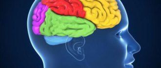
Functions are distributed not only between the hemispheres, but also across different areas of the brain. The cerebral cortex can be divided into four paired lobes: occipital, parietal, temporal and frontal.
- The frontal lobes can be called the command post of the brain. Here are the centers that ensure a person’s independence and initiative, as well as his ability to critically self-assessment. The frontal lobes are also responsible for mastering skills. It is thanks to them that initially complex work becomes automatic and does not require much effort.
- The temporal lobes in the upper regions process auditory sensations, turning them into images. It is in this part of the brain that the words addressed to a person are recognized and filled with meaning, as well as the selection of words to express one’s thoughts. The anterior and middle parts of the temporal lobes are associated with the sense of smell. It is the temporal lobes that store memories. The dominant temporal lobe deals with verbal memory and object names, while the non-dominant temporal lobe deals with visual memory.
- The parietal lobes are responsible for the ability to put parts together into a whole. For example, to read you need to be able to put letters into words and words into phrases. Same with numbers and numbers. This part of the brain is also responsible for sensing your body and distinguishing between the right and left sides. The parietal lobes are the center of the three-dimensional image. It provides a three-dimensional perception of the surrounding world. This side is also involved in spatial orientation and the perception of heat, cold and pain.
- The occipital lobes are responsible for processing visual information. Everything we see, we do not see with our eyes, which only record light and translate it into electrical impulses. We “see” with the occipital lobes, which interpret signals from the eyes.
USEFUL CONNECTIONS
The human brain contains about 100 billion neurons. This value is approximate. After all, it is almost impossible to count all microscopic cells without missing a single one.
The channels or nerve pathways along which signals travel are called synapses. Two cells participate in their formation - transmitting and receiving. Synapses are located in those areas of nerve cells where they contact each other. They are also present where nerve cells connect with muscles or glands.
STORE YOUR MEMORIES IN TERABYTES
A piece of brain the size of a grain of sand contains 100,000 neurons. Each of them is connected to its “neighbors” using 40,000 synapses. It turns out that there are more connections in the brain than there are stars in the Universe. This organ has enough space for 1000 terabytes of information.
The middle part of the synapse is the synaptic cleft—the space between two interacting nerve cells. An electrical impulse passes through this gap.
The final part of the synapse is the postsynaptic terminal. This cell fragment comes into contact with many sensitive receptors.
Frontal part
The most anterior part of the brain is called the frontal lobe. Its boundaries are delineated by two grooves: the central one at the back, separating it from the parietal lobe (this groove is also called the Rolandic groove), the lateral one at the bottom, the structure of which is described in detail above. Anterior to the central recess are the precentral grooves. One is located above, and the second is below. These grooves limit the central gyrus.
The frontal lobe is divided into three frontal gyri: superior, middle and inferior. They are delimited from each other by the superior and inferior frontal grooves. We can say that it is in the frontal lobe that the largest grooves and convolutions of the brain are located.
Formation of the cortex in embryogenesis
The grooves and gyri in neuroanatomy that give the brain its wrinkled appearance serve two critical functions. They help increase the surface area of the cortex, which allows more neurons to pack into it and enhance the brain's ability to process information. The sulci and convolutions of the brain form divisions, creating boundaries between the lobes of the brain, dividing it into two hemispheres.
Main grooves:
- The interhemispheric fissure is a deep groove in the center of the brain that contains the corpus callosum.
- The Sylvian fissure (lateral sulcus) separates the parietal and frontal lobes.
- Roland's fissure (central sulcus), separating the fusiform gyrus and the hippocampal gyrus on the inferior surface of the temporal lobes.
- Parieto-occipital - separates the parietal and occipital lobes.
- The calcarine fissure (spur-like groove or prominent fissure) is located in the occipital lobes and divides the visual cortex.
The main convolutions of the brain:
- The angular gyrus of the parietal lobe helps in processing auditory and visual recognition.
- Broca's gyrus (Broca's center) is an area of the brain located in the left frontal lobe in most people that controls functions related to speech production.
- The cingulate gyrus, an arched fold located above the corpus callosum, is a component of the limbic system and processes sensory input regarding emotions and regulates aggressive behavior.
- The fusiform gyrus is located in the temporal and occipital lobes and consists of lateral and medial parts. It is thought to play a role in word and face recognition.
- The hippocampal gyrus lies on the inner surface of the temporal lobe, which borders the hippocampus. Plays an important role for memory.
- The lingual gyrus in the occipital lobe is involved in visual processing. It is limited by the collateral groove and calcarine fissure. In front it is in contact with the pararpopampal gyrus, and together they form the medial part of the fusiform gyrus.
It is followed by the central one, separating the motor cortex (precentral gyrus) from the somatosensory cortex (postcentral gyrus). Most of the cortical sulci and gyri of the brain, the anatomy of which begins to take shape between 24 and 38 weeks of pregnancy, continue to grow and develop after the newborn is born.
The early state of the brain has a strong influence on the final level of gyrification. In particular, there is an inverse relationship between cortical thickness and gyrification. Brain regions with low thickness values have a higher level of gyrification. The opposite is also true: brain regions with a high thickness value (for example, thickening of the hippocampal gyrus cortex) have a low level of gyrification.
Embryogenesis is the intrauterine development of the fetus from the moment of conception to birth. First, uneven depressions form on the cerebral cortex, which give rise to furrows. The primary grooves are formed first. This occurs around the 10th week of intrauterine development. After this, secondary and tertiary depressions are formed.
The deepest groove is the lateral one; it is one of the first to form. It is followed in depth by the central one, which separates the motor (motor) and sensory (sensitive) zones of the cerebral cortex.
Parietal part
This lobe of the brain is limited from other structures by four sulci: central, lateral, parieto-occipital and transverse occipital. Behind the central, by analogy with the frontal lobe, there is a postcentral sulcus, which in some textbooks is further divided into two parts: upper and lower. The two recesses listed above limit the postcentral gyrus.
The parietal part of the brain is divided into two lobes (upper and lower) by the interparietal groove. The inferior lobule includes the supramarginal and angular gyri.
Temporal part
The temporal part of the cerebral hemispheres is limited by the lateral sulcus from above, and from behind by a conditional line drawn from this sulcus to the posterior occipital one. The structure of this lobe of the brain is easy to remember: three parallel convolutions are separated by three parallel grooves. The grooves and convolutions of the brain in the temporal part are called the same name: superior, middle and inferior temporal.
Medial surface
The sulcus of the corpus callosum is located most medially, which then passes into the sulcus of the hippocampus, which borders the hippocampus itself. Next to the callosal sulcus are the subparietal and callosal-marginal sulci. The rhinal sulcus runs parallel to the hippocampus.
The recesses of the brain listed above limit a specific system, which is called limbic. It, in turn, consists of the cingulate and hippocampal gyri.
In addition to the limbic system itself, on the inner surface of the brain there are also structures that continue their course from the outer part of the cerebral cortex. In this way, the parieto-occipital groove extends, behind which the precuneus is located (a gyrus resembling a trapezoid in shape). Next to this depression there is also a calcarine groove, which extends from the back of the head and forward all the way to the corpus callosum. Between the two recesses mentioned above is the sphenoid gyrus.
Bottom surface
The lower, or basal, surface of the brain is formed by parts of the frontal, temporal and occipital lobes. However, in addition to these structures, the so-called olfactory brain is also located on the basal surface. It consists of the olfactory sulcus, surrounded by the straight gyrus and orbital sulci.
The temporal lobe, based on the brain, contains the inferior temporal and occipitotemporal sulci, between which the gyrus of the same name is located. The lingular gyrus is also detailed nearby.
Lobes of the brain and their functions
Each hemisphere is divided into four lobes: frontal, parietal, temporal and occipital. Most brain functions rely on different regions throughout the brain working together, but each lobe performs the bulk of relatively specific functions.
The frontal lobe is located in the most anterior region of the cerebral cortex, separated from the parietal lobe by the central sulcus, and from the temporal lobe by the lateral sulcus. The region typically contains the most important executive functions for a person, including emotion regulation, planning, reasoning, and problem solving.
The parietal lobe is responsible for the integration of sensory information, including contact, temperature, pressure, and pain. Because of the processing that occurs in the parietal lobe, it is possible to distinguish between the touch of two objects at nearby points (rather than as a single object). This process is called two-point.
The temporal lobe also contains areas involved in sensory processing, especially important for hearing, language recognition, and memory formation. The primary auditory cortex receives audio information through the ears and secondary areas and processes the data so that a person understands what he hears (words, laughing, crying, etc.). The medial (closer to the center of the brain) part contains the hippocampus, an area important for memory, learning, and perception of emotions. Certain areas of the temporal lobe process complex visual information, including faces and scenes.