Structural features
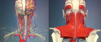
The spinal cord and brain are a single whole, an integral part of the nervous system. All mental functions, control of vital processes (activity, touch, sensitivity of the limbs) are carried out with their help. They are covered with protective structures that work harmoniously to provide nutrition and remove metabolic products.
The membranes of the spinal cord and brain are largely similar in structure. They continue the spine and envelop the spinal cord, preventing damage to it. This is a kind of “clothing” of the most important human organ, characterized by increased sensitivity. All layers are interconnected and they function as one, although their tasks are slightly different. There are three shells in total, and each has its own characteristics.
Dura shell
It is a fibrous formation with increased density, consisting of connective tissue. In the spine, it envelops the brain along with nerves and roots, spinal ganglia, as well as other membranes and fluid. The outer part is separated from the bone tissue by the epidural space, which consists of venous bundles and a fatty layer.
The hard shell of the spinal cord is inextricably linked with the same structure of the brain. In the head, the latter is fused with the periosteum, therefore it fits tightly to the inner surface of the skull, without forming an epidural space, which is its characteristic feature. The space between the dura mater and the arachnoid membrane is called the subdural; it is very narrow and filled with fluid similar to tissue.
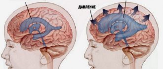
The main functions of the hard shell are to create natural shock absorption, which reduces pressure and eliminates mechanical impact on the brain structure during movement or injury. In addition, there are a number of other tasks:
- synthesis of thrombin and fibrin - important hormones in the body;
- ensuring normal metabolic processes in tissues and lymph movement;
- normalization of blood pressure in the body;
- suppression of inflammatory processes;
- immunomodulation.
In addition, the shell has such an anatomy that it takes part in the blood supply. Tight closure with the vertebral bones allows it to reliably fix soft tissues in the ridge. This is important to ensure their safety during movement, physical exercise, falling, or injury.
Important! Connective tissue is attached to the periosteum by several types of ligaments: anterior, lateral, dorsal. If it is necessary to remove the dura mater, they present a serious obstacle for the surgeon due to the peculiarities of their structure.
No ads 1
Arachnoid
The arachnoid membrane of the human spinal cord is located on the outer part of the soft tissue, but deeper than the hard tissue. It covers the structure of the central nervous system and is devoid of color and blood vessels. In general, it is a connective tissue covered by endothelial cells. Connecting with the hard shell, it forms a space where the cerebrospinal fluid functions, but does not enter the grooves or depressions, passes by them, forming something like bridges. It is this cerebrospinal fluid that protects the nerve structures from various adverse effects and maintains water balance in the system.
Its main functions are:
What is spinal myelography
- formation of hormones in the body;
- maintaining natural metabolic processes;
- transportation of cerebrospinal fluid into the venous blood;
- mechanical protection of the brain;
- formation of nervous tissue (in particular, cerebrospinal fluid);
- generation of nerve impulses;
- participation in metabolic processes in neurons.
The middle shell has a complex structure and looks like a mesh fabric, with a small thickness but high strength. It was its resemblance to a spider's web that gave it its name. Some experts believe that it is devoid of nerve endings, but this is only a theory that has not been proven to date.
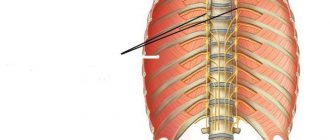
Visual structure and location of the spinal cord membranes
Soft shell
Closest to the brain is the soft shell, which has a loose structure and consists of connective tissue. It contains blood vessels and plexuses, nerve endings and small arteries, all of which are responsible for providing the brain with enough blood for normal functioning. Unlike the arachnoid, it goes into all the cracks and grooves.
But, despite their close location, the brain is not covered by it, since between them there is a small space called subpial. It is separated from the subarachnoid space by many blood vessels. Its main functions include supplying the brain with blood and nutrients, normalizing metabolism and metabolism, as well as maintaining the natural performance of the body.
The functioning of all membranes is interconnected and the structure of the spine as a whole. Various malfunctions, changes in the amount of cerebrospinal fluid or inflammatory processes at any level lead to serious consequences and disorders and diseases of internal organs.
[node:field_similarlink]
Spaces between shells
All the membranes of the spinal cord and brain, although they are close to each other, do not touch tightly. Spaces are formed between them, which have their own characteristics and functions.
- Epidural. It is located between the hard shell and the bone tissue of the spinal column. It is filled predominantly with fat cells to eliminate nutritional deficiencies. Cells become a strategic reserve for neurons in extreme situations, which ensures the control and functioning of processes in the body. This space reduces the load on the deep layers of the spinal cord, eliminating their deformation, due to its loose structure.
- Subdural. Located between the dura mater and the arachnoid membrane. It contains liquor, the amount of which always changes. On average, an adult has 150–250 ml. Cerebrospinal fluid provides the brain with nutrients (minerals, proteins), protects it from falls or impacts, maintaining pressure. Thanks to the movement of cerebrospinal fluid and its constituent lymphocytes and leukocytes, infectious processes are suppressed in the central nervous system and bacteria and microorganisms are absorbed.
- Subarachnoid. Located between the arachnoid and soft membrane. It constantly contains most of the cerebrospinal fluid. This allows you to most effectively protect the central nervous system, brainstem, cerebellum and medulla oblongata.
In case of tissue damage, the first step is to analyze the cerebrospinal fluid, as it allows you to determine the extent of the pathological process, predict the course, and choose effective control tactics. An infection or inflammation that appears in one area quickly spreads to neighboring ones. This is due to the constant movement of cerebrospinal fluid.
No ads 2
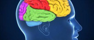
Large hemispheres
- the largest part of the brain, accounting for approximately 70% of its weight in adults. Normally, the hemispheres are symmetrical. They are connected to each other by a massive bundle of axons (corpus callosum), which ensures the exchange of information.
Each hemisphere consists of four lobes: frontal, parietal, temporal and occipital. The frontal cortex contains centers that regulate motor activity, as well as, probably, centers for planning and foresight. In the cortex of the parietal lobes, located behind the frontal lobes, there are zones of bodily sensations, including touch and joint-muscular sensation. Adjacent to the parietal lobe is the temporal lobe, in which the primary auditory cortex, as well as the centers of speech and other higher functions, are located. The posterior parts of the brain are occupied by the occipital lobe, located above the cerebellum; its cortex contains areas of visual sensation.
Areas of the cortex not directly associated with the regulation of movements or the analysis of sensory information are called the associative cortex. In these specialized zones, associative connections are formed between different areas and parts of the brain and the information coming from them is integrated. The association cortex supports complex functions such as learning, memory, language, and thinking.
Diseases
The meninges can be injured or suffer from damage of an infectious nature. Increasingly, problems are associated with the development of oncology. They are recorded in patients of different ages and health conditions. In addition to infectious processes, there are other malfunctions:
- Fibrosis. It represents a negative consequence of the surgical intervention. It leads to an increase in the volume of the membrane, characteristic tissue scarring, and an inflammatory process that occurs immediately in all intershell spaces. The disease is also often provoked by cancer or spinal injuries.
- Meningitis. Severe pathology of the spinal cord, which occurs as a result of the penetration of a viral infection into the body (pneumococcus, meningococcus). It is accompanied by a number of characteristic symptoms and, if left untreated, can lead to serious complications and even death of the patient.
- Arachnoiditis. An inflammatory process develops in the lumbar region of the spinal cord, which also affects the membranes. All three levels suffer. Clinically, the disease manifests itself with focal symptoms and neurasthenic disorders.
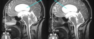
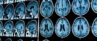
The shells or the space between them can also be damaged as a result of injury. Usually these are bruises or fractures that cause compression of the spinal cord. Acute disruption of cerebrospinal fluid circulation causes paralysis or hydrocephalus. Many malfunctions of the membranes in the clinical picture can be confused with other infectious diseases, therefore an MRI is always prescribed to clarify the diagnosis.
Features of treatment
Inflammatory processes in the membranes of the spinal cord or brain require immediate treatment in a hospital setting. Self-medication of any disease at home often leads to death or serious complications. Therefore, when the first signs of illness appear, you should consult a doctor and follow all recommendations.
Features of treatment of possible pathologies:
- Viral infection. Monitor body temperature and take enough fluids. If a person cannot drink a lot of water, droppers with saline solution are prescribed. If cysts form or the volume of cerebrospinal fluid increases, then medications are required to normalize the pressure. The chosen tactics to combat inflammation are adjusted as the patient’s condition improves.
- Injury. The membranes of the spinal cord provide its normal nutrition and blood circulation, therefore, when scars, adhesions and other damage form, this function is disrupted, the movement of cerebrospinal fluid is hampered, which leads to the appearance of cysts and intervertebral hernia. Treatment in this case includes taking a set of medications to improve metabolic processes. If traditional therapy is ineffective, surgical intervention is prescribed.
- Infectious processes. The entry of pathogenic bacteria into the organ requires the prescription of antibiotics. In most cases, this is a broad-spectrum drug. An important point is also monitoring water balance and body temperature.
The consequences of diseases of the membranes can be unpredictable. Inflammatory processes cause disturbances in the functioning of the body, fever, vomiting, seizures, and convulsions. Often hemorrhages lead to paralysis, which makes a person disabled for life.
The spinal membranes form a single system and are directly connected to the hypothalamus and cerebellum. Violation of their integrity or inflammatory processes lead to a deterioration in the general condition. Usually accompanied by seizures, vomiting, and fever. Modern medicine has reduced the mortality rate due to such diseases to 10–15%. But the risk still exists. Therefore, when you notice the first signs, you should immediately consult a doctor.
ELECTRICAL ACTIVITY OF THE BRAIN
Using electrodes placed on the surface of the head or inserted into the brain, it is possible to record the electrical activity of the brain caused by the discharges of its cells. Recording the electrical activity of the brain using electrodes on the surface of the head is called an electroencephalogram (EEG). It does not allow recording the discharge of an individual neuron. Only as a result of the synchronized activity of thousands or millions of neurons do noticeable oscillations (waves) appear in the recorded curve.
With continuous recording of the EEG, cyclic changes are revealed that reflect the general level of activity of the individual. In a state of active wakefulness, the EEG records low-amplitude, non-rhythmic beta waves. In a state of relaxed wakefulness with eyes closed, alpha waves predominate at a frequency of 7–12 cycles per second. The onset of sleep is indicated by the appearance of high-amplitude slow waves (delta waves). During periods of dreaming sleep, beta waves reappear on the EEG, and the EEG may give the false impression that the person is awake (hence the term “paradoxical sleep”). Dreams are often accompanied by rapid eye movements (with the eyelids closed). Therefore, dreaming sleep is also called rapid eye movement sleep (see also SLEEP). EEG allows you to diagnose some brain diseases, in particular epilepsy (see EPILEPSY).
If you record the electrical activity of the brain during the action of a certain stimulus (visual, auditory or tactile), then you can identify the so-called. evoked potentials are synchronous discharges of a certain group of neurons that occur in response to a specific external stimulus. The study of evoked potentials made it possible to clarify the localization of brain functions, in particular, to associate speech function with certain areas of the temporal and frontal lobes. This study also helps to assess the state of sensory systems in patients with sensory impairment.