Location and anatomy of the back of the head in humans
Blood circulation, innervation, muscles of the occipital part
The occiput is the back part of the head, located above the cervical region and under the crown. The bone that forms it borders on the sphenoid, parietal and temporal bones. It is convex on the outside and concave on the inside. Consists of four different elements:
- Scales. It looks like a triangular plate. It has a concave part on one side and a convex part on the other side. Due to the attachment of muscle fibers and ligaments to its surface, it has a rough texture.
- Articular condyles located in the lateral parts. Responsible for the fusion of the skull with the spine.
- Main body. Fuses tightly with the sphenoid cranial bone by the age of 17-20. The shape resembles a quadrangle, the back of which forms one of the sides of the occipital foramen. In children, it has clefts filled with cartilaginous tissue. Gradually they harden.
- Big hole. It is localized between the three listed parts, forming a passage between the spinal column and the cranial cavity. The perfect combination of its shape with the first cervical vertebra results in a strong interaction between two important skeletal segments.
If in humans the occipital bone acts as a single system, then in animals it can consist of several interconnected bone structures.
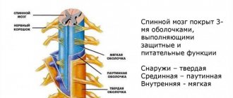
The blood supply to the occipital part of the head is carried out by the posterior auricular artery. Innervation occurs due to the nerve endings of the greater occipital nerve, which is formed from the posterior branch of the second cervical spinal nerve. The skin is also innervated by the greater occipital nerve, which arises from the cervical nerve plexus.
Head skeleton
Chapter 4. BONES AND THEIR COMPOUNDS
The skeleton of the head is represented by bones, which, tightly connected by sutures, protect the brain and sensory organs from mechanical influences. It provides support to the face, the initial parts of the respiratory and digestive systems.
The skull (cranium) is divided into two sections - the brain and the face. The bones of the cranium form a cavity for the brain and partially a cavity for the sensory organs. The bones of the facial skull make up the bony base of the face and the skeleton of the initial parts of the respiratory and digestive systems. Some skull bones have air-filled cavities inside and connect to the nasal cavity. This bone structure significantly reduces the mass of the skull and at the same time maintains its necessary strength. The bones of the brain skull include eight bones: two paired - temporal and parietal and four unpaired - frontal, ethmoid, sphenoid and occipital. Part of the bones of the facial skull makes up the skeleton of the masticatory apparatus: the paired maxillary bone and the unpaired lower jaw. Other facial bones are smaller in size. These are paired bones: palatine, nasal, lacrimal, zygomatic, inferior nasal concha; unpaired bones include the vomer and hyoid bone. They are part of the cavities of the facial skull and determine its configuration.
Bones of the brain section of the skull. The frontal bone (os frontale) is located in front of the paired parietal bones and participates in the formation of the anterior part of the cranial vault and the anterior cranial fossa (Fig. 21).
Rice. 21. Frontal bone:
1 - scales; 2 - frontal tubercle; 3. - temporal line; 4—zygomatic process; 5—supraorbital margin; b—supraorbital foramen; 7—nose; S—glabella (glabella); 9 - brow ridge
The frontal bone consists of the frontal squama, orbital and nasal parts. The frontal scales are involved in the formation of the cranial vault. On the convex outer surface of the frontal bone there are paired protrusions - the frontal tubercles, and below - the brow ridges. The flat surface between the brow ridges is called the glabella. Inferiorly, the brow ridges pass into the supraorbital margin, which laterally ends with the zygomatic process and connects to the zygomatic bone. The right orbital part is separated from the left by a deep ethmoid notch, in which the perforated plate of the ethmoid bone is located.
The orbital parts of the frontal bone, with their lower orbital surface, take part in the formation of the upper wall of the orbit, and with their inner surface, in the formation of the anterior cranial fossa. In the lateral parts of the orbital surface there is a shallow fossa of the lacrimal gland, and medially there is a trochlear fossa.
The nasal part is located between the orbital parts and limits the ethmoidal notch in front and on the sides. On the nasal part there is a nasal spine, which participates in the formation of the nasal septum, on the sides of which there are openings into the frontal sinus.
The parietal bone (os parietale) is a paired plate that forms the middle part of the cranial vault. It has a convex (outer) and concave (inner) surface (Fig. 22, 23), four edges and four corners.
Rice. 22. Right parietal bone (inner surface):
1 - sagittal edge; 2—parietal foramen; 3 - groove of the superior sagittal sinus; 4 - occipital angle; 5—occipital edge; b—mastoid angle; 7— groove of the sigmoid sinus; 8-10— groove of the middle meningeal artery; 11 — wedge-shaped angle; 12—frontal edge; 13 - inner surface; 14 - frontal angle
The upper (sagittal) edge connects with the opposite parietal bone, the anterior (frontal) and posterior (occipital) - with the frontal and occipital bones, respectively. The scales of the temporal bone (squamosal bone) are superimposed on the lower edge of the parietal bone. The relief of the inner surface of the parietal bone is determined by the adjacent dura mater and its vessels. Thus, along the upper edge of the parietal bone there is a groove of the superior sagittal sinus, in addition, the branches of the meningeal arteries are visible on the inner surface.
Rice. 23. Left parietal bone (outer surface):
1 - sagittal edge; 2 - parietal foramen; 3—occipital angle; 4 - occipital edge; 5 - superior temporal line; 6 - mastoid angle; 7— scaly edge; 8— wedge-shaped angle; 9—inferior temporal line; 70—frontal edge; 11 - parietal tubercle; 12— outer surface; 13 - frontal angle
The occipital bone (os occipitale) consists of a basilar and two lateral parts, the occipital scales (Fig. 24, 25).
Rice. 24. Occipital bone (inner surface):
1 - groove of the superior sagittal sinus;. 2 - medullary fossa; 3— occipital scales; 4 - cruciform elevation; 5—internal occipital protrusion; 6— groove of the transverse sinus; 7—internal occipital crest; 8 - cerebellar fossa; 9— condylar canal; 1st; 10 - jugular process; 11 - large hole; 12 - jugular tubercle; 13 - basilar part; 14—pharyngeal tubercle; 15 - occipital condyle; 16 - lateral part; 17—mastoid edge; 18 - lambdoid edge
They surround the foramen magnum, through which the cranial cavity connects to the spinal canal. Anterior to the foramen magnum is the main (basilar) part of the occipital bone, which, fused with the body of the sphenoid bone, forms a slightly inclined surface - the slope.
Rice. 25. Occipital bone (posterior and ventral view):
1 - occipital scales; 2 - jugular tubercle; 3 - slope; 4 - large hole; 5 - condyle canal; 6 - occipital condyle; 7— condylar fossa; 8—lower nuchal line; 9 - external occipital crest; 10—upper nuchal line; 11—highest nuchal line; 12 - occipital platform; 13 - external occipital protuberance
On the lower surface of the lateral (lateral) parts there is an occipital condyle, which serves to connect with the first cervical vertebra. The basilar and lateral parts and lower parts of the occipital scales are involved in the formation of the base of the skull (posterior fossa), where the cerebellum and other brain structures are located.
The occipital scales participate in the formation of the cranial vault. In the center of its inner surface there is a cruciform eminence, which forms the internal occipital protrusion. Somewhat inferior to it is the internal occipital crest. Up from the occipital eminence to the connection with the parietal bones there is a groove of the superior sagittal sinus, to the sides there is a groove of the transverse sinus, down there is a groove of the sigmoid sinus. On the outer surface of the occipital scales, the external occipital protrusion, external nuchal crest and other formations are distinguished. The serrated edge of the scales is connected to the lambdoid suture. parietal and temporal bones.
The ethmoid bone (os ethmoidale), together with other bones, takes part in the formation of the anterior part of the base of the skull, the walls of the orbits and the nasal cavity of the facial part of the skull.
The bone consists of a cribriform plate, from which a perpendicular plate extends downward, participating in the formation of the septum of the nasal cavity. On both sides of the perpendicular plate there are lattice labyrinths consisting of air cells. There are three pairs of cells of the ethmoid bone that connect to the nasal cavity: anterior, middle and posterior. The superior and middle turbinates hang from the ethmoid bone into the nasal cavity, forming three nasal passages in each half of the nasal cavity. On the lateral side, the ethmoidal labyrinths are covered with a thin orbital plate, which participates in the formation of the inner wall of the orbit.
The sphenoid bone (os sphenoidale) is located between the frontal and occipital bones and is located in the center of the base of the skull (Fig. 26).
Rice. 26. Sphenoid bone (top view):
1 — small wing (left); 2 - body; 3 - pre-cross groove; 4 - pituitary fossa; 5—visual channel; 6—superior orbital fissure; 7— round hole; 8, 12—large wings; 9— oval hole; 10—foramen spinosum; 11— saddle back
This bone is shaped like a butterfly. It consists of a body and three paired processes: large and small wings and pterygoid processes. On the upper surface of the body of the bone there is a depression (sella turcica), in which the main endocrine gland, the pituitary gland, is located. In the body of the sphenoid bone there is a sinus that connects to the nasal cavity. Two small wings extend to the sides from the anterosuperior surface of the sphenoid bone; at the base of each there is a large opening of the optic canal, through which the optic nerve passes into the orbit. Between the lesser and greater wings there is the superior orbital fissure, through which the oculomotor, lateral, abducens and ophthalmic nerves, the first branch of the trigeminal nerve, pass from the cranial cavity into the orbit.
The large wings contain the orbital, temporal, maxillary and medullary surfaces. At the base of the greater wing there are the round, oval and spinous foramina, through which branches of the trigeminal nerve and vessels pass. Finger-shaped depressions, elevations and arterial grooves are clearly visible on the brain surface.
The pterygoid process extends downward in pairs from the body of the sphenoid bone and consists of lateral and medial plates, between which there is a pterygoid fossa on the posterior surface. The medial plate forms the nasal cavity, the lateral plate forms the infratemporal fossa. At the base of the pterygoid process there is a narrow canal of the same name, through which vessels and nerves pass.
The temporal bone (os temporale) is a paired bone (Fig. 27, 28), part of the base of the skull and the lateral part of the cranial vault, connected in front with the sphenoid, in the back with the occipital and above with the parietal bones. The temporal bone is a container for the organs of hearing and balance; blood vessels and nerves pass through its canals. The temporal bone is divided into squamosal, petrous and tympanic parts. With the lower jaw, the temporal bone forms a joint, and with the zygomatic bone, the zygomatic arch.
Rice. 27. Right temporal bone (inner surface):
1 - upper edge of the pyramid; 2 - groove of the superior petrosal sinus; 3 - groove of the sigmoid sinus; 4 - internal process; 5 - jugular notch; 6 - styloid process; 7—internal auditory opening;.?—internal auditory canal; 9 - groove of the inferior petrosal sinus; 10—rear edge of the pyramid; 11— back surface of the pyramid
On the inner surface of the scaly part there are finger-like impressions and cerebral elevations, and a trace of the middle meningeal artery is visible.
On the outer convex surface of the scaly part, slightly higher and anterior to the external auditory opening, a horizontally located zygomatic process begins. At the base of the latter there is a mandibular fossa, with which the condylar process of the mandible forms a joint.
Rice. 28. Right temporal bone (outer surface):
1 - tympanic-squamosal fissure; 2 - petrotympanic fissure; 3—styloid process; 4 - tympanomastoid fissure
The pyramid (stony part) of the temporal bone has a triangular shape. It has anterior, posterior and inferior surfaces. The anterior surface of the pyramid participates in the formation of the middle cranial fossa, the posterior surface - the posterior cranial fossa, and the lower one is part of the outer base of the skull. On the anterior surface at the apex of the pyramid there is a trigeminal depression, in which lies the trigeminal nerve ganglion. On the posterior surface of the pyramid is the internal auditory opening, which passes into the internal auditory canal. At the bottom of the latter there are several openings for the facial, vestibular-cochlear cranial nerves and vessels of the vestibular-cochlear organ. On the posterior surface of the pyramid there is the external opening of the vestibule aqueduct, the cochlear canaliculus, and the groove of the sigmoid sinus. A long styloid process extends from the lower surface of the pyramid; near it there is a stylomastoid foramen through which the facial nerve exits. In the center of the lower surface of the pyramid there is a rounded hole, which gives rise to the carotid canal. The internal carotid artery passes through it into the cranial cavity. Posterior to the external opening of the carotid canal, the jugular fossa is visible, which in the region of the posterior edge of the pyramid passes into the jugular notch.
The jugular notches of the temporal and occipital bones, when connected, form the jugular foramen on the whole skull, through which the internal jugular vein and three cranial nerves pass: glossopharyngeal, vagus and accessory.
In the thickness of the stony part there are the tympanic cavity and the bony labyrinth. The tympanic cavity is connected to the air cells of the mastoid process, as well as to the cavity of the nasopharynx through the auditory tube.
In the pyramid of the temporal bone there are the carotid and facial canals, as well as the canaliculus of the chorda tympani, the tympanic canaliculus, the mastoid canaliculus, the carotid-tympanic canals, in which the vessels, nerves and muscle that tensor the tympanic membrane are located.
Bones of the facial part of the skull. They are represented by paired bones (upper jaw, palatine, zygomatic, nasal, lacrimal and inferior nasal concha) and unpaired bones (lower jaw, hyoid bone and vomer).
The upper jaw (maxilla) consists of a body and four processes: frontal, zygomatic, palatine and alveolar.
There are four surfaces in the body: anterior, infratemporal, orbital and nasal. In the body of the upper jaw there is a rather large maxillary (maxillary) sinus (Fig. 29, 30).
Rice. 29. Upper jaw (lateral view):
1 - orbital surface; 2 - infraorbital groove; 3 - zygomatic process; 4— alveolar openings; 5 - infratemporal surface; 6 — front surface; 7— canine fossa; 8 - anterior nasal spine; 9 - body of the upper jaw; 10—nasal notch; 11 - infraorbital canal; 22 - infraorbital foramen; 13 - zygomaticomaxillary suture; 14 - frontal process; 15 - lacrimal edge; 16—infraorbital margin
The upper jaw participates in the formation of the nasal cavity, orbit, oral cavity, infratemporal and pterygopalatine fossa. The alveolar process has cells for the eight upper teeth.
The palatine bone (os palatinum) is paired, consists of two bone plates - perpendicular and horizontal, which form part of the wall of the nasal cavity and the hard palate.
The zygomatic bone (os zygomaticum) is paired, has lateral, temporal, orbital surfaces, frontal and temporal processes. With its size, this spine determines the width and shape of the face.
Rice. 30. Left upper jaw (view from the medial side):
1 - frontal process; 2 - nasal surface; 3 - anterior nasal spine; 4—pterygopalatine groove; 5 - maxillary (maxillary) sinus
The lacrimal bone (os lacrimale) is paired, participates in the formation of the inner wall of the orbit and limits the fossa of the lacrimal gland.
The inferior turbinate (concha nasalis inferior) is a paired bone. With one edge it connects to the upper jaw and palatine bone, and with the other it hangs into the nasal cavity, limiting the lower nasal passage.
The lower jaw (mandibula) is the only movable bone in the human skull; it consists of a body and two branches (Fig. 31).
On the outer surface of the body there is a mental protuberance, and on its sides there is a mental tubercle and mental foramen. Along the upper edge of the body of the lower jaw there are dental alveoli, separated by septa. This edge is called the alveolar edge. Each branch of the bone ends at the top with an anterior coronoid and posterior condylar (articular) process. On the inner surface of the branch there is a hole that leads into the canal of the lower jaw.
Rice. 31. Lower jaw:
I - head of the lower jaw; 2 - pterygoid fossa; 3 - neck of the lower jaw; 4, 5 — branches of the lower jaw; b—angle of the lower jaw; 7— canal of the lower jaw; 8 - temporal crest; 9 - opening of the lower jaw; 10— coronoid process; II - mandibular notch; 12— condylar process
The hyoid bone (os hyoideum) consists of a body, a pair of large and a pair of small horns and is located in the neck, between the lower jaw and larynx.
The vomer (vomer), connecting to the ethmoid bone, participates in the formation of the nasal septum and separates the paired openings of the exit from the nasal cavity - the choanae.
The bones of the skull are connected to each other using continuous connections - synarthrosis. The latter include bone fusions - synostoses; cartilaginous - synchondrosis and fibrous - syndesmosis. The predominant type of fibrous joints of the skull are sutures. Depending on the shape, the following types of seams are distinguished: jagged, scaly and flat. Thus, the dentate includes the coronal (between the frontal and parietal bones), sagittal (along the midline between the paired parietal bones) and lambdoid (between the occipital scales and the parietal bones) sutures.
The scaly part of the temporal bone and the parietal bone on the lateral surface of the cranial vault are connected by a scaly suture. The bones of the face are connected by flat sutures.
The cartilaginous junction is located between the occipital and sphenoid bones only until the age of 20, and then it ossifies (synostosis).
The temporomandibular joint (articulatio temporoman-dibularis) is paired, complex in structure, elliptical in shape. Formed by the head of the condylar process of the mandible and the articular fossa of the temporal bone. Inside the joint there is an articular disc, which divides the joint cavity into two floors: upper and lower, isolated from each other. The two temporomandibular joints function together (movement in one joint moves the other) and are considered a single combined joint. In the joint, lowering and raising of the lower jaw, lateral movements (right and left) and pushing it forward and backward are possible.
Skull as a whole. The skeleton of the skull (Fig. 32, 33) is conventionally divided into a vault, or roof, and a base.
Rice. 32. Human skull (front view, according to R. D. Sinelnikov):
1 - coronal suture; 2 - parietal bone; 3 - orbital part of the frontal bone; 4 - orbital surface of the greater wing, sphenoid bone; 5—zygomatic bone; 6— inferior nasal concha; 7—upper jaw; 8—mental protuberance of the lower jaw; 9 - nasal cavity; 10— opener; 11—perpendicular plate of the ethmoid bone; 12—orbital surface of the upper jaw; 13 - inferior orbital fissure; 14 - lacrimal bone; 15 - orbital plate of the ethmoid bone; 16 - superior orbital fissure; 17— scaly part of the temporal bone; 18—zygomatic process of the frontal bone; 19—visual channel; 20—nasal bone; 21 - frontal tubercle
The cranial vault is formed by the scaly parts of the frontal, temporal, occipital bones and parietal bones. The base of the skull consists of the frontal, ethmoid, sphenoid, temporal and occipital bones. There are internal and external bases of the skull.
Rice. 33. Human skull (side view, according to R. D. Sinelnikov):
1 - parietal bone; 2— coronal suture; 3 - frontal tubercle; 4 - temporal surface of the large wing of the sphenoid bone; 5 - orbital plate of the ethmoid bone; b - lacrimal bone; 7—nasal bone; 8—temporal fossa; 9—anterior nasal spine; 10—body of the upper jaw; 11 - lower jaw; 12—zygomatic bone; 13 - zygomatic arch; 14 - styloid process; 15 — condylar process of the lower jaw; 16—mastoid process; 17— external auditory canal; 18— lambdoid suture; 19 - scales of the occipital bone; 20 - superior temporal line; 21 - scaly part of the temporal bone
The internal base of the skull (basis cranii interna) has three cranial fossae: anterior, middle and posterior (Fig. 34). In the anterior cranial fossa there are the lobes of the cerebral hemispheres, in the middle - the temporal lobes of the cerebral hemispheres, and in the posterior - the cerebellum and parts of the brain stem: the cerebral peduncles and the medulla oblongata.
The anterior cranial fossa is formed by the orbital part of the frontal bone, the ethmoid bone (ethmoid plate) and the lesser wings of the sphenoid bone and communicates with the nasal cavity through the holes in the cribriform plate. These openings serve as the passage for the olfactory nerves (1st pair).
The walls of the middle cranial fossa are formed by the body and large wings of the sphenoid bone, the anterior surface of the pyramids, and the squamous part of the temporal bones. The middle cranial fossa communicates with the orbit and the pterygopalatine fossa. From this fossa, the optic nerve (II pair), the orbital artery and vein pass into the cavity of the orbit through the optic canal. Through the superior orbital fissure, the oculomotor (III pair), trochlear (IV pair), abducens (VI pair) and ophthalmic (first branch of the trigeminal nerve (V pair)) nerves pass into the orbit. Somewhat posterior to the superior orbital fissure there is a round foramen through which the the maxillary nerve (second branch of the V pair), and the mandibular nerve (third branch of the V pair) emerges from the skull through the foramen ovale. In the pituitary fossa of the sella turcica there is an endocrine gland - the pituitary gland.
Rice. 34. Inner base of the skull:
1 - orbital part of the frontal bone; 2 - cockscomb; 3 - cribriform plate; 4—visual channel; 5 - pituitary fossa;. 6— back of the saddle; 7 - round hole; 8 - oval hole; 9 — torn hole; 10—foramen spinosum; 11 - internal auditory opening; 12 - jugular foramen; 13 - sublingual canal; 14 - lambdoid seam; 75 - slope; 16 - groove of the transverse sinus; 77—internal occipital protrusion; 18 - large (occipital) foramen; 19 - occipital scales; 20 - groove of the sigmoid sinus; 21 - pyramid (stony part) of the temporal bone; 22 - scaly part of the temporal bone; 23 - large wing of the sphenoid bone; 24 - lesser wing of the sphenoid bone
The occipital bone, posterior surfaces of the pyramids, and temporal bones take part in the formation of the posterior cranial fossa.
Between the back of the sella turcica and the foramen magnum there is a clivus.
The internal auditory foramen (right and left) opens into the posterior cranial fossa, from which the vestibulocochlear nerve (VIII pair) emerges, and from the facial nerve canal - the facial nerve (VII pair). The lingual pharyngeal (IX pair), vagus (X pair) and accessory (XI pair) nerves exit through the jugular foramen of the base of the skull. The nerve of the same name, the XII pair, passes through the canal of the hypoglossal nerve. In addition to the nerves, the internal jugular vein emerges from the cranial cavity through the jugular foramen, which passes into the sigmoid sinus. The formed foramen magnum connects the cavity of the posterior cranial fossa with the spinal canal, at the level of which the medulla oblongata passes into the spinal cord.
The outer base of the skull (basis cranii extema) in its anterior section is covered by the facial bones (it contains a bony palate, limited in front by the alveolar process of the upper jaw and teeth), and the posterior section is formed by the outer surfaces of the sphenoid, occipital and temporal bones (Fig. 35).
Rice. 35. External base of the skull:
1 - palatine process of the upper jaw; 2—incisal hole; 3 - median palatal suture; 4 - transverse palatal suture; 5—choana; b—inferior orbital fissure; 7— zygomatic arch; 8 — opener wing; 9— pterygoid fossa; 10 - lateral plate of the pterygoid process; 77— pterygoid process; 12 - oval hole; 13 - mandibular fossa ; 14—styloid process; 15 - external auditory canal; 16—mastoid process; 77— mastoid notch; 18— occipital condyle; 19 - condylar fossa; 20— large (occipital) foramen; 27 - lower nuchal line; 22—external occipital protrusion; 23 - pharyngeal tubercle; 24 - condylar canal; 25—jugular foramen; 26— occipital-mastoid suture; 27— external carotid foramen; 28— stylomastoid foramen; 29—torn hole; 30 - petrotympanic fissure; 31 - foramen spinosum; 32 - articular tubercle; 33 - wedge-squamous suture; 34 - pterygoid hook; 35—greater palatine foramen; 36— zygomaticomaxillary suture
This area has a large number of openings through which vessels and nerves pass, providing blood supply to the brain. The central part of the external base of the skull is occupied by the foramen magnum, on the sides of which are the occipital condyles. The latter connect to the first vertebra of the cervical spine. The exit from the nasal cavity is represented by paired openings (choanae), which pass into the nasal cavity. In addition, on the outer surface of the base of the skull there are the pterygoid processes of the sphenoid bone, the external opening of the carotid canal, the styloid process, the stylomastoid foramen, the mastoid process, the myotubal canal, the jugular foramen and other formations.
In the skeleton of the facial skull, the central place is occupied by the nasal cavity, orbits, oral cavity, infratemporal and pterygopalatine fossa.
The nasal cavity (cavitas nasi) is the initial section of the respiratory tract and contains the organ of smell. It has one entrance pyriform opening and two exit openings - choanae.
The nasal cavity is divided into two halves by a bone plate. The nasal cavity is divided into upper, lower and lateral (lateral) walls. The upper wall is formed by the nasal bones, the ethmoid bone, the nasal part of the frontal and the body of the sphenoid bones. The lower wall is represented by the upper palatine processes of the upper jaw and the horizontal plates of the bones of the palate. The lateral wall consists of the frontal process of the maxilla, the lacrimal bone, the ethmoid labyrinth, the perpendicular plate of the palatine bone, the middle (medial) plate of the pterygoid process of the sphenoid bone.
The turbinates divide the lateral part of the cavity into three nasal passages: upper, middle and lower. The sinuses of the sphenoid bone and the posterior cells of the ethmoid bone open into the upper nasal passage; in the middle nasal passage - the sinuses of the upper jaw and frontal bone, as well as the cells of the ethmoid bone; in the lower nasal passage - the nasolacrimal canal, which begins in the orbit.
The orbit (orbita) is a paired cavity, has the shape of a tetrahedral pyramid with rounded edges, the apex of which is directed backward and medially. The optic canal passes through this area. The orbital cavity contains the eyeball with muscles, the lacrimal gland and other formations. It has an entrance and four walls: upper, lower, medial and lateral. The upper wall is formed by the orbital part of the frontal bone and the lesser wings of the sphenoid bone; lower - zygomatic bone and upper jaw; medial - the frontal process of the maxilla, the lacrimal bone, the orbital plate of the ethmoid bone, the body of the sphenoid bone and part of the frontal bone; lateral - the zygomatic bone and the greater wing of the sphenoid bone. Between the lateral and inferior walls there is the inferior orbital fissure, which opens into the pterygopalatine and infratemporal fossa. The superior orbital fissure and optic foramen open into the middle cranial fossa; The nasolacrimal duct connects to the nasal cavity.
The oral cavity (cavitas oris) is formed by the bony (hard) palate, the palatine processes of the right and left upper jaws and the horizontal plates of the palatine bones; the lateral and anterior walls are formed by the alveolar processes of the upper jaws, which together make up the upper alveolar arch. The bony palate serves as the hard (bone) basis of the upper wall of the oral cavity. The upper and lower alveolar arches, together with the teeth and the body of the lower jaw, make up the skeleton of the anterior and lateral walls of the oral cavity.
The infratemporal fossa is located behind the upper jaw, inward from the zygomatic bone and zygomatic arch and externally from the pterygoid process of the sphenoid bone, and forms part of the outer base of the skull.
The pterygopalatine fossa is located between the bones of the brain and facial skull and has four walls: anterior, superior, posterior and medial. The anterior wall is formed by the tubercle of the upper jaw, the upper - by the part of the body and the base of the greater wing of the sphenoid bone, the posterior - by the base of the pterygoid process of the sphenoid bone, the medial - by the perpendicular plate of the palatine bone. Canals and openings open into the pterygopalatine fossa, through which it communicates with neighboring cavities.
Age-related features of the skull. The skull of a newborn has a number of significant features. As a result of active brain growth, its cerebral part is 8 times larger than the facial part, and in adults it is only 2 times larger. A characteristic feature of a newborn is the presence of fontanelles: anterior, posterior and paired lateral ones - sphenoid and mastoid. The anterior fontanel is the largest and has a quadrangular shape. The lateral fontanelles, if present, are overgrown by the second or third month, and the anterior one - in the second year of the child’s life.
The sutures of the skull are formed before 3-5 years of age, and the growth of the skull ends by 25-30 years of a person’s life.
The next feature is the presence of cartilaginous layers between the bones of the base of the skull. In addition (third feature), the newborn has no developed air sinuses, tubercles, processes, no teeth, and poorly developed jaws. In old age, changes occur in the facial skull as a result of tooth loss: the alveolar processes of the jaws decrease, the facial part of the skull shortens somewhat, and the bones of the skull become thinner and more fragile.
Types and shape of the back of the head
Shapes of the back of the head
When assessing the shape of the skull, experts pay attention to the structure of the forehead, crown, occipital part and the tubercles located on them. The anatomy of the back of the human head is the same for all people, regardless of race, gender, mental and physical abilities. However, the types of napes may differ. This part of the skull can be:
- round, in which the occipital line smoothly curves, and its protruding part is located in the middle;
- flat (straight, vertical), when the occipital line is closest to a straight line;
- oblique, in which the occipital line goes in an obliquely straight direction;
- angular, where the nuchal line is slightly curved in the middle at an angle;
- protruding, noticeable in people whose nuchal line arches sharply, protruding at the top.
The position of the occipital line is often determined in profile relative to a vertical line. It passes through the most prominent point of the back of the head along an inclination when the contour of the back of the head is tilted forward, vertically if the contour coincides with the vertical, with a protrusion when the contour extends beyond the vertical.
It is believed that a highly developed forehead in combination with a cut off (underdeveloped) back of the head is found in people prone to self-centeredness and a craving for loneliness. They have poor contact with society and lose their connection with the Cosmos. And, conversely, adult men and women with a well-developed occiput are distinguished by their education and sociability.
Anomalies and damage to the occipital region
If significant deviations are noticed, for example, missing bone fragments, this condition is attributed to a genetic pathology, which is always associated with serious diseases. Based on the disturbed structure and anatomy of the cranial bones, the doctor can make a diagnosis. For example, during examination, you can identify:
- partial or complete fusion of the condyles with the first vertebra;
- change in the weight of the occipital protrusion;
- formation of extra sutures, condyles, bones.
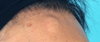
When examining newborns, dolichocephaly is diagnosed based on the shape of the back of the head (protruding or flat). Also considered an anomaly is a low border of hair growth in the occipital part of the skull.
The integrity of the occipital bone can be damaged due to an accident, a fall from a height, a gunshot or knife wound, or injuries to adjacent bone structures. Swelling and hematomas form at the fracture site. In this case, the victim experiences the following symptoms:
- vascular spasm;
- severe pain in the damaged area;
- nausea, vomiting;
- impaired pupil reaction to light;
- difficulty breathing;
- fainting, loss of orientation in space.
Fractures of any skull bones can be fatal, so the patient needs emergency medical care and proper treatment.
If the examination does not reveal the presence of hematomas or signs of brain damage, surgery will not be necessary. The victim is treated with antiseptics, painkillers and sedatives are administered. If X-ray images reveal bone displacement, the patient undergoes surgery, since fragments can damage brain tissue and provoke the development of epilepsy and other serious diseases.
Diseases that cause pain in the back of the head
Pain in the back of the head is often associated with overexertion, overwork of the neck muscles, and increased intracranial pressure. Unpleasant symptoms are observed against the background of psycho-emotional tension, stress, anxiety, and depression. This problem can be dealt with by self-massage, taking sedatives, painkillers.
The back of the head can also hurt for other reasons related to diseases of the spinal column:
- Osteochondrosis. It is characterized by the destruction of the cervical vertebrae, which disrupts their depreciation and compresses the artery supplying the brain. In addition to headaches, patients complain of frequent attacks of dizziness, nausea, ringing in the ears, and numbness of the extremities.
- Neuralgia of the occipital nerve. Develops as a result of severe hypothermia or against the background of other diseases of the spine. A person experiences severe shooting pain, which intensifies when turning or tilting the head.
- Cervical spondylosis. The growth of bone tissue due to the ossification of salts, taking the form of spines. They put pressure on healthy tissues and blood vessels, impairing neck mobility, causing dull pain that worsens with exercise.
The back of the head reacts to pathological processes as acutely as other parts of the body. It can hurt due to vascular spasms, surges in blood pressure, stroke, meningitis, tumor formations, and concussion. Diseases of the ENT organs, teeth, and eyes make themselves felt by pain in the occipital area.
Causes of pain in the back of the head
A severe headache in the back of the head never occurs without a reason. It can be a signal of diseases:
- spine;
- vascular system;
- neurological system.
Depending on the cause of the headache in the back of the head, it can be of a different nature and be accompanied by certain clinical manifestations, which must be clearly described to the doctor.
Pain in the back of the head due to osteochondrosis
If the neck and back of the head hurt, the reasons may be the presence of a disease such as osteochondrosis of the cervical spine. The disease manifests itself in the destruction of the discs of the cervical vertebrae, and pain appears constantly and is felt not only in the neck and back of the head, but also in the temples. They become more intense with head movements and may be accompanied by:
- tinnitus;
- nausea;
- coordination disorders;
- veil before the eyes and double vision.
More about osteochondrosis
Pain in the back of the head with hypertension
Hypertensive attacks are characterized by the appearance of bursting pain, which is accompanied by pulsation. They may appear when waking up after a night's sleep. In addition, it is observed:
- general weakness;
- dizziness;
- cardiopalmus;
- increased pain when trying to tilt your head;
- reduction of pain after sudden vomiting.
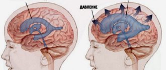
Pain in the back of the head with increased intracranial pressure
Increased intracranial pressure is characterized by:
- pressing, bursting pain in the occipital region or throughout the head;
- increased pain in bright light and loud sounds;
- heaviness in the head and pain in the eyeballs;
- vomiting, which does not reduce pain syndromes.
Pain in the back of the head due to cervical myositis
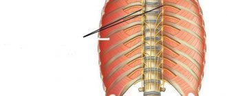
Inflammatory processes in the neck muscles caused by hypothermia or injury are characterized by pain symptoms that spread from the neck to the occipital, shoulder and interscapular areas. It appears when you move your head and is asymmetrical.
More about myositis
Pain in the back of the head due to occipital neuralgia
Neuralgia of the occipital nerve, resulting from hypothermia or accompanying osteochondrosis, is characterized by very strong shooting pains. They occur periodically, like attacks with any attempt to change the position of the head.
During rest, a slight pressing pain is felt in the occipital region.
Read more about occipital neuralgia
Pain in the back of the head due to vascular diseases
Spasms of the cranial arteries cause throbbing pain, which becomes stronger when trying to move the head and subsides somewhat at rest. The pain begins in the back of the head and eventually spreads to the frontal area. It is accompanied by a feeling of heaviness in the head and begins in the morning after waking up.