Fourth ventricle of the brain
The fourth ventricle (ventriculus quartus) develops from the cavity of the rhombencephalon. At the top it communicates with the III ventricle using the cerebral aqueduct, and at the bottom - with the central canal of the spinal cord, through the median aperture (apertura mediana ventriculi quarti), paired lateral apertures (aperturae laterales) of the IV ventricle communicate with the subarachnoid space of the brain (cisterna cerebello-medullaris) . The lateral openings of the fourth ventricle are located near the flocculus of the cerebellum in the lateral corners of the roof of the rhomboid fossa (Fig. 466). The fourth ventricle is filled with cerebrospinal fluid and the choroid plexus. Consists of top and side walls; its bottom is formed by a diamond-shaped pit. The upper wall of the fourth ventricle is limited by the superior medullary velum, which is attached to the cerebellar uvula (lingula cerebelli). The lower medullary velum (velum medullare inferius), which is a thin epithelial plate, begins here (Fig. 467). Adjacent to it is the vascular base of the fourth ventricle (tela choroidea ventriculi quarti) - the tegmentum. The junction of the superior and inferior medullary sails forms the apex of the tent (fastigium). The lateral border of the fourth ventricle is the superior cerebellar peduncle.
466. Rhomboid fossa and cranial nerve nuclei. 1 - nucl. n. oculomotorii; 2 - nucl. n. trochleares; 3 - nucl. tractus mesencephalic! n. trigemini; 4 - n. motorius n. trigemini; 5 - nucl. sensorius principalis n. trigemini; 6 - nucl. n. abducentis; 7 - nucl. n. facialis; 8 - nucl. n. vestibularis; 9 - nucl. cochleares; 10 - n. facialis; 11 - nucl. salivatorii superior et inferior; 12 - nucl. n. hypoglossi; 13 - nucl. ambiguus; 14 - n. tractus spinalis n. trigemini; 15 - n. tractus solitarii; 16 - n. accessorius; 17 - nucl. dorsalis n. trigemini; 18 - nucl. spinalis n. accessorii; 19 - tuberculum n. gracilis; 20 - tuberculum n. cuneati; 21 - trigonum n. vagi; 22 - colliculus facialis; 23 - eminentia medialis; 24 - trigonum lemnisci; 25 - colliculus inferior; 26 - colliculus superior
The rhomboid fossa represents the floor of the fourth ventricle. Develops from the isthmus of the rhombencephalon, hindbrain and medulla oblongata. It has upper, lower and lateral angles. The rhomboid fossa is divided by brain stripes (striae medullares), which are located in the middle of the bottom of the rhomboid fossa and extend transversely to the lateral angles. The superior angle of the rhomboid fossa is bounded by the superior cerebellar peduncles, the inferior angle by the inferior cerebellar peduncles, which diverge to the sides at an angle of 50-85°, and the lateral angles by the inferior and middle cerebellar peduncles.
467. The brain on a sagittal section. 1 - sulcus centralis; 2 - lobus paracentralis; 3 - precuneus; 4 - sulcus parietooccipitalis; 5 - cuneus; 6 - sulcus calcarinus; 7 - gyrus occipi totem poralis; 8 - corpus pineale; 9 - cerebellum; 10 - medulla oblongata; 11 - ventriculus quartus; 12 - pons; 13 - colliculi superior et inferior; 14 - pedunculi cerebri; 15 - aqueductus cerebri; 16 - hypophysis; 17 - chiasma opticum; 18 - commissura anterior; 19 – fornix
The diamond-shaped fossa is divided into two symmetrical parts by the median groove (sulcus medianus). In the upper corner of the fossa, above the brain stripes, which are lighter in color, lies the paired medial eminence (eminentia medialis). Its lower part ends with the facial tubercle (colliculus facialis). The facial tubercle is represented by the convexity of the facial nerve, which in this place bends around the nucleus of the abducens nerve (nucl. abducens). Closer to the ventral surface from the nucleus of the abducens nerve lie the solitary tract (nucl. tr. solitarius), the parasympathetic nucleus - the superior salivary nucleus (nucl. salivatorius superior), the motor nucleus (nucl. motorius) of the facial nerve (VII pair). The medial eminence contains the motor nucleus of the trigeminal nerve (V pair).
Lateral and above the medial eminence there is a bluish place (locus ceruleus), where the sensitive nucleus (nucl. sensorius) and the spinal tract of the trigeminal nerve (tr. spinalis n. trygemini) (V pair) are located.
In the lateral corners of the rhomboid fossa there is a vestibular area (area vestibularis). In this area there are 4 nuclei of the vestibular nerve (nucl. vestibulares): medial, lateral, superior, inferior and two nuclei of the auditory nerve: anterior cochlear (nucl. cochlearis anterior) and posterior cochlear (nucl. cochlearis posterior) (VIII pair). In the lower corner of the rhomboid fossa near the median sulcus there is a triangle of the hypoglossal nerve (trigonum n. hypoglossi), onto which the nucleus of this nerve is projected (XII pair). Lateral to the triangle of the hypoglossal nerve there is a darker triangular hillock (ala cinerea), which is the location of the parasympathetic nucleus of the vagus nerve (nucl. dorsalis n. vagi). Somewhat higher, on the line ala cinerea, lies the large motor double nucleus (n. ambiguus), belonging to the IX and X pairs of cranial nerves. Lateral to this nucleus is the lower salivary nucleus - parasympathetic (nucl. salivatorius inferior), from which part of the fibers of the glossopharyngeal nerve emerges. Next to it is the sensitive nucleus of the IX and X pairs (nucl. tr. solitarii).
Thus, summing up the topography of the nuclei of the bottom of the rhomboid fossa, it should be noted that the motor nuclei are located closer to the midline, the sensitive ones lie more laterally, and the autonomic ones are distributed between them.
What diseases can affect the ventricles
Despite the small size of the ventricular system, the process of liquor circulation is extremely important for the functioning of the central nervous system and the whole organism. Like any organ, the cavities in the brain are often susceptible to various diseases. Hydrocephalus of the brain is considered the most common disease that has no age restrictions.
The lateral cisterns are characterized by asymmetry, which means that the cause of the anomaly lies in the malfunction of the choroid plexuses. The accumulation of cerebrospinal fluid in the cavities is not always caused by increased secretion production; blockage of the conductor channels often leads to a pathological condition.
Hydrocephalus syndrome
The expansion of the ventricles of the brain manifests itself in a bright clinic, since the central nervous system ceases to receive balanced nutrition and oxygen. Neurological symptoms appear when the brain is unable to perform its functions productively.
Pathologies in the ventricular system have primary or secondary causes and treatment is aimed at restoring all stages of liquor circulation. Diseases of the ventricles require immediate treatment, since delays threaten degenerative changes in the brain matter.
Enlargement of the ventricles of the brain in the fetus is considered a congenital disorder; early diagnosis helps to carry out emergency neurosurgical intervention immediately after the birth of the child.
Suspicion of enlargement of the lateral ventricles of the brain is always subject to a thorough diagnostic examination. Visualization of brain structures allows you to evaluate the entire ventricular system and identify the cause of the failure.
Diagnostic methods:
- Physical examination by a neurologist - neurological status is assessed;
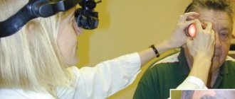
- Consultation with an ophthalmologist – examination of the fundus;
- Neurosonography - the study is carried out through the central fontanelle for children under 1 - 1.5 years old;
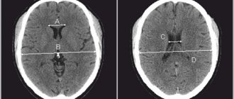
- MRI or CT with contrast – layer-by-layer visualization of structures, performed on adults and children from 2 years of age;
- Doppler ultrasound – evaluates the vessels of the head and neck.
Highly accurate methods allow the assessment of all parameters in the ventricular system. An index is calculated for each cavity; if the percentage values are higher than normal, then a diagnosis of hydrocele is made. The degree of pathology is determined by the compliance of the ventricles with age criteria and the individual index.
The absorption of cerebrospinal fluid in the subarachnoid space into the vascular network directly depends on the constancy of intracranial pressure. Failure of the cerebrospinal fluid circulation leads to unstable pressure; when it decreases, fluid absorption is impossible.
Liquid is continuously produced and if there is no free outflow, then the cisterns grow, increasing in volume and width; the pressure on nearby structures depends on how enlarged the ventricles of the brain are.
Thus, during diagnosis it is revealed at what stage in the cerebrospinal fluid circulation the failure occurred and what was the cause. The primary causes always relate to congenital anomalies.
Ventricular dilatation (MRI image)
Secondary disorders in the ventricular system are a consequence of the influence of various negative factors and associated pathologies:
- Traumatic brain injuries (including birth injuries);
- Cystic formations;
- Intracranial neoplasms - tumors inside the cavity can be of intraventricular origin, that is, the primary focus, and the origin of cancer cells occurs precisely in the storage area. When atypical cells grow through the walls of the ventricle, we are talking about a secondary focus (metastasis). Ependymoma and choroidpapilloma are formed by abnormal cell division in the choroid plexus;
- Penetration of viruses and bacteria - the entry of pathogenic agents causes inflammation of the meninges and a suppurative process, as a result of which a structural change in blood vessels occurs;
- Vascular thrombosis;
- Cerebral hemorrhage – more common in older people.
Bleeding in the ventricles is a life-threatening condition. The pathology has a high mortality rate, since the clinical picture develops rapidly and the patient cannot always be transported to the hospital on time.
The disease more often affects older people (atraumatic hemorrhage), as age-related changes occur in the structure of blood vessels.
In the presence of concomitant diseases (atherosclerosis, cardiovascular pathology, blood diseases), this leads to various internal damage to the blood vessels, resulting in rupture and effusion of blood into the cavity. Intraventricular hemorrhage can also be traumatic.
A burst vessel leads to acute cerebrovascular accident. If the hemorrhage occurs in the lateral cisterns, then the blood flows through the channels to the lower ventricles. Blood clots form in the cavities, thus causing ventricular tamponade and rapidly increasing cerebral edema.
Blockage of the rhomboid cistern quickly leads to death, since the work of the vegetative centers that control vital mechanisms is disrupted.
With a timely neurosurgical operation, a drainage system is installed, with the help of which it is possible to remove intracerebral hematomas.
Poor circulation of the cerebrospinal fluid causes the accumulation of fluid in the cavities, as a result of which the ventricles enlarge and put pressure on neighboring structures. An increase in the total volume of fluid in the brain leads to an increase in ICP and the development of intracranial hypertension.
As a result of increased pressure, nerve cells die, which is dangerous for the development of encephalopathy - dystrophic damage to brain tissue is irreversible.
In children, there are cases where the lateral ventricles are dilated, but parents do not notice changes in behavior or other pathological symptoms. Perhaps this will be a variant of the norm (individual) and we are not talking about a congenital disease (ventriculomegaly), but provided that the CMD is also normal.
To do this, ICP is measured; there are several methods - non-invasive measurements using CT or MRI diagnostics, a minimally invasive technique in the form of a puncture in the lumbar spinal canal.
But neurosurgeons more often resort to the technique of puncturing cavities; when puncturing the brain, a special needle is inserted and the anterior horns of the lateral ventricles are punctured. Direct collection of cerebrospinal fluid from cavities is considered a highly informative technique.
If the ICP exceeds the indicators, then in all cases of failure of the cerebrospinal fluid circulation, emergency surgery is indicated. Installation of shunt systems allows you to restore fluid circulation, which leads to stabilization of cerebral pressure and relief of pathological symptoms.
A shunt in the brain is a silicone conductor - a tube. Through the drainage system, cerebrospinal fluid from the ventricles flows into the abdominal (or thoracic) cavity; the pump in the system takes on the function of regulating drainage and CMD.
How not to miss ventricular diseases? The causes of CNS pathologies are different, but all of them are manifested by general cerebral symptoms, which should be paid special attention to and promptly seek medical help.
In infants, liquorodynamic disturbances are often visible visually. Unossified vaults in the skull provide additional space for the accumulation of excess fluid, thus making the skull voluminous and asymmetrical. There is a delay in psychomotor development and a number of neuralgic syndromes.
But quite often hydrocephalus occurs in a compensatory manner and does not have pronounced symptoms; the signs appear gradually as fluid accumulates.
General cerebral symptoms in cases of impaired cerebrospinal fluid circulation in children and adults:
- Severe headaches. In children they manifest themselves in the form of incessant crying and anxiety, the baby refuses to eat;
- Vomiting that does not bring relief. Infants experience excessive regurgitation after feeding;
- Dizziness, nausea;
- Decreased performance. Children are characterized by lethargy or, conversely, hyperactivity;
- Loss of coordination - unsteadiness of gait;
- Decreased vision;
- Constant feeling of fatigue, drowsiness;
- Sleep disturbance;
- Violations in the psycho-emotional sphere – mood swings, up to the development of mental illness.
When a certain brain structure is compressed, additional symptoms appear - focal ones. With timely diagnosis of the pathology, hydrocephalus can be successfully treated with conservative methods (except for congenital forms).
Severe forms of dropsy or advanced cases are accompanied by convulsions and seizures. Pressure on the brain stem causes paralysis.
In case of chronic disturbance of liquor circulation, the prognosis for life is often unfavorable. Installing a shunt may be ineffective because the brain tissue already has degenerative changes.
Since various pathologies of the central nervous system have similar symptoms, a comprehensive diagnosis is important to differentiate diseases of the ventricular system.
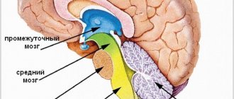
Features of the third ventricle
The 3rd ventricle of the brain is part of the cerebrospinal fluid circulation system and is the connecting link between the lateral cisterns and the rhomboid fossa. In a frontal section of the brain, the cistern looks like a narrow slit, which communicates with the 4th cavity through the Sylvian aqueduct.
A feature of the cavity is the dense surrounding of various structures, as well as the presence of vegetative centers. Excessive accumulation of cerebrospinal fluid or obstructed outflow from the cavity leads to pressure on the nerve nodes that maintain internal homeostasis.
The width of the tank in an adult should not exceed 6 mm; for children under one year of age, the norm is 3–4 mm. The severity of the process is determined by the presence and severity of neurological symptoms. Diagnostic measurement of ventricular parameters allows emergency measures to be taken and, if necessary, surgical intervention.
Disturbances in liquorodynamics always require emergency treatment, which is aimed at restoring the functionality of the central nervous system and eliminating the cause of the disturbance. Degenerative changes in brain tissue cannot be restored, but installing a shunt helps improve the patient’s quality of life.