Anatomy of Human Spinal Nerves - Information:
Spinal nerves, nn. spinales, are located in the correct order (neuromeres), corresponding to the myotomes (myomeres) of the body and alternating with the segments of the spinal column; Each nerve has a corresponding skin area (dermatome).
A person has 31 pairs of spinal nerves, namely: 8 pairs of cervical, 12 pairs of thoracic, 5 pairs of lumbar, 5 pairs of sacral and 1 pair of coccygeal.
Each spinal nerve leaves the spinal cord with two roots: posterior (sensitive) and anterior (motor); both roots are connected into one trunk, truncus n. spinalis, emerging from the spinal canal through the intervertebral foramen. Near and somewhat outward from the junction, the dorsal root forms a node, ganglion spinale, in which the anterior motor root does not participate. Due to the connection of both roots, spinal nerves are mixed nerves: they contain sensory (afferent) fibers from the cells of the spinal ganglia, motor (efferent) fibers from the cells of the anterior horn, as well as vegetative fibers from the cells of the lateral horns, emerging from the spinal cord as part of the anterior root. Vegetative fibers are also present in the dorsal root.
Autonomic fibers entering the animal nerves through the roots provide processes in the soma such as trophism, vasomotor reactions, etc. In cyclostomes (lampreys), both roots continue into separate nerves - motor and sensory. In the further course of evolution, starting with transverse fish, the roots come closer together and merge, so that the separate course is maintained only for the roots, and the nerves become mixed.
Each spinal nerve, upon exiting the intervertebral foramen, is divided into two branches, respectively, into two parts of the myotome (dorsal and ventral):
- posterior, ramus dorsalis, for the autochthonous muscles of the back and the skin covering it, developing from the dorsal part of the myotome;
- anterior, ramus ventralis, for the ventral wall of the trunk and limbs, developing from the ventral parts of the myotomes.
In addition, two more types of branches depart from the spinal nerve:
- for the innervation of the viscera and blood vessels - connecting branches to the sympathetic trunk, rr. communicantes;
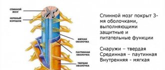
- for innervation of the spinal cord membranes - r. meningeus, going back through the intervertebral foramen.
Posterior branches of the spinal nerves. The posterior branches, rami dorsales, of all spinal nerves go back between the transverse processes of the vertebrae, bending around their articular processes. All of them (with the exception of the I cervical, IV and V sacral and coccygeal) are divided into ramus medialis and ramus lateralis, which supply the skin of the back of the head, the back of the neck and back, as well as the deep spinal muscles.
Posterior branch of the 1st cervical nerve, n. suboccipitalis emerges between the occipital bone and the atlas and then divides into branches supplying mm. recti capitis major et minor, m. semispinalis capitis, mm. obliqui capitis. To skin n. suboccipitalis does not produce branches. Posterior branch of the II cervical nerve, n. occipitalis major, emerging between the posterior arch of the atlas and the second vertebra, then pierces the muscles and, becoming subcutaneous, innervates the occipital region of the head. The rami dorsales of the thoracic nerves are divided into medial and lateral branches, giving branches to the autochthonous muscles; the cutaneous branches of the upper thoracic nerves arise only from the rami mediales, and those of the lower ones - from the rami laterales.
The cutaneous branches of the three upper lumbar nerves go to the upper part of the gluteal region called the nn. clunium superiores, and the cutaneous branches of the sacral ones are called nn. Clunium mediai. Anterior branches of the spinal nerves. The anterior branches, rami ventrales, of the spinal nerves innervate the skin and muscles of the ventral wall of the body and both pairs of limbs. Since the skin of the abdomen in its lower part takes part in the development of the external genitalia, the skin covering them is also innervated by the anterior branches. The latter, except for the first two, are much larger than the hind ones.
The anterior branches of the spinal nerves retain their original metameric structure only in the thoracic region (nn. intercostales). In the remaining sections associated with the limbs, during the development of which segmentation is lost, the fibers extending from the anterior spinal rami are intertwined. This is how nerve plexuses, plexus, are formed, in which fibers of different neuromeres are exchanged. A complex redistribution of fibers occurs in the plexuses: the anterior branch of each spinal nerve supplies its fibers to several peripheral nerves, and, therefore, each of them contains fibers from several segments of the spinal cord.
It is therefore clear that damage to one or another nerve is not accompanied by dysfunction of all muscles receiving innervation from the segments that gave rise to this nerve. Most of the nerves arising from the plexuses are mixed; therefore, the clinical picture of the lesion consists of motor disturbances, sensory disturbances and autonomic disorders. There are three large plexuses: cervical, brachial and lumbosacral. The latter is divided into lumbar, sacral and coccygeal.
Spinal nerves
Spinal nerves (nn. spinales) extend from the spinal cord in the form of two roots: the anterior (ventral), consisting of motor fibers, and the posterior (dorsal), which forms sensory fibers. In the area of the intervertebral foramen they unite into one trunk - the mixed spinal nerve. At the junction, the dorsal root forms a nerve spinal ganglion (ganglion spinale), consisting of false unipolar (pseudo-unipolar) cells with a T-shaped branching process. Each spinal nerve upon exiting the intervertebral foramen is divided into four branches: 1) anterior (ventral) - for the anterior wall of the trunk and limbs; 2) posterior (dorsal) - for the muscles and skin of the back and neck; 3) connective - to the node of the sympathetic trunk; 4) meningeal (meningeal), heading back into the spinal canal to innervate the membranes of the spinal cord (Fig. 125).
Rice. 125. Diagram of the formation and branching of the spinal nerve (thoracic). 1 - anterior root; 2 - shell branch; 3 - node of the sympathetic trunk; 4 - branching of the anterior branch to the skin; 5 - anterior branch (intercostal nerve); 6 - connecting branch to the sympathetic trunk; 7 - posterior branch; 8 - spinal node; 9 - posterior root
Together with each pair of spinal nerves, the embryo develops a certain area of muscle (myotome) and skin (dermatome). Based on this, segmental innervation of muscles and skin is distinguished. In an adult, such correct distribution of the peripheral branching of the spinal nerves is not observed due to the loss of the initial segmentation of the muscles and areas of the skin that they supply. This is especially pronounced in the area around the limbs. In humans, there are 8 pairs of cervical, 12 pairs of thoracic, 5 pairs of lumbar, 5 pairs of sacral and a pair of coccygeal spinal nerves.
The posterior branches of the spinal nerves contain sensory and motor fibers and are directed to the skin and muscles of the back and neck. Among them, the posterior branch of the first cervical nerve stands out - the suboccipital nerve, consisting only of motor fibers, innervates the short muscles of the back of the head, and the second cervical nerve - the greater occipital nerve, innervates most of the skin of the back of the head. Sensory fibers of the posterior branches of the lumbar and sacral nerves innervate the skin of the gluteal region and are called the superior and middle nerves of the buttocks. The remaining posterior branches of the spinal nerves do not have special names.
The anterior branches of the spinal nerves contain sensory and motor fibers intended for the muscles and skin of the neck, the anterior and lateral surfaces of the torso, and the upper and lower extremities. The anterior branches of adjacent nerves are connected to each other in the form of loops, exchanging fibers and forming plexuses. The exception is the anterior branches of the thoracic nerves, which run segmentally in the intercostal spaces. The anterior branches of the remaining nerves form four plexuses: cervical, brachial, lumbar and sacral.
The cervical plexus is formed by the anterior rami of the four superior cervical spinal nerves. It lies on the side of the transverse processes of the upper cervical pores between the muscles and is covered by the sternocleidomastoid muscle. The branches of the cervical plexus emerge from under the posterior edge of this muscle approximately in its middle. Among them there are cutaneous, muscular and mixed branches.
The sensory branches of the cervical plexus are:
1) the lesser occipital nerve, innervating the lateral part of the skin of the back of the head; 2) the great auricular nerve, innervating the auricle and external auditory canal;
3) transverse cervical nerve, innervating the skin of the neck;
4) supraclavicular nerves - a bundle of nerves going down and innervating the skin above the collarbone, pectoralis major and deltoid muscles.
The muscular (motor) branches innervate the deep muscles of the neck and, connecting with the hypoglossal nerve (XII pair of cranial nerves), form a cervical loop, due to which the anterior muscles of the neck are innervated below the hyoid bone.
The mixed branch of the cervical plexus is the phrenic nerve. It descends along the anterior scalene muscle into the chest cavity, passes in the middle mediastinum between the pericardium and the mediastinal pleura and approaches the thoraco-abdominal barrier. Innervates the diaphragm (motor fibers), pleura and pericardium (sensory fibers) and penetrates the abdominal cavity, innervating the peritoneal ligaments of the liver there.
The brachial plexus is formed by the anterior branches of the four lower cervical and part of the first thoracic spinal nerves. It exits through the space between the anterior and middle scalene muscles into the supraclavicular fossa and is located next to the subclavian artery. Then behind the clavicle it descends into the axillary cavity and here forms three main bundles located around the axillary artery (Fig. 126). From these bundles begin the long nerves of the brachial plexus, which innervate the upper limb. Short nerves that innervate the muscles of the shoulder girdle extend from the upper part of the brachial plexus. The largest of these is the axillary nerve, which goes to the deltoid and teres minor muscles, the skin over them and to the shoulder joint bursa. The remaining nerves innervate the pectoralis major and minor, serratus anterior, subclavian, supraspinatus, infraspinatus, subscapularis, latissimus dorsi, teres major, rhomboids, and levator scapulae muscles.
Rice. 126. Branches of the brachial plexus. 1 - axillary artery; 2 - axillary vein; 3 - brachial plexus; 4 - short branches of the brachial plexus to the pectoralis major and minor muscles; 5 - musculocutaneous nerve; 6 - median nerve; 7 - cutaneous medial nerve of the forearm; 8 - ulnar nerve; 9 - radial nerve; 10 - axillary nerve; 11 - cutaneous medial nerve of the shoulder; 12 - serratus anterior muscle; 13 - short branch to the latissimus dorsi muscle; 14 - short branch to the serratus anterior muscle; 15 - short branch to the subscapularis muscle
The long branches of the brachial plexus include the following:
1. Medial cutaneous nerve of the shoulder; innervates the skin of the inner surface of the shoulder.
2. Medial cutaneous nerve of the forearm; innervates the skin of the inner surface of the forearm.
3. Musculocutaneous nerve; supplies three muscles of the shoulder with motor branches: biceps, brachialis and coracobrachialis, and then passes to the forearm, where it innervates the skin of the outer side.
The median nerve in the shoulder passes along with the brachial artery and veins in the medial groove; does not give branches. On the forearm it gives branches to all the muscles of the anterior group (flexors), with the exception of the flexor carpi ulnaris and part of the deep flexor digitorum. Together with the flexor tendons of the fingers, it passes through the carpal canal to the palm, where it innervates the muscles of the eminence of the thumb, except for the adductor and part of the short flexor pollicis, and two lateral lumbrical muscles. The cutaneous branches form common and then proper palmar digital nerves, which innervate the skin of the thumb, index, middle and half of the ring finger.
5. The ulnar nerve runs along the inner surface of the shoulder; does not give branches. It goes around the medial epicondyle of the humerus and passes to the forearm, where in the groove of the same name it runs next to the ulnar artery. On the forearm, it innervates the flexor carpi ulnaris and part of the deep flexor digitorum; in the lower third of the forearm it is divided into dorsal and palmar branches. The palmar branch gives rise to cutaneous and muscular branches. The cutaneous branches are represented by the common and proper palmar digital nerves, innervating the skin of the little finger and the medial side of the ring finger. The muscular branch is deep, passes to the muscles of the eminence of the little finger, all interosseous, two medial lumbricals, adductor pollicis and to the deep head of the short flexor pollicis. The dorsal branch gives rise to the dorsal digital nerves, which innervate the skin of 21/2 fingers, starting with the little finger.
6. The radial nerve is the thickest nerve of the brachial plexus. On the shoulder, it passes in the brachiomuscular canal between the humerus and the heads of the triceps muscle, giving off muscle branches to this muscle and skin branches to the posterior surface of the shoulder and forearm. In the lateral groove, the ulnar fossa is divided into deep and superficial branches. The deep branch innervates all the muscles of the posterior surface of the forearm (extensors), and the superficial branch runs in the groove along with the radial artery, passes to the back of the hand, where it innervates the skin of 2 1/2 fingers, starting from the thumb.
Anterior branches of the thoracic spinal nerves. These branches do not form a plexus and run in the intercostal spaces. They are called intercostal nerves, innervate the intrinsic chest muscles, participate in the innervation of the muscles of the anterior abdominal wall and give off anterior and lateral cutaneous branches that innervate the skin of the chest and abdomen.
Lumbar plexus. Formed by the anterior branches of the three upper lumbar spinal nerves, partly the twelfth thoracic and the fourth lumbar. It lies in the thickness of the psoas major muscle, its branches come out from under it from the outside, piercing the muscle from the front or from the inside. Among the short branches there are: iliohypogastric, ilioinguinal, femoral-genital nerves, innervating the lower parts of the muscles and skin of the anterior abdominal wall, external genitalia and upper thigh. Long branches extend to the lower limb. These include the following.
1. Lateral cutaneous nerve of the thigh; comes out from under the lateral edge of the psoas major muscle and descends to the thigh; innervates the skin of the outer surface of the thigh.
2. Obturator nerve; lies on the lateral wall of the pelvis, passes through the obturator canal, giving branches to the hip joint; innervates the adductor muscles of the thigh and the skin of the inner thigh.
3. The femoral nerve is the largest nerve of the lumbar plexus; passes between the iliacus and psoas major muscles, passes to the thigh under the inguinal ligament; innervates the anterior group of muscles of the thigh and the skin of its anterior surface. Its longest sensory branch, the saphenous nerve, goes to the medial surface of the leg; innervates the skin of the anteromedial surface of the leg and dorsum of the foot.
Sacral plexus. Formed by the anterior branches of the fourth (part) and fifth lumbar, all sacral and coccygeal nerves. It is located in the small pelvis on the anterior surface of the sacrum and piriformis muscle and exits through the greater sciatic foramen above and below the piriformis muscle into the gluteal region. Short branches of the sacral plexus innervate the muscles of the pelvis (except for the iliopsoas) and gluteal region (superior and inferior gluteal nerves). The long branches are represented by two nerves: 1) the posterior cutaneous nerve of the thigh innervates the skin of the perineum, gluteal region and posterior surface of the thigh; 2) the sciatic nerve (p. ischiadicus) is a direct continuation of the sacral plexus. Coming out of the pelvis, it moves to the back of the thigh and here passes between the muscles to which it gives off motor branches (the posterior group of thigh muscles). In the popliteal fossa it divides into the tibial nerve and the common peroneal nerve. The tibial nerve, having given off the medial cutaneous nerve of the calf, passes in the ankle-popliteal canal between the muscles of the posterior group of the leg, innervating them, passes to the foot behind the medial malleolus and is divided into the medial and lateral plantar nerves, innervating the skin and muscles of the sole of the foot. The common peroneal nerve runs laterally, giving off a branch to innervate the skin of the posterolateral surface of the leg and. divided into superficial and deep. The superficial peroneal nerve innervates the muscles of the lateral group of the leg and passes to the dorsum of the foot, participating in the innervation of the skin of the dorsum of the foot. The deep peroneal nerve passes between the muscles of the anterior group, giving branches to them, passes to the foot, innervates the short muscles of the dorsum of the foot and the skin of the first interdigital space.
The cutaneous branches of the tibial and common peroneal nerves, connecting on the posterior surface of the leg, form the sural nerve, which innervates the skin of the lateral edge of the foot.
The pudendal nerve departs from the sacral plexus, which branches in the area of the ischiorectal fossa, innervating the skin and muscles of the perineum; its terminal branches innervate the external genitalia.