The complex structure of the human body allows distantly located organs to interact harmoniously to ensure internal balance - for example, the connection of the brain with underlying structures occurs through pairs of cranial nerves. One part of them provides sensitive perception of information, while others are responsible for motor activity. The smaller group has a mixed structure and is therefore ready for complex functioning. Understanding the characteristics of cranial nerves allows doctors to select the correct treatment for their diseases.
Cranial nerve pairs and their nuclei
The formation and development of nervous structures occurs already at the stage of intrauterine fetal formation - individual structures, for example, cranial nerves, begin their activity even before the moment of birth. Their final maturation takes more time, but, in general, most of the central nervous system of the newborn ensures its full interaction with the external environment.
In total, it is customary to distinguish 12 pairs of nerves. Their central part in the form of nuclei remains inside the brain. Whereas impulses reach the innervated organs through special fibers - a collection of nerve cell processes. Depending on what function these structures perform, experts traditionally distinguish several subgroups of pairs of cranial nerves:
- motor;
- sensitive;
- mixed.
The location of the exit of the nerve fiber is also of great importance - above the brain stem or below it. Doctors consider this criterion when diagnosing various pathologies of the cranial nerves.
Sensory cranial nerves
Sensory nerves transmit information from the senses to the brain. These include the olfactory, optic and vestibulocochlear nerves.
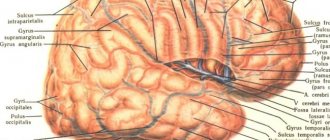
Olfactory nerve Olfactory nerves transmit information to the brain from receptor cells located in the mucous membrane of the nasal cavity. Thin threads of the nerve (15–20) penetrate into the cranial cavity, the olfactory bulbs lying on the lower surface of the frontal lobes of the cerebral hemispheres. This is where the olfactory tracts begin, along which information is sent to the subcortical centers and to the cerebral cortex. If the frontal region is damaged, smell disturbances may occur.
Optic Nerve The optic nerve is formed by processes of nerve cells in the retina that emerge near the posterior pole of the eyeball. Inside the skull, the fibers of the optic nerve cross and pass into the visual tract, which ends in the subcortical centers. Next, the pathways go to the higher center in the cortex of the occipital lobe of the hemispheres. At the site of the optic chiasm, only the nerve fibers coming from the inner halves of the retina cross, creating the conditions for binocular vision (receiving the same image in both eyes). When the optic nerve, optic chiasm or optic tract is damaged, visual impairment will vary, which makes it possible to diagnose their localization.
The vestibular-cochlear nerve The vestibular-cochlear nerve consists of 2 parts: the cochlear and the vestibular. The first conducts impulses from the organ of hearing, the second - from the organ of balance. The hearing and balance receptors are located inside the temporal bone. Both parts of the nerve connect in the internal auditory canal, from there they enter the cranial cavity. In the brain, the pathways for auditory and vestibular information are different: the auditory center is located in the temporal lobe of the cerebral hemispheres, and the vestibular center is in the cerebellum. If the temporal bone is damaged, not only hearing loss and balance disorders are possible, but also impaired salivation and facial expressions, since next to the vestibulocochlear nerve in the internal auditory canal there is a nerve (facial), which is involved in the innervation of the salivary glands and facial muscles.
Sensitive nerve structures
All brain nerves are important for the full functioning of the human body. If the predominant activity of a couple is the perception and transmission of information, then they are called sensory nerve fibers.
Thus, the olfactory nerve originates from the cells of the nasal mucosa. It then passes through the cribriform plate of the skull and moves deep into the brain to a structure called the olfactory bulb. Next, the nerve fiber forms a kind of tract, which passes into the olfactory triangle. The sensory structure ends in the tubercle of the cerebral cortex.
Another important sensory unit is the optic nerve. Its ganglion cells from the retina rush into the cranium, where the pair forms a kind of cross, from which the fibers run to the lateral geniculate structure of the brain. From here the optic nerve will continue to its final destination - the occipital part of the cerebral cortex.
Cells of the eighth pair of cranial nerves - auditory or vestibulocochlear. It is inherent not only to perceive and transmit the sounds of the surrounding world, but also to be responsible for the balance of the human body in space. Both parts of the nerve fiber - from the cochlear root and from the vestibular root, which begins from the vestibular ganglion, are directed to their final targets - the cerebellum, as well as the quadrigeminal cortex and the median geniculate body. The auditory nerve ends in the temporal gyrus.
Where cranial nerves exit the brain
List of nerves
I pair - olfactory nerve (lat. nervus olfactorius
)
II pair - optic nerve (lat. nervus opticus
)
III pair - oculomotor nerve (lat. nervus oculomotorius
)
IV pair - trochlear nerve (lat. nervus trochlearis
)
V pair - trigeminal nerve (lat. nervus trigeminus
)
VI pair - abducens nerve (lat. nervus abducens
)
VII pair - facial nerve (lat. nervus facialis
)
VIII pair - vestibulocochlear nerve (lat. nervus vestibulocochlearis
)
IX pair - glossopharyngeal nerve (lat. nervus glossopharyngeus
)
X pair - vagus nerve (lat. nervus vagus
)
XI pair - accessory nerve (lat. nervus accessorius
)
XII pair - hypoglossal nerve (lat. nervus hypoglossus
)
Mnemonic rules
• Onegin Knew Where Tatyana Was, He Loved to Listen to the Language of his Infinitely Dear Friend.
• Onegin Knew Where Tatyana Was, He Was Flying Like a Bullet, His Tongue Lolling Out to His Waist.
• To memorize a pair of cranial nerves in Latin: About Oryasin, the Donkey sharpens the axe, And the Fakir, having kicked out the Guests, wants to howl like a Shark.
• Smell, see, move your eyes, move the trigeminal block, face, hearing, tongue and pharynx. Don't fornicate in vain. Add under tongues.
• I smelled, I saw, I moved my eye, and the trigeminal block was abducted. Face and hearing, and glossopharynx, wandering, walked with an additional gait, finding all the nerves under the tongue.
Development of cranial nerves in embryogenesis
The olfactory and optic nerves develop from protrusions of the anterior medullary bladder and consist of axons of neurons that are located in the mucous membrane of the nasal cavity (the organ of smell) or in the retina of the eye. The remaining sensory nerves are formed by the eviction of young nerve cells from the developing brain, the processes of which form sensory nerves or sensory (afferent) fibers of mixed nerves. Motor cranial nerves are formed from motor (efferent) nerve fibers, which are processes of cells of the motor nuclei located in the brain stem. The formation of cranial nerves in phylogenesis is associated with the development of visceral arches and their derivatives, sensory organs and reduction of somites in the head region.
(Somite ( primary segment
,
dorsal segment
; from ancient Greek soma - “body”) is a paired metameric formation in the embryos of some invertebrates (annelids, insects) and all chordates (including humans).)
Where cranial nerves exit the brain
It is impossible to say about the first (olfactory) nerve that it “comes out” of the brain, since it carries only afferent (sensitive) information. Olfactory nerve
is the processes of olfactory cells of the nasal mucosa collected in olfactory filaments. The olfactory filaments reach the olfactory bulb through the openings of the cribriform plate of the ethmoid bone.
It is also impossible to say about the second (optic) nerve that it “comes out” of the brain, for the same reason. It originates from the optic nerve head, located at the posterior pole of the eye. The optic nerve passes into the cranial cavity through the optic canal formed by the lesser wing of the sphenoid bone. In the cranial cavity, the optic nerves of both eyes form a chiasm, and only part of the fibers intersect. Further, the fiber paths are called the “optic tract”.
The third (oculomotor) nerve exits from the ventral (“facial”) side of the trunk next to the interpeduncular fossa (fossa interpeduncularis).
The IV (trochlear) nerve is the only one emerging from the dorsal (“dorsal”) side of the trunk, from the upper edge of the rhomboid fossa, bending and emerging to the ventral side from under the cerebral peduncles.
The V (trigeminal) nerve exits the ventral side of the pons.
Nerves VI to VIII also exit on the ventral side of the brain stem between the medulla oblongata and the pons, from the edges to the center in a row, with VII and VIII lying close to each other at the “corner” of the medulla oblongata, and VI (abducens) at the level of the anterolateral sulcus .
Nerves IX to XII emerge from the medulla oblongata on the ventral side. The XI (accessory) nerve stands somewhat apart - it combines, in addition to the head part, some roots of the spinal cord. Nerves IX to XI emerge from the lateral surface of the medulla oblongata, from bottom to top in a row.
The XII (hypoglossal) nerve emerges from the anterolateral groove (lat. sulcus ventrolateralis
).
Motor nerve structures
In the nuclei of the midbrain, just below its aqueduct, the oculomotor nerve begins its journey. As it progresses, it forms a branched system of fibers - the upper parts of the pair are responsible for the functioning of the eyelids. While its lower three parts innervate the rectus muscle groups of the orbit.
Several more pairs of cranial nerve structures provide full movement of the eyeballs and adjacent tissues:
- trochlear - the nuclei of the pair are localized anterior to the cerebral aqueduct, and the fibers stretch to the superior oblique muscle of the orbit;
- abducens - the pair originates from the tegmental nuclei of the pons, the fibers extend to the lateral rectus muscle of the orbit;
- accessory nerve - its upper part begins in the region of the medulla oblongata, while the lower part in the upper part of the spinal cord, after exiting the cranial cavity through the foramen magnum, it again splits into two parts, which go to the fibers of the vagus nerve, as well as to the muscle structures of the brachial nerve belts;
- hypoglossal - the pair is localized mainly in the rhomboid fossa with its core, then its fibers move through the medulla oblongata and leave the cranial cavity to innervate the muscle structures of the tongue.
A person immediately learns about damage to the fibers of intracranial pairs - deviations in the functioning of the muscles of the orbit and oral cavity.
CRANIAL NERVES. PERIPHERAL NERVOUS SYSTEM
The doctrine of the nervous system (neurology)
12 pairs of cranial nerves depart from the brain stem, the nuclei of which lie in the gray matter of the brain. The somatosensitive nuclei of these nerves correspond to the posterior horns of the spinal cord, the somatomotor nuclei to the anterior horn, and the autonomic nuclei to the lateral horns. The bodies of sensory neurons, the processes of which enter the brain as part of some cranial nerves, are located in the corresponding nodes (ganglia) outside the brain.
Cranial nerves have their own names and serial numbers, indicated by Roman numerals (Table 22). Among the cranial nerves there are sensory, motor and mixed nerves. The sensory nerves include the olfactory (I pair), optic (II), vestibulocochlear (VIII pair). The motor nerves are trochlear (VI pair), abducens (VI pair), accessory (XI pair), hypoglossal (XII pair). The mixed nerves include the oculomotor (III pair), trigeminal (VII pair), facial (VII pair), glossopharyngeal (IX pair) and vagus (X pair).
Olfactory nerves (I), sensitive, are formed by axons of olfactory (receptor) cells, the bodies of which are located in the mucous membrane of the olfactory region of the nasal cavity. 15–20 pairs of thin olfactory nerves that pass through the openings of the cribriform plate are directed to the olfactory bulb of the brain.
The optic nerve (II), sensitive, consists of axons of ganglion neurocytes of the retina of the eyeball. After leaving the eyeball, the optic nerve travels through the optic canal to the base of the brain, where both optic nerves approach each other and form an incomplete optic chiasm. Posteriorly, the chiasm passes into the right and left optic tracts, running on each side to the superior colliculus and the lateral geniculate body (Fig. 189).
The oculomotor nerve (III), mixed, has a motor and parasympathetic nuclei. This nerve exits the medial surface of the cerebral peduncle (in the interpeduncular fossa), passes through the lateral wall of the cavernous sinus, then penetrates through the superior orbital fissure into the orbit, where it divides into superior and inferior branches (Fig. 190). The superior branch of the oculomotor nerve innervates the levator palpebrae superioris muscle and the superior rectus muscle of the eye. The inferior branch innervates the inferior and medial rectus muscles of the eye, as well as the inferior oblique muscle of the eye. Autonomic fibers form the oculomotor (parasympathetic) root, which goes to the ciliary ganglion adjacent to the lateral surface of the optic nerve.
The trochlear nerve (IV), motor, leaves the midbrain behind the quadrigeminal plate, bends around the cerebral peduncle on the lateral side. Next, the nerve goes forward in the thickness of the lateral wall of the cavernous sinus and through the superior orbital fissure penetrates into the orbit, where it innervates the superior oblique muscle of the eye (see Fig. 190).
Trigeminal nerve (V), mixed, innervates the skin of the face, the mucous membrane of the nose and its sinuses, the oral cavity, the anterior 2/3 of the tongue, teeth, conjunctiva of the eye, muscles of mastication, muscles of the floor of the mouth (mylohyoid, anterior belly of the digastric muscle) , muscles that strain the eardrum and velum palatine.
The trigeminal nerve has a motor and three sensory nuclei: the mesencephalic, pontine and spinal. The trigeminal nerve leaves the brain at the beginning of the middle cerebellar peduncle. The sensitive root is formed by the central processes of pseudounipolar cells, the bodies of which are located in the trigeminal ganglion, which is located on the anterior surface of the pyramid of the temporal bone, in the splitting of the dura mater of the brain (Fig. 191). The central processes of the cells of this node go into the brain to the neurons of the sensitive nuclei of this nerve, and the peripheral processes end with receptors in the skin and mucous membranes of the head organs. The motor root of the trigeminal nerve is formed by processes of cells of the motor nucleus located in the upper part of the pons.
Three major branches arise from the trigeminal nerve: the ophthalmic nerve, the maxillary nerve and the mandibular nerve. The ophthalmic and mandibular nerves contain only sensory nerve fibers; the mandibular nerve consists of sensory and motor nerve fibers.
The ophthalmic nerve , the first branch of the trigeminal nerve, passes into the orbit through the superior orbital fissure along with the oculomotor, trochlear, and abducens nerves. The ophthalmic nerve gives off a branch to the dura mater of the brain, and in the orbit it is divided into the frontal, nasociliary and lacrimal nerves.
The frontal nerve passes under the upper wall of the orbit and is divided into the supraorbital nerve, which leaves the orbit through the supraorbital notch and ends in the skin of the forehead, and the supratrochlear nerve, which branches in the skin of the root of the nose, lower forehead, medial corner of the eye, as well as in the skin and conjunctiva of the upper century.
The nasociliary nerve in the orbit divides into its terminal branches, heading to the conjunctiva of the eye, the skin of the upper eyelid and the mucous membrane of the nasal cavity, and also gives off a long root to the ciliary ganglion, long ciliary nerves that go to the posterior surface of the eyeball. The posterior ethmoidal nerve goes to the mucous membrane of the posterior cells of the ethmoid bone and the sphenoid sinus, the anterior ethmoidal nerve - to the dura mater of the brain in the region of the anterior cranial fossa, to the mucous membrane of the nose and frontal sinus, as well as to the skin of the tip of the nose. The subtrochlear nerve goes to the lacrimal sac, lacrimal caruncle, skin of the upper eyelid and dorsum of the nose.
The lacrimal nerve gives off branches to the lacrimal gland, the conjunctiva of the upper eyelid and to the skin in the area of the outer corner of the eye. The maxillary nerve, the second branch of the trigeminal nerve, gives off a meningeal branch in the cranial cavity to the dura mater of the brain, then exits through the foramen rotundum into the pterygopalatine fossa, where it divides into the infraorbital and zygomatic nerves, and also gives off nodal branches to the parasympathetic pterygopalatine ganglion.
The infraorbital nerve enters the orbit through the inferior orbital fissure, lies in the infraorbital groove, in the infraorbital canal, and gives off the superior alveolar nerves (anterior, middle and posterior) to the teeth and gums of the upper jaw. Upon exiting the infraorbital foramen, the nerve gives off branches (forms a “lesser crow’s foot”) to the lower eyelid, external nose, and upper lip.
The zygomatic nerve arises from the maxillary nerve in the pterygopalatine fossa and passes into the orbit through the superior orbital fissure. In the orbit, the nerve gives off a parasympathetic (secretory) branch to the lacrimal gland, then passes through the zygomatic bone and branches in the skin of the upper cheek, lower eyelid, anterior temporal region and lateral forehead.
Nodal branches containing sensory fibers extend from the maxillary nerve to the pterygopalatine ganglion, which belongs to the parasympathetic part of the autonomic nervous system. It is located in the pterygopalatine fossa, medial and inferior to the maxillary nerve. Preganglionic parasympathetic fibers (in the form of the greater petrosal nerve from the facial nerve) and postganglionic sympathetic fibers from the nerve of the pterygoid canal also pass to the node. From the pterygopalatine node, the medial and lateral superior posterior nasal branches depart to the mucous membrane of the nasal cavity, the greater palatine nerve goes to the mucous membrane of the gums, hard palate, the small palatine nerves go to the mucous membrane of the soft palate and the palatine tonsil.
The mandibular nerve is the third branch of the trigeminal nerve. It exits the cranial cavity through the foramen ovale and immediately divides into its motor and sensory branches. The motor branches are the masticatory nerve, deep temporal nerves, lateral and medial pterygoid nerves, which go to the masticatory muscles of the same name. The motor branches also include the nerve of the tensor tympani muscle and the nerve of the tensor palatine muscle. The sensory branches of the mandibular nerve include the meningeal branch, buccal, auriculotemporal, lingual nerves going to the dura mater of the brain, the mucous membrane of the cheek and the skin of the corner of the mouth, the auricle, the external auditory canal, the temporomandibular joint and the skin of the temporal region . The major branches of the mandibular nerve are the lingual and inferior alveolar nerves.
The lingual nerve , to which the chorda tympani, which carries taste fibers, approaches, innervates the mucous membrane of the anterior two-thirds of the tongue. Parasympathetic fibers of the chorda tympani go to the submandibular and sublingual nodes of the autonomic part of the nervous system.
The inferior alveolar nerve gives off the mylohyoid nerve (to the muscle of the same name and to the anterior belly of the digastric muscle), then enters the mandibular canal on the inner surface of the lower jaw, where it gives branches to the teeth of the lower jaw, to the gum. After exiting through the mental foramen, the inferior alveolar nerve becomes the mental nerve, which innervates the skin of the chin and lower lip.
The abducens nerve leaves the brain at the posterior edge of the pons, pierces the dura mater of the brain, passes lateral to the internal carotid artery in the cavernous sinus and passes through the superior orbital fissure into the orbit, where it innervates the lateral rectus muscle of the eye.
The facial nerve (VII ) unites two nerves - the facial (motor) itself and the intermediate nerve, which contains sensory gustatory and autonomic (parasympathetic) fibers.
Motor fibers begin from the motor nucleus, autonomic fibers begin from the superior salivary nucleus. Sensory fibers are processes of the cells of the genu ganglion, located in the canal of the facial nerve. The facial nerve leaves the brain at the posterior edge of the pons, lateral to the abducens nerve, enters the internal auditory canal, goes into the canal of the facial nerve of the temporal bone, which leaves through the stylomastoid foramen at the base of the skull. After leaving the foramen, the facial nerve gives off the posterior auricular nerve to the occipital, posterior and superior auricular muscles, as well as to the posterior belly of the digastric muscle and the stylohyoid muscle (Fig. 192).
In the thickness of the parotid salivary gland, the branches of the facial nerve are located, forming the parotid plexus. From this plexus, the temporal, zygomatic, buccal branches, the marginal branch of the mandible and the cervical branch go up, forward and down to the facial muscles.
The temporal branches (2–3) go up to the auricular muscles, the frontal belly of the supracranial muscle, the orbicularis oculi muscle, and the corrugator muscle. The zygomatic branches (3–4) are directed to the orbicularis oculi muscle and the zygomaticus major muscle. The buccal branches (3–4) go forward to the zygomatic major and minor muscles, the levator labii superioris and angle of the mouth muscles, the orbicularis oris muscle, the buccal and nasal muscles, and the laughter muscle. The marginal branch of the mandible goes forward and down to the muscles that depress the lower lip and the angle of the mouth, to the mental muscle.
The cervical branch runs down to the subcutaneous muscle of the neck and joins the transverse cervical nerve (from the cervical plexus), forming the cervical loop.
In the canal of the facial nerve, the greater petrosal and stapedial nerves and the chorda tympani depart from it. The greater petrosal nerve leaves the canal through the cleft of the same name, then passes through the foramen lacerum and enters the pterygoid canal, where, together with the deep petrosal nerve (sympathetic), it forms the nerve of the pterygoid canal, which approaches the pterygopalatine ganglion. The greater petrosal nerve consists of preganglionic parasympathetic fibers, which are axons of neurons of the superior salivary nucleus.
The stapedius nerve goes into the tympanic cavity to the stapedius muscle. The chorda tympanum, formed by parasympathetic (preganglionic) and sensory (gustatory) fibers, is separated from the facial nerve before it exits the canal of the same name, passes through the tympanic cavity in transit, and exits it through the petrotympanic fissure. The chorda tympani then joins the lingual nerve.
The vestibulocochlear nerve (VIII) exits the brain behind the pons, lateral to the facial nerve, and goes into the internal auditory canal, where it divides into the vestibular and cochlear parts. The vestibular part, formed by the processes of neurons of the vestibular ganglion, located at the bottom of the internal auditory canal, innervates the walls of the membranous labyrinth of the inner ear. The central processes of these neurons are directed to the vestibular nuclei of the brain stem.
The cochlear part, formed by the processes of neurons of the cochlear ganglion, located in the spiral canal of the cochlea, begins with receptors in the spiral organ of the cochlea of the inner ear. The central processes go to the cochlear (auditory) nuclei.
The glossopharyngeal nerve (IX) contains sensory, motor and parasympathetic (secretory) fibers. Sensitive fibers, which are processes of the cells of the superior and inferior ganglia of the glossopharyngeal nerve, located in the jugular foramen and in the petrous fossa of the temporal bone, end on the neurons of the nucleus of the solitary tract.
Motor fibers emerge from the nucleus ambiguus, autonomic fibers from the inferior salivary nucleus. The glossopharyngeal nerve leaves the medulla oblongata behind the olive, passes to the neck through the jugular foramen and branches in the mucous membrane of the posterior third of the back of the tongue (lingual branches) (Fig. 193). On the way to the tongue, the tympanic nerve and other branches depart from the glossopharyngeal nerve.
The tympanic nerve, containing sensory and parasympathetic (secretory) fibers, extends from the lower node of this nerve into the tympanic canal of the temporal bone and innervates the mucous membrane of the tympanic cavity, the cells of the mastoid process and the auditory tube. The fibers of the tympanic nerve continue into the lesser petrosal nerve, which exits the tympanic cavity through the cleft of the same name, then goes through the foramen lacerum into the auricular ganglion, which lies near the beginning of the mandibular nerve.
The sinus branch, sensitive, goes down to the carotid glomerulus, located in the bifurcation zone of the common carotid artery, and innervates the carotid sinus and the carotid glomerulus.
Pharyngeal branches (2–3) enter the wall of the pharynx, participating in the formation of the pharyngeal plexus. The branch of the stylopharyngeal muscle goes to the muscle of the same name. The tonsil branches, sensitive, innervate the mucous membrane of the palatine arches and the palatine tonsil.
The vagus nerve (X), mixed, innervates the membrane of the brain, organs of the neck, chest cavity, and most of the abdominal organs. Sensory fibers are processes of neurons of the superior and inferior ganglia of the vagus nerve, located in the region of the jugular foramen. Motor fibers originate from the nucleus ambiguum located in the medulla oblongata. Autonomic parasympathetic fibers begin from the posterior nucleus of the vagus nerve.
The vagus nerve leaves the medulla oblongata behind the olive, goes through the jugular foramen to the neck, passes next to the internal jugular vein and carotid artery, and penetrates below into the chest cavity, into the posterior mediastinum (Fig. 194). The vagus nerves then pass through the diaphragm along with the esophagus into the abdominal cavity. The left vagus nerve with its branches is located on the anterior wall of the stomach, the right one on the posterior wall. The vagus nerve has cephalic, cervical, thoracic and abdominal sections.
In the head, from the vagus nerve (to the level of the jugular foramen), the meningeal branch departs, which goes to the dura mater of the brain, and the auricular branch, which innervates the skin of the external auditory canal and the auricle.
In the cervical region, the vagus nerve gives off several branches. The pharyngeal branches (2–3), together with the branches of the glossopharyngeal nerve and the superior sympathetic ganglion, form the pharyngeal plexus. The superior cervical cardiac branch descends and is part of the cardiac plexus. The superior laryngeal nerve innervates the mucous membrane of the root of the tongue and larynx above the glottis, as well as the inferior pharyngeal constrictor and cricothyroid muscle. The recurrent laryngeal nerve, which arises from the vagus nerve at the level of the subclavian artery on the right and at the level of the aortic arch on the left, goes up and innervates the mucous membrane of the larynx below the glottis and all the muscles of the larynx (except the cricothyroid).
In the thoracic region, the cardiac, bronchial and esophageal branches depart from the vagus nerve. In the abdominal region, the anterior gastric branches and hepatic branches depart from the left vagus nerve, which go to the liver between the leaves of the lesser omentum.
From the right vagus nerve the posterior gastric and celiac branches depart to the celiac plexus, which, together with the sympathetic fibers of the celiac plexus, are directed to the liver, spleen, pancreas, small intestine and colon (to the level of the sigmoid colon).
The accessory nerve (XI) is formed by processes of motor nuclei located in the tegmentum of the medulla oblongata and in the spinal cord. The nerve emerges from the medulla oblongata behind the olive and from the posterior lateral sulcus of the spinal cord. From the cranial cavity, the nerve exits through the jugular foramen and goes to the trapezius and sternocleidomastoid muscles.
The hypoglossal nerve (XII) is formed by fibers of the motor nucleus of the same name and innervates the muscles of the tongue and some muscles of the neck. The nerve leaves the brain in the groove between the pyramid and the olive, passes through the canal of the hypoglossal nerve of the occipital bone, goes down and forward, into the thickness of the tongue, to its muscles (see Fig. 193).
Mixed nerve fibers
Their complex structure—the presence of both sensory and motor nerve cells—allows these cranial nerves to perform many important functions for humans.
Thus, the exit point of the trigeminal nerve can be called the area between the middle cerebellar peduncle and the pons. The sensory fibers of the pair - the orbital with the maxillary, as well as the mandibular - connect with the motor ones, and then branch into many smaller branches. In total, their number reaches 15, and their activity covers almost the entire facial part of the cranial muscles.
The nucleus of the seventh cranial pair is located in the pons, or rather its tire. It is also called the facial nerve. It rushes through the layers of the pons towards the base of the brain, where the pair will be divided into several branches:
- large rocky;
- stirrup;
- drum string.
The glossopharyngeal nerve is no less complex in structure. Experts trace the pair from the inferior olive to the pharyngeal plexus and the thickness of the tongue. Many branches and interweavings with other cranial bundles of neurocytes allow it to perform a variety of functions. Whereas the vagus pair, intertwining with the fibers of the glossopharyngeal and accessory intracranial formation, as well as vessels, creates a complex neurovascular structure. Its functioning affects the organs of the abdomen and solar plexus.
Functions of the cranial nerves
All 12 pairs of cranial nerves have their own functional characteristics. They arise from their anatomical structure and location in the hierarchy of the central nervous system. Some of their reactions can be observed visually - for example, the movement of the eyeballs in bright light or the asymmetry of facial muscles during paresis of the facial segment.
The functions of the cranial nerves are to innervate certain areas of muscle groups or entire organs - the generation of an impulse in the cells of the nuclei and their movement along the fibers of paired nerve structures to the final goal. For example, the first pair is inherently responsible for a person’s perception of odors. While the second pair is the completeness and quality of transmission of visual information.
The function of the oculomotor pair - fibers to the nuclei of cranial nerves 3, 4 and 6, is to coordinate the movements of the eyelids and orbital muscles. So, if the nerve fiber that is responsible for the movement of the eyeball is damaged, the person develops strabismus, ptosis.
The activity of the trigeminal nerve - the 5th pair of cranial fibers - is judged by the completeness of perception of sensitive and motor information from facial tissues and masticatory muscles. Transmitting information from taste buds is the task of the 7th and 9th pairs of cranial nerves. But from the auditory receptors, retina and upper/lower limbs about the location of the body in space - 8 pairs of cranial nerves. The condition and activity of the pulmonary-gastric structures is monitored by the 10th and 12th pairs of cranial nerves.
What are nerves?
Nerves are bundles of fibers (nerve cell processes) surrounded by special sheaths. The number of nerve fibers in one nerve reaches tens and hundreds of thousands, so the diameter of the nerve varies from fractions of a millimeter to a centimeter. The length of the nerves and the number of their branches are determined by the structural features and development of the organs to which the nerves are directed.
From the spinal cord there are 31 pairs of spinal nerves (to the right and left sides of the body), which serve the organs, muscles and skin of the trunk and limbs. 12 pairs of cranial nerves depart from the brain, regulating the activity of mainly the organs of the head and neck. The longest of all cranial nerves, the vagus nerve, is involved in regulating the functioning of the organs of the thoracic and abdominal cavities. Cranial nerves have serial numbers and their own names. They originate primarily from the brain stem - there are the nuclei (nerve centers) of the cranial nerves. There are sensory, motor and mixed cranial nerves.
Classification of cranial nerve formations
To facilitate understanding of the structural features, location and functional load of cranial-cerebral pairs, specialists created a brief classification of them:
| Numbering | Pair name | Function in the body |
| 1 | Olfactory | Human perception of a wide variety of odors |
| 2 | Visual | Transmission of perceived visual images to the occipital cortex of the brain |
| 3 | Oculomotor | Reaction of the pupils to light stimuli, movement of the eyeball in the socket |
| 4 | Block | Moving the eyes downwards or to the sides |
| 5 | Trigeminal | Sensitivity of the facial, oral and pharyngeal structures, as well as the transmission of commands to the muscle groups responsible for chewing food |
| 6 | Abductor | Moving the eyes outward |
| 7 | Facial | Activity of facial and stapedius muscles, performance of the salivary glands, as well as perception of information from the tip of the tongue |
| 8 | Auditory | Transmission of auditory signals from the structures of the inner ear to the auditory cortex |
| 9 | Glossopharyngeal | Detail of the levator muscle in the pharynx, the work of paired salivary glands, sensitivity of the throat, structures of the middle ear and auditory tube |
| 10 | Wandering | Innervation of structures from the pharynx to the intra-abdominal space and solar plexus |
| 11 | Additional | Ability to move the head in different directions - from shrugging the shoulders to bringing the angle of the scapula to the spine |
| 12 | Sublingual | Tongue movements, responsibility for the completeness of the act of swallowing and chewing |
The entire number of cranial nerves - twelve pairs of them - makes it possible to create a complex system of relationships between the brain and distant organs dependent on it.
Pathologies of sensitive cranial nerves
In most cases, lesions of sensitive neurocytes of cranial nerves are associated with their hypothermia or damage, sometimes with neuroinfections suffered by a person.
For example, hearing impairment is expressed in people’s complaints about a persistent deterioration in their perception of sounds from the outside world, up to persistent deafness. At the same time, patients may experience noise in the head of varying intensity. If disorders occur in the vestibular pairs, symptoms of dizziness, disorientation in space, nausea and even vomiting will appear.
Visual disturbances are no less common - due to stagnation, swelling or injury of the nerve fiber, a significant decrease in visual acuity occurs for patients. There may be areas of no image on the retina of the eyeballs and excessive light sensitivity.
Lesions of the olfactory pair are less common in medical practice, since they are the shortest among the cranial nerve fibers and are located deep in the brain. People may experience olfactory hallucinations or deterioration/loss of the ability to distinguish odors.
Pathologies of mixed cranial-cerebral formations lead to a decrease in a person’s quality of life - disruptions in cardiac and respiratory activity, food absorption, distortion of taste perception, unpleasant sensations in the facial part of the skull, up to persistent pain syndrome due to chronic inflammation, for example, of the trigeminal pair. Such changes in the innervation of the cranial nerves can only be dealt with by competently selected complex treatment, which will be selected by a doctor taking into account the etiology of the disorder.