Anatomy of the Dura mater of the human brain - information:
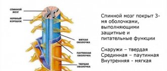
The dura mater encephali is a dense whitish connective tissue membrane lying outside the other membranes. Its outer surface is directly adjacent to the cranial bones, for which the hard shell serves as the periosteum, which is how it differs from the same shell of the spinal cord.
The inner surface facing the brain is covered with endothelium and, as a result, is smooth and shiny. Between it and the arachnoid membrane of the brain there is a narrow slit-like space, spatium subdurale, filled with a small amount of fluid. In some places the hard shell splits into two sheets. This splitting occurs in the area of the venous sinuses, as well as in the area of the fossa at the apex of the pyramid of the temporal bone (impressio trigemini), where the trigeminal ganglion lies.
The hard shell gives off several processes from its inner side, which, penetrating between the parts of the brain, separate them from each other. Falx cerebri, the falx cerebri, is located in the sagittal direction between both hemispheres of the cerebrum. Attached along the midline of the cranial vault to the edges of the sulcus sinus sagittalis superioris, its anterior narrow end grows into the crista galli, and its wide posterior end fuses with the upper surface of the cerebellar tentorium.
Tentorium cerebelli, the tentorium of the cerebellum, is a horizontally stretched plate, slightly convex upward, like a gable roof. This plate is attached along the edges of the sulcus sinus transversa of the occipital bone and along the upper edge of the pyramid of the temporal bone on both sides to the processus clinoideus posterior of the sphenoid bone. The tentorium separates the occipital lobes of the cerebrum from the underlying cerebellum. Falx cerebelli, the falx of the cerebellum, is located, like the falx cerebellum, in the midline along the crista occipitalis interna to the foramen magnum of the occipital bone, covering the latter on the sides with two legs; this low process projects into the posterior notch of the cerebellum.
Diaphragma sellae, sellae diaphragm, a plate that bounds above the receptacle for the pituitary gland at the bottom of the sella turcica. In the middle it is pierced by a hole for the passage of a funnel, infundibulum, to which the hypophysis is attached.
The blood vessels of the hard shell also feed the bones of the skull and form depressions, sulci meningei, on the inner plate of the latter. Of the arteries, the largest is a. meningea media, branch a. maxillaris, passing into the skull through the foramen spinosum of the sphenoid bone. In the anterior cranial fossa a small branch branches from a. ophthalmica, and in the back - branches from a. pharyngea ascendens, from a. vertebralis and from a. occipitalis, penetrating through the foramen mastoideum. The veins of the dura mater accompany the corresponding arteries, usually two at a time, and flow partly into the sinuses, partly into the plexus pterygoideus.
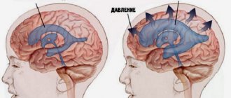
Nerves. The dura mater is innervated by the trigeminal nerve. In addition to its own veins, the dura mater contains a number of receptacles that collect blood from the brain and are called the sinuses of the dura matris, sinus durae matris. The sinuses are venous channels without valves (triangular in cross section), lying in the thickness of the hard shell itself at the places where its processes are attached to the skull and differing from the veins in the structure of their walls. The latter are formed by tightly stretched sheets of the hard shell, as a result of which they do not fall off when cut and gape when wounded. The inflexibility of the walls of the venous sinuses ensures the free outflow of venous blood when intracranial pressure changes, which is important for the uninterrupted activity of the brain, which explains the presence of such venous sinuses only in the skull.
There are the following sinuses: Sinus transversus - the largest and widest, located along the posterior edge of the tentorium cerebelli in the sulcus sinus transversi of the occipital bone, from where it descends as sinus sigmoideus into the sulcus sinus sigmoidei and then at the foramen jugulare it passes into the mouth of v. jugularis interna. Due to this, the transverse sinus with the sigmoid sinus serves as the main collector for all venous blood of the cranial cavity. All other sines flow into it, partly directly and partly indirectly. They fall directly into it:
Sinus sagittalis superior runs along the upper edge of the falx cerebri along the entire sulcus sinus sagittalis superioris from crista galli to protuberantia occipitalis interna; On the sides of the sinus sagittalis superior, in the thickness of the dura mater, there are so-called blood lakes - small cavities communicating on one side with the sinus and diploic veins, and on the other with the veins of the dura mater and brain.
Sinus occipitalis is, as it were, a continuation of the previous one along the place of attachment of the falx cerebelli to the crista occipitalis interna and further (after bifurcation) along both edges of the foramen magnum of the occipital bone. Sinus rectus on the line of attachment of falx cerebri to tentorium cerebelli. It receives in front the sinus sagittalis inferior, running along the lower free edge of the falx cerebri, as well as v. cerebri magna, through which blood flows from the deep parts of the brain. In the place where the named sinuses converge (sinus transversus, sinus sagittalis superior, sinus rectus and sinus occipitalis), a general expansion is formed, known as the drain of the sinuses, confluens sinuum. At the base of the skull, on the side of the sella turcica, there is a cavernous sinus, sinus cavernosus, which looks like either a venous plexus or a wide lacuna surrounding the internal carotid artery. It is connected to the same sinus on the other side by two transverse anastomoses, sinus intercavernosi, passing in front and behind the fossa hypophysialis, as a result of which a venous ring is formed in the area of the sella turcica.
The cavernous sinus is a complex anatomical complex, which, in addition to the sinus itself, includes the internal carotid artery, nerve trunks and surrounding connective tissue. All these formations constitute, as it were, a special device that plays an important role in regulating the intracranial flow of venous blood. In front, v. flows into the cavernous sinus. ophthalmica superior, passing through the superior orbital fissure, as well as the lower end of the sinus sphenoparietalis, running along the edge of the alae minoris. The outflow of blood from the sinus cavernosus occurs in two sinuses lying behind: sinus petrosus superior et inferior, located in the grooves of the same name, sulcus sinus petrosi superioris et inferioris.
Both sinus petrosi inferiores are connected to each other by several venous canals, which lie in the thickness of the hard shell on the basilar part of the occipital bone and are collectively called plexus basilaris. Plexus basilaris communicates with the venous plexuses of the spinal canal, through which blood thus flows from the cranial cavity. The main route for the outflow of blood from the sinuses is the internal jugular veins, but in addition, the venous sinuses are connected to the veins of the outer surface of the skull through the so-called emissary veins, vv. emissariae, passing through openings in the cranial bones (foramen parietale, foramen mastoideum, canalis condylaris).
The same role is played by small veins that leave the skull along with the nerves through the foramen ovale, foramen rotundum and canalis hypoglossal. The venae diploicae, the veins of the spongy substance of the bones of the skull, also flow into the sinuses of the dura mater; at the other end they may have a connection with the external veins of the head. Venae diploicae are canals that anastomose with each other, lined from the inside with a layer of endothelium and passing through the spongy substance of the flat bones of the skull.
Meninges of the brain
The membranes of the brain, meninges, are a direct continuation of the membranes of the spinal cord - hard, arachnoid and soft.
The dura mater encéphali is a dense whitish connective tissue membrane lying outside the other membranes. Its outer surface is directly adjacent to the cranial bones, for which the hard shell serves as the periosteum, which is how it differs from the same shell of the spinal cord. The inner surface facing the brain is covered with endothelium and, as a result, is smooth and shiny. Between it and the arachnoid membrane of the brain there is a narrow slit-like space, spátium subdurále, filled with a small amount of fluid. In some places the hard shell splits into two sheets. This splitting occurs in the area of the venous sinuses (see below), as well as in the area of the fossa at the apex of the pyramid of the temporal bone (impressio trigemini), where the trigeminal ganglion lies. The hard shell gives off several processes from its inner side, which, penetrating between the parts of the brain, separate them from each other (Fig. 303).
Rice. 303 . The dura mater of the brain and its venous sinuses.
1 , 18 - sinus petrosus superior (dexter et sinister); 2 - sinus petrosus inferior; 5 - falx cerebri; 4 - sinus sagittalis superior; 5 - sinus sagittalis inferior; 6 - infundibulum; 7 - a. carotis interna; 8 - n. opticus; 9 - crista galli; 10 , 14 - sinus intercavernosus: 11 - sinus sphenoparietalis; 12 - cerebri media; 13 - diaphragma sellae; 15 - dorsum sellae; 16 - sinus cavernosus; 17 - plexus basillaris; 19 - bulbus superior v. jugular is internae; 20 - sinus sigmoideus; 21 - tentorium cerebelli; 22 - vv. cerebri inferiores; 23 - sinus transversus; 24 - confluence sinuum; 25 - sinus rectus; 26 - v. cerebri magna; 27 - vv. cerebri superiores.
Falx cerebri, falx cerebri
, located in the sagittal direction between both hemispheres of the cerebrum. Attached along the midline of the cranial vault to the edges of the sinus sagittalis superióris, its anterior narrow end grows into the crista gálli, and its wide posterior end fuses with the upper surface of the cerebellar tentorium.
Tentorium cerebelli, tentorium cerebellum
, represents a horizontally stretched plate, slightly convex upward, like a gable roof. This plate is attached along the edges of the sinus transvérsi of the occipital bone and along the upper edge of the pyramid of the temporal bone on both sides to the procéssus clinoideus posterior of the sphenoid bone. The tentorium separates the occipital lobes of the cerebrum from the underlying cerebellum.
Falx cerebelli, falx cerebellum
, is located, like the falx cerebri, in the midline along the crista occipitalis intérna to the foramen magnum of the occipital bone, covering the latter on the sides with two legs; this low process projects into the posterior notch of the cerebellum.
Diaphrágma sellae, saddle diaphragm
, a plate that bounds above the receptacle for the pituitary gland at the bottom of the sella turcica. In the middle it is pierced by a hole for the passage of a funnel, infundibulum, to which the hypnophysis is attached.
The blood vessels of the hard shell also feed the bones of the skull and form depressions, sulci meningei, on the inner plate of the latter. Of the arteries, the largest is a. meningea media, branch a. maxillaris, passing into the skull through the foramen spinosum of the sphenoid bone. In the anterior cranial fossa a small branch branches from a. ophthalmica, and in the back - branches from a. pharýngea ascendens, from a. vertebralis and from a. occipitalis, penetrating through the foramen mastoideum. The veins of the dura mater accompany the corresponding arteries, usually two at a time, and flow partly into the sinuses, partly into the plexus pterygoideus.
Nerves . The dura mater is innervated by the trigeminal nerve.
In addition to its own veins, the dura mater contains a number of receptacles that collect blood from the brain and are called the sinuses of the dura mater.
(see Fig. 303).
The sinuses are venous channels without valves (triangular in cross section), lying in the thickness of the hard shell itself at the places where its processes are attached to the skull and differing from the veins in the structure of their walls. The latter are formed by tightly stretched sheets of the hard shell, as a result of which they do not fall off when cut and gape when wounded. The inflexibility of the walls of the venous sinuses ensures the free outflow of venous blood when intracranial pressure changes, which is important for the uninterrupted activity of the brain, which explains the presence of such venous sinuses only in the skull.